
Adaptation of different types of exercises ( aerobic, strengthening and cardiovascular)
physiological adaptations aerobic strengthening cardiovascular exercise types
exercise training adaptation aerobic resistance cardiovascular
exercise physiology adaptation aerobic cardiovascular training diagram
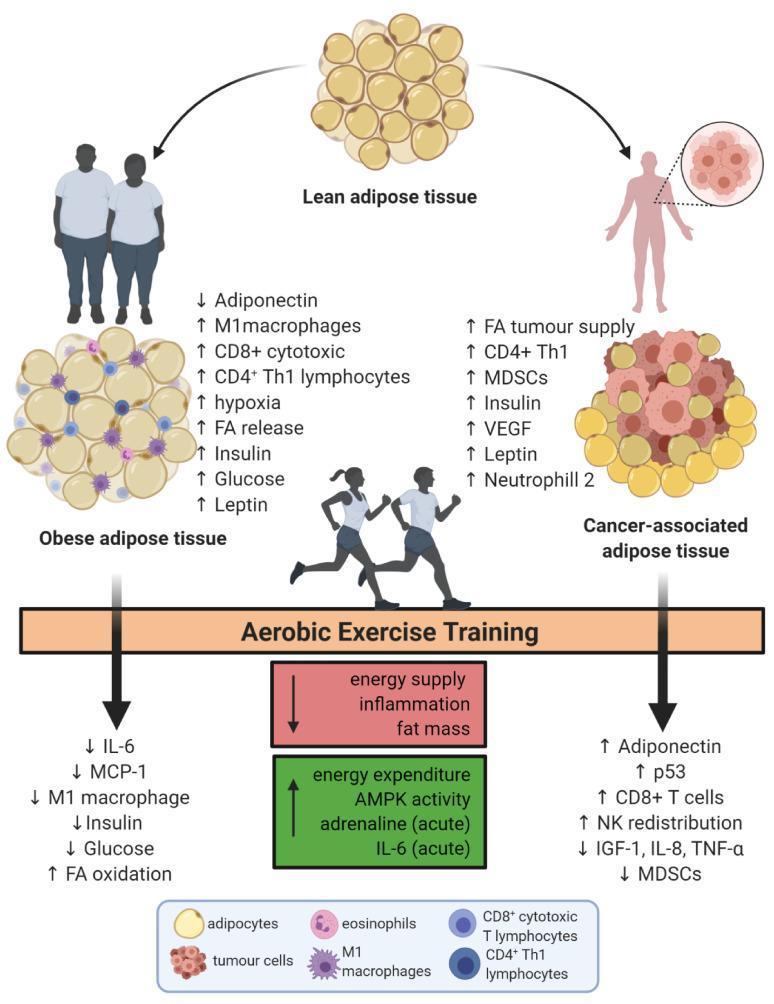
This pathophysiology diagram illustrates the metabolic and immunological effects of aerobic exercise training on adipose tissue in the context of obesity and cancer. The top section displays 'Lean adipose tissue' transitioning into two diseased states: 'Obese adipose tissue' and 'Cancer-associated adipose tissue.' Obese adipose tissue is characterized by hyperplastic adipocytes, increased M1 macrophages, hypoxia, and elevated levels of insulin, glucose, and leptin, with decreased adiponectin. Cancer-associated adipose tissue shows infiltration of tumor cells, increased MDSCs, VEGF, and fatty acid tumor supply. The central horizontal bar labeled 'Aerobic Exercise Training' acts as a metabolic modifier. Downward pathways from exercise indicate systemic improvements: reduced inflammation (IL-6, MCP-1, TNF-α), decreased fat mass and insulin/glucose levels, and increased energy expenditure and AMPK activity. Immunologically, exercise promotes anti-tumor responses through NK cell redistribution and increased CD8+ T cells. The diagram synthesizes complex interactions between lifestyle intervention, endocrine signaling, and the immune microenvironment in oncology and metabolic health.
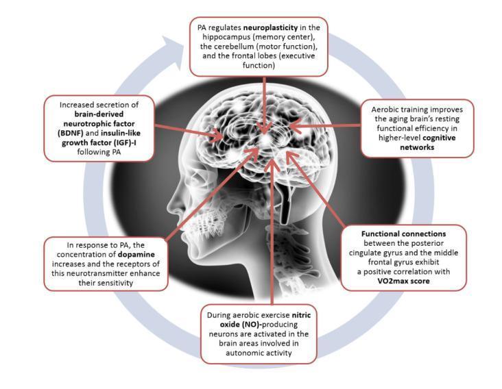
This medical infographic illustrates the pathophysiology and neuroplastic effects of aerobic exercise on the human central nervous system. Centered on a lateral profile of the human head with the brain visible, the diagram uses call-out boxes to detail biochemical and structural changes. Key educational concepts include the regulation of neuroplasticity in the hippocampus (memory), cerebellum (motor function), and frontal lobes (executive function). The diagram highlights molecular markers, specifically the increased secretion of Brain-Derived Neurotrophic Factor (BDNF) and Insulin-like Growth Factor (IGF)-1 following physical activity. It also depicts neurotransmitter modulation, noting increased dopamine concentration and receptor sensitivity. Functional adaptations shown include the activation of nitric oxide (NO)-producing neurons in autonomic regions and enhanced connectivity between the posterior cingulate gyrus and middle frontal gyrus correlated with VO2max. The content summarizes how aerobic training improves resting functional efficiency in cognitive networks, particularly relevant to geriatric medicine and neuro-rehabilitation.
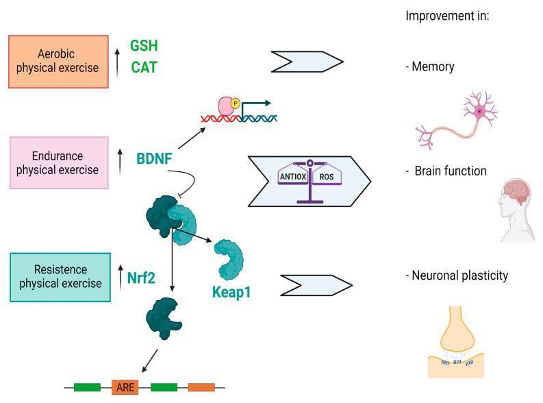
This pathophysiology diagram illustrates the molecular signaling pathways through which physical exercise influences brain function and neuroplasticity. The flowchart categorizes three exercise modalities: aerobic, endurance, and resistance training. Aerobic exercise is linked to the upregulation of endogenous antioxidants glutathione peroxidase (GSH) and catalase (CAT). Endurance exercise is shown to increase Brain-Derived Neurotrophic Factor (BDNF) levels. Resistance exercise triggers the activation of the Nrf2 (nuclear factor erythroid 2-related factor 2) pathway. The central mechanism depicts the dissociation of Nrf2 from its inhibitor Keap1, followed by Nrf2 translocation to the nucleus where it binds to Antioxidant Response Elements (ARE) on DNA. This molecular cascade mediates a balance between antioxidant defenses (ANTIOX) and reactive oxygen species (ROS). The educational focus highlights the clinical outcomes of these pathways, specifically improvements in memory, general brain function, and neuronal plasticity, represented by icons of a neuron, a human profile with a highlighted brain, and a synaptic cleft.
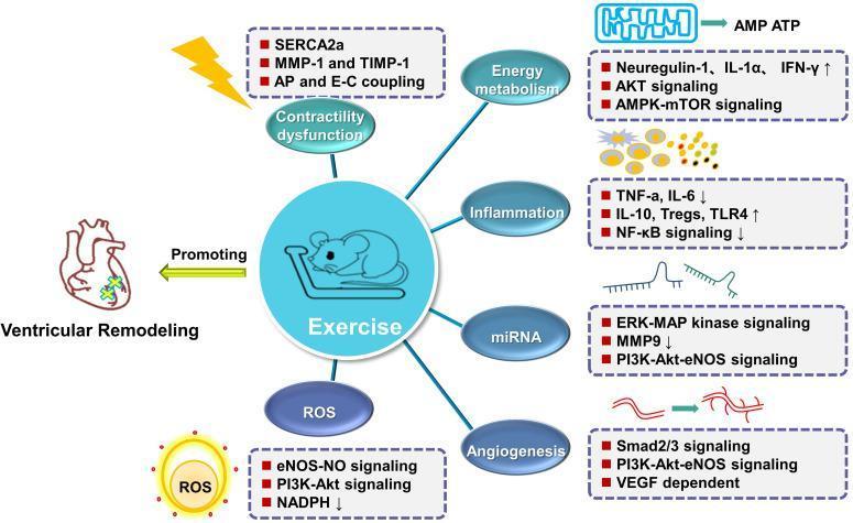
This pathophysiology diagram summarizes the molecular mechanisms by which exercise training mitigates ventricular remodeling and restores cardiac function post-myocardial infarction. The central figure depicts a laboratory mouse on a treadmill, symbolizing exercise intervention. Radiating from this center are six key physiological domains: Contractility dysfunction, Energy metabolism, Inflammation, miRNA expression, Angiogenesis, and ROS (oxidative stress). For each domain, specific signaling pathways and biomarkers are detailed. Key pathways include AKT, AMPK-mTOR, and PI3K-Akt-eNOS signaling. Notable inflammatory changes shown include decreased TNF-̑a, IL-6, and NF-̑̑kB signaling, alongside increased IL-10 and Tregs. The angiogenesis section highlights Smad2/3 and VEGF-dependent pathways. The diagram illustrates how these multi-faceted interventions collectively inhibit reactive oxygen species (ROS) and cellular dysfunction to counteract adverse ventricular remodeling, represented by a heart illustration. This graphic serves as a summary of cardioprotective exercise physiology at the cellular and molecular levels.
Adaptation to Different Types of Exercise
1. Aerobic (Endurance) Exercise Adaptation
Cardiovascular Adaptations
- Increased maximal cardiac output - this is the single most important adaptation, rising up to ~40% with training. It results from:
- Expansion of plasma volume and blood volume - this increases cardiac preload
- Cardiac hypertrophy (eccentric/volume-load hypertrophy) - ventricular walls thicken and chamber dimensions increase, increasing filling capacity
- Increased stroke volume - via Starling's law; a larger preload drives a greater stroke volume
- Resting and submaximal heart rate decreases (bradycardia of training), allowing a given cardiac output with fewer beats
- Improved O2 delivery - enhanced by capillary proliferation in skeletal muscle, increasing O2 extraction per unit of blood
- Sports anemia - a slight drop in [hemoglobin] reflecting plasma expansion (not true anemia); this actually reduces viscosity and favors flow - Medical Physiology, p. 1782
Skeletal Muscle Adaptations
- Mitochondrial biogenesis - driven by PGC-1α transcription, aerobic training progressively increases mitochondrial number and enzyme content (citric acid cycle, oxidative phosphorylation, β-oxidation). This is the basis of "enzyme adaptation" seen over weeks of training. - Medical Physiology, p. 1783
- Capillary density increases - more capillaries per muscle fiber improves O2 and CO2 diffusion
- Oxidative capacity increases - shifts the muscle toward preferential use of fat oxidation at a given VO2, sparing glycogen and reducing lactate/H+ production (glycogen sparing = basis of endurance in marathon runners)
- Fiber type adaptation - primarily affects type I (slow-twitch) and type IIa fibers; these undergo hypertrophy of slow-twitch fibers with increased oxidative enzymes - Miller's Review of Orthopaedics, p. 71
- Fatigue resistance - the cumulative effect of all the above is dramatically reduced susceptibility to fatigue
Key Outcome: VO₂max
Detraining: A significant decline in aerobic fitness begins after only 2 weeks of inactivity, underlining that adaptation is a dynamic and reversible process. - Miller's Review of Orthopaedics, p. 71
2. Strength (Resistance) Training Adaptation
Neuromuscular Adaptations (Early - weeks 1-6)
- Neural recruitment - early strength gains are predominantly neurological; the nervous system learns to activate more motor units simultaneously, reduce co-contraction of antagonists, and synchronize motor unit firing
- Improved motor patterns - with repeated practice, movements become more efficient and coordinated, reducing superfluous muscle activity and energy expenditure - Medical Physiology, p. 1765
Structural / Hypertrophic Adaptations (Weeks 6+)
- Myofibrillar hypertrophy - the primary long-term adaptation; synthesis of actin and myosin contractile proteins increases, enlarging muscle fiber cross-sectional area
- Type II fiber hypertrophy - strength training selectively hypertrophies fast-twitch (type IIx) fibers, which produce more force and contract at greater velocity against a load - Medical Physiology, p. 1168; Miller's Review of Orthopaedics, p. 71
- Increased myofibrillar protein synthesis - driven by mechanical tension activating mTOR and satellite cell pathways
- Connective tissue strengthening - tendons and ligaments thicken and increase in tensile strength
- Bone density - mechanical loading stimulates osteoblast activity, increasing bone mineral density
Cardiovascular Effects (Moderate)
- Resistance training produces concentric cardiac hypertrophy (increased wall thickness with relatively normal chamber size), distinct from the eccentric hypertrophy of endurance athletes
- It improves resting blood pressure and insulin sensitivity, but to a lesser degree than aerobic training for most cardiovascular endpoints
3. Cardiovascular Exercise Adaptation
Heart Adaptations
| Adaptation | Mechanism | Functional Result |
|---|---|---|
| Increased stroke volume | Eccentric hypertrophy + ↑ preload | Higher cardiac output at maximal exercise |
| Resting bradycardia | Increased vagal tone | Lower resting heart rate (athletes: 40-60 bpm) |
| Increased heart size | Volume overload remodeling | More efficient pump |
| Improved contractility | Calcium handling and myosin isoforms | Faster, stronger contractions |
Vascular Adaptations
- Arterial remodeling - conduit and resistance arteries enlarge in diameter (structural dilation), minimizing vascular resistance as cardiac output is redistributed to muscles. Arterial wall thickness decreases, improving compliance. - PMID 26756625
- Endothelial function - nitric oxide (NO) production increases, improving vasodilation responses and reducing arterial stiffness
- Capillary proliferation (angiogenesis) - new capillary networks form in trained muscles, matching perfusion to metabolic demand
- Reduced peripheral resistance - trained arterioles dilate more effectively, lowering total vascular resistance at rest
Metabolic Adaptations
- Increased insulin sensitivity - skeletal muscle GLUT4 transporters increase, improving glucose uptake
- Improved lipid profiles - raises HDL, lowers triglycerides and LDL
- Reduced resting blood pressure
- AMPK activation - AMP-activated protein kinase acts as a cellular energy sensor, upregulating oxidative metabolism and mitochondrial biogenesis
Comparing the Three Types: A Summary
| Feature | Aerobic Training | Strength Training | Cardiovascular Training |
|---|---|---|---|
| Primary fiber type | Type I, IIa | Type IIx | Type I, IIa |
| Cardiac adaptation | Eccentric hypertrophy | Concentric hypertrophy | Eccentric hypertrophy |
| Muscle change | Oxidative capacity ↑, capillaries ↑ | Cross-sectional area ↑, force ↑ | Oxidative capacity ↑ |
| Key metabolic shift | Fat oxidation ↑, glycogen sparing | Protein synthesis ↑ | Glucose uptake ↑, lipids ↓ |
| VO₂max | ↑↑↑ | Minimal change | ↑↑ |
| Neural adaptation | Moderate | Significant (early) | Moderate |
| Bone/connective tissue | Moderate | Significant | Moderate |
| Reversibility | Rapid (2 weeks) | Slower (weeks-months) | Rapid |
Key Principle: Specificity of Training
- Medical Physiology (Boron & Boulpaep), Ch. 60 - Aerobic and Strength Training Adaptations
- Miller's Review of Orthopaedics, 9th Ed. - Athletic Training, Injury, and Adaptation (p. 71)
- Firestein & Kelley's Textbook of Rheumatology - Exercise in Rheumatic Disease (p. 730)
- Cardiovascular Adaptations to Exercise Training, Hellsten & Nyberg (2016), PMID 26756625
- Combined aerobic and resistance training meta-analysis, Al-Mhanna et al. (2024), PMID 38887616