
Management of keratosis
Which type of keratosis are you asking about?
keratosis pilaris treatment management
keratosis pilaris skin lesions follicular papules
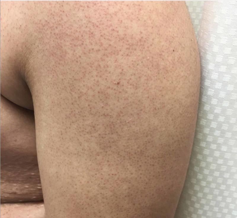
This clinical photograph displays a close-up view of the skin, likely on the extensor surface of an arm or leg, demonstrating classic features of Keratosis pilaris. The primary findings consist of numerous, densely distributed, pinpoint erythematous papules that are distinctly follicular in origin. These conical, slightly raised lesions give the skin a characteristic 'goose-flesh' or sandpaper-like rough texture. The background skin exhibits subtle reddish hyperpigmentation and erythema concentrated around the hair follicles. No secondary changes such as scaling, blistering, or pustules are present. This image serves as a high-quality educational example of a common benign dermatological condition involving keratinous plugging of the hair follicles, typically seen in the pediatric and adolescent populations on the proximal extremities and trunk.
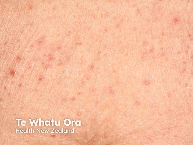
This is a clinical photograph of the dorsal back demonstrating numerous tiny follicular papules consistent with keratosis pilaris–like eruption. The lesions are typically 1–3 mm, skin-colored to pink, with a fine keratotic keratin plug at the follicular ostium. Distribution is diffuse over the thoracic–upper lumbar back, with greater concentration along hair-bearing regions and sparing of the trunk folds. No confluent plaque or significant surface desquamation is evident. The appearance resembles folliculitis but lacks pustulation or obvious infectious inflammation. The presentation is temporally associated with Bruton tyrosine kinase (BTK) inhibitor administration, a class effect previously reported as a follicular keratosis pilaris–like eruption. The image captures an early to mid-stage manifestation; individual lesions show discrete elevation and mild erythema on light-skinned skin. Imaging modality is clinical photography; technique includes close-up, high-resolution digital capture with neutral lighting to preserve true color. Anatomic context: integumentary system; posterior trunk (dorsal back); no deep structure assessment. Clinically, this finding may represent a drug eruption rather than classic keratosis pilaris and should prompt evaluation of therapy continuation, dose modification, or dermatology consultation. Differential diagnoses include keratosis pilaris, idiopathic follicular hyperkeratosis, mild folliculitis, and pityriasis rubra pilaris variants. Histology would show hyperkeratosis and follicular plugging if biopsied, confirmatory.
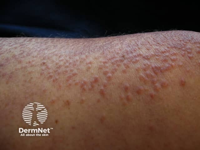
This is a high-resolution clinical photograph of the skin on the forearm, showing diffuse, numerous small keratotic follicular papules on a pink-to-skin-toned background. Imaging modality: clinical photography with macro close-up. Lesions are 1–3 mm, sessile, follicular papules with central keratotic plugs and surface roughness; mild perifollicular erythema; no pustules. The pattern and morphology are classic for keratosis pilaris (follicular hyperkeratosis), a common benign dermatologic condition often associated with atopy or dry skin. It typically affects extensor surfaces such as arms and thighs. Distinguishing surface features include a rough, sandpaper-like texture and discrete, dome-shaped papules surrounding hair follicles. Differential considerations include folliculitis, eczema with follicular involvement, pityriasis rubra pilaris, and less likely lichen planus or papular syphilids. Clinical significance: primarily cosmetic, generally benign; management emphasizes emollients and keratolytics (urea, lactic acid, salicylic acid) with cautious use of irritants. Potential clinical applications: dermatology education, differential diagnosis practice, baseline documentation, and monitoring therapeutic response. No imaging beyond surface photography; no biopsy performed. The forearm distribution supports extensor-site involvement and is helpful for educational comparisons with similar papular presentations. This image can support patient counseling, track treatment outcomes, and serve as a reference in clinical quizzes highlighting keratotic follicular lesions and benign cutaneous disorders.
PMID: 41277649
Keratosis Pilaris (KP) - Management
Overview
General Principles
- KP is not curable - it is a genetic condition and any improvement from treatment is temporary; lesions return if treatment is stopped.
- Patient education is essential: set realistic expectations about management vs. cure.
- Treatment goals are symptom relief and cosmesis - reducing roughness and erythema.
- Most patients have a variable response to therapy.
Step-by-Step Management
Step 1: Emollients (First-Line)
- Regular moisturizers to hydrate and reduce dryness.
- Best used after bathing while skin is still damp (occlusion effect).
- Reduces rough texture, but has minimal effect on erythema.
- Examples: petrolatum-based creams, ceramide-containing moisturizers.
Step 2: Keratolytic Agents (First-Line)
| Agent | Concentration | Notes |
|---|---|---|
| Ammonium lactate | 5-12% (AmLactin, Lac-Hydrin) | Most commonly used; smooths lesions but rarely improves erythema |
| Urea | 10-40% creams/lotions | Effective for softening; higher concentrations more effective |
| Lactic acid | 5-12% | AHA; preferred topical in recent systematic review (2025) |
| Glycolic acid | 10-15% creams | AHA; effective for texture improvement |
| Salicylic acid | 2-6% | BHA; exfoliates follicular plugs |
Note: Therapies that improve the keratotic component often exacerbate redness - a common therapeutic challenge.
Step 3: Topical Retinoids
- Tretinoin (retinoic acid) cream 0.025-0.1% applied at night.
- Tazarotene 0.05-0.1% gel or cream.
- Mechanism: normalizes follicular keratinization.
- May cause local irritation and worsen erythema initially.
- Some patients respond well; results are inconsistent.
Step 4: Additional Topical Options
- Topical calcipotriene (calcipotriol): vitamin D analogue shown effective in some patients (Andrews' Diseases of the Skin).
- Topical corticosteroids: limited role; used short-term if inflammation/erythema is prominent; not recommended long-term.
Managing Erythema/KP Rubra
- Pulsed dye laser (PDL): targets oxyhemoglobin; most useful for facial erythema in KP rubra. Well established as first-line laser for the erythematous component.
- 810-nm diode laser: randomized trial (JAMA Dermatology 2015) showed efficacy for KP.
- Intense Pulsed Light (IPL): helpful for erythema in some cases.
Laser & Device Therapies (Moderate-Severe / Refractory Cases)
| Laser | Evidence |
|---|---|
| Nd:YAG (1064 nm) | Consistently favourable outcomes |
| CO2 laser (ablative) | Efficacious; more adverse effects (dyspigmentation risk) |
| Erbium:YAG | Promising; fewer adverse effects than CO2 |
| Pulsed dye laser | Best for erythema component |
| Diode 810 nm | RCT evidence available |
- Microdermabrasion can provide mechanical exfoliation and temporary smoothing.
KP Atrophicans (Severe Variant)
- Topical emollients, corticosteroids, tretinoin, and keratolytics have no consistent benefit.
- Systemic isotretinoin can stop progression and induce remission in some cases.
- Dermabrasion and CO2 laser resurfacing used for scarring.
Practical Summary Table
| Severity | Treatment |
|---|---|
| Mild | Emollients + ammonium lactate 12% or urea cream |
| Moderate | Add topical retinoids (tretinoin) ± AHA (lactic/glycolic acid) |
| Erythema-dominant | Pulsed dye laser or Nd:YAG laser |
| Refractory/severe | Combination approach: keratolytics + laser; consider calcipotriene |
| Atrophicans variant | Systemic isotretinoin; CO2 laser/dermabrasion for scarring |
Key Points for Patient Education
- KP is genetic - it cannot be permanently cured.
- Improvement occurs in many during late adolescence/adulthood.
- Regular, consistent moisturization is the cornerstone.
- Treatments must be continued to maintain results.
- Avoid harsh scrubbing which can worsen erythema.
- Humid climates/summer months often bring natural improvement.
- Fitzpatrick's Dermatology, 8e - Chapter 49 (KERATOSIS PILARIS)
- Andrews' Diseases of the Skin, p. 3898-3903
- Textbook of Family Medicine, 9e, p. 924
- Beyron A. Keratosis pilaris: a systematic review... Eur J Dermatol 2025 (PMID 41277649) - 27 studies (2011-2024); proposes stepwise stratified therapy