
Provide image of pdl1 and pd1 pathway
The PD-1/PD-L1 pathway is a key immune checkpoint: PD-1 (programmed death-1) is a receptor on T cells, and when it binds its ligand PD-L1 (expressed on tumor cells or antigen-presenting cells), T cell activity is suppressed - allowing tumors to evade immune destruction. Blocking this pathway with checkpoint inhibitors restores T cell killing.
Searching sources...
Reading File
Searching the Web
PD-1 PD-L1 pathway diagram image immune checkpoint
Now let me fetch the best images and search the medical library simultaneously:
Loading Image
Loading Image
Finding Sources
Here are two clear diagrams of the PD-1/PD-L1 pathway:
PD-1 / PD-L1 Immune Checkpoint Pathway
Diagram 1 - Clinical Mechanism (NCI)
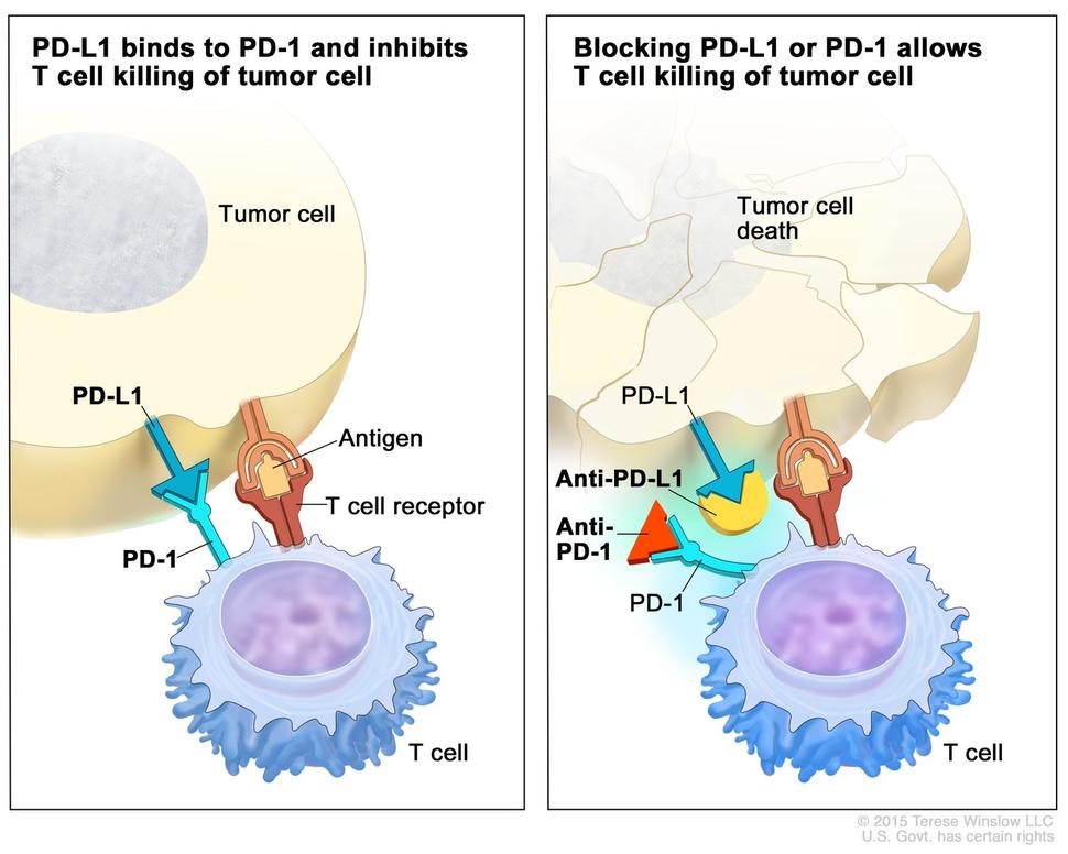
Left panel: PD-L1 on the tumor cell surface binds PD-1 on the T cell. This interaction sends an inhibitory signal that shuts down the T cell - the tumor escapes immune killing.
Right panel: Anti-PD-L1 or Anti-PD-1 antibodies block the binding. The T cell remains active and kills the tumor cell.
Diagram 2 - Intracellular Signaling Cascade
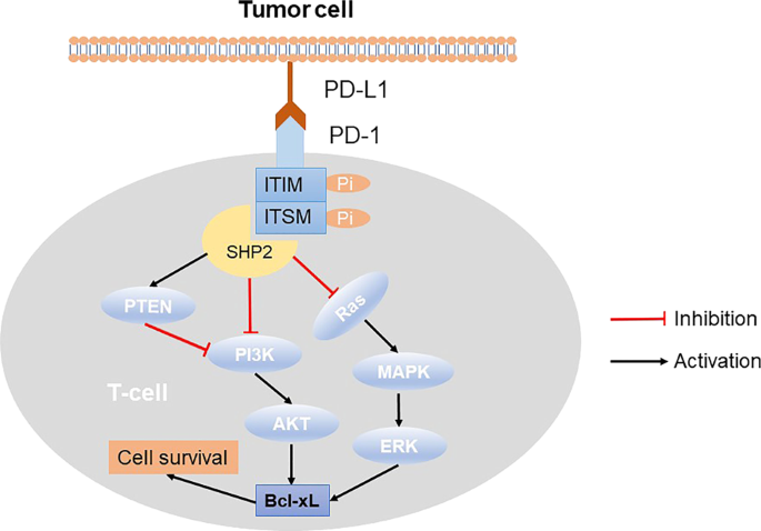
When PD-L1 (tumor cell) binds PD-1 (T cell), the cytoplasmic tail of PD-1 contains two key motifs:
- ITIM (immunoreceptor tyrosine-based inhibitory motif)
- ITSM (immunoreceptor tyrosine-based switch motif)
These get phosphorylated and recruit SHP-2 (a phosphatase), which then:
- Inhibits PI3K → AKT signaling (reduces cell survival)
- Inhibits Ras → MAPK → ERK signaling (reduces proliferation)
- Inhibits PTEN indirectly
Net result: T cell exhaustion, reduced cytokine production, and impaired cytotoxic function - the tumor survives.
Key Takeaways
| Component | Location | Role |
|---|---|---|
| PD-1 | T cell surface | Inhibitory receptor |
| PD-L1 (B7-H1) | Tumor cells, APCs | Ligand that silences T cells |
| PD-L2 (B7-DC) | Dendritic cells | Second ligand for PD-1 |
| Anti-PD-1 drugs | - | Nivolumab, Pembrolizumab |
| Anti-PD-L1 drugs | - | Atezolizumab, Durvalumab, Avelumab |
The PD-1/PD-L1 axis is upregulated by IFN-γ in the tumor microenvironment, which is the tumor's adaptive mechanism to resist immune attack. Checkpoint inhibitors targeting this pathway are now standard of care in many cancers (NSCLC, melanoma, urothelial cancer, and others) as covered in PMC review literature.
This is a shared conversation. Sign in to Orris to start your own chat.