

IMG_3602.jpg
What is this tiny hole in the foot
analyse_media
Reading File
Finding Sources
Finding Sources
Reading File
Reading File
Loading Image
The tiny holes/pits in your foot image are most consistent with Pitted Keratolysis - a superficial bacterial skin infection of the sole. Here's a full breakdown:
Pitted Keratolysis
The image shows multiple small, pinpoint dark pits scattered across the weight-bearing heel area. This pattern - tiny crater-like depressions in thick plantar skin - is the classic presentation of pitted keratolysis.
Here is what a more advanced case looks like for comparison (your image appears to be an early/mild stage):
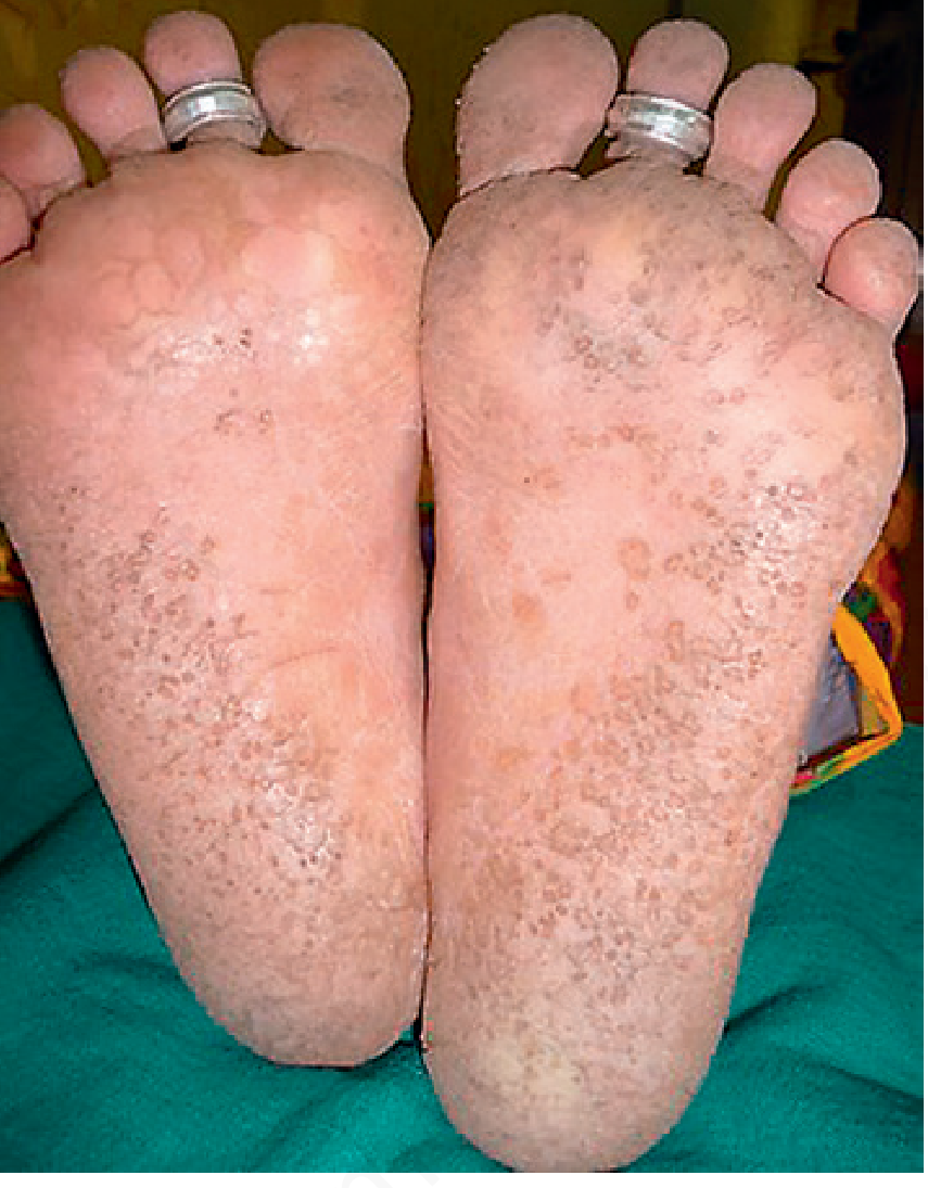
Fig. 14.26 Pitted keratolysis - Andrews' Diseases of the Skin
What causes it?
It is a non-inflammatory bacterial infection of the stratum corneum (outer skin layer). The main culprit is Kytococcus sedentarius, though Corynebacterium spp., Dermatophilus congolensis, and Actinomyces spp. can also cause it. These bacteria produce serine proteases that literally digest and pit the keratin in the thick foot skin. Sulfur-containing compounds released during this process cause a characteristic bad smell. - Dermatology 2-Volume Set 5e
Who gets it?
- Adult males with sweaty feet (~90% of cases, male-to-female ratio strongly skewed)
- People in hot/humid climates, or those who wear occlusive footwear for long hours (athletes, soldiers, workers)
- Predisposing factors: hyperhidrosis (excessive sweating), prolonged occlusion, elevated skin surface pH - Fitzpatrick's Dermatology
Clinical features
- Small (1-7 mm), crater-like pits on weight-bearing soles - heel, ball of foot, toes
- Pits may coalesce into larger craters or rings
- Usually no pain or itching
- Often noticed because of bad foot odor rather than visible lesions
- No redness or inflammation around the pits
How to distinguish it from plantar warts
| Feature | Pitted Keratolysis | Plantar Wart |
|---|---|---|
| Skin lines | Intact - run through the lesion | Disrupted - lines go around it |
| Dots | Shallow dark pits | Thrombosed capillaries (bleed when pared) |
| Cause | Bacteria | HPV virus |
| Number | Multiple, scattered | Often single or clustered |
| Wood's lamp | No fluorescence | No fluorescence |
Treatment
Good news - it responds quickly to simple topical treatments (Andrews' Diseases of the Skin):
- First-line topicals: Erythromycin, clindamycin, or mupirocin cream/gel applied twice daily
- Alternatives: 5% benzoyl peroxide gel, miconazole/clotrimazole cream
- For sweating: 20% aluminum chloride solution to control hyperhidrosis
- Hygiene: Keep feet dry, use antibacterial soap, change socks frequently, avoid occlusive footwear when possible
- Stubborn cases: Oral clarithromycin (single dose), or botulinum toxin if hyperhidrosis is severe
Most cases resolve within 1-2 weeks of topical treatment.
Note: This is a visual assessment only - a dermatologist or podiatrist can confirm the diagnosis in person. If the spots are painful when pressed, cause bleeding when pared, or disrupt the normal skin ridge pattern, a plantar wart should be considered instead.
Create a detailed treatment guide I can reference
Asking for Preferences
What format do you want the treatment guide in? · What should the guide cover?
Finding Sources
Finding Sources
Reading File
Reading File
Reading File
Reading File
Reading File
Reading File
Reading File
I now have comprehensive material from both Andrews' and Fitzpatrick's. Here is the full treatment guide:
Foot Lesion Treatment Guide
Pitted Keratolysis & Plantar Warts
How to Tell Them Apart First
Before treating, confirm which condition you are dealing with - they look similar but have very different causes and treatments.
| Feature | Pitted Keratolysis | Plantar Wart |
|---|---|---|
| Cause | Bacterial (Kytococcus sedentarius, Corynebacterium spp.) | HPV types 1, 2, 27, 57 |
| Appearance | Multiple tiny crater-like pits, may coalesce | Firm, raised papule with central pulpy core |
| Skin ridge lines | Intact - lines run through the lesion | Disrupted - lines go around the lesion |
| Black dots | Shallow dark pits (bacterial activity) | Thrombosed capillaries (bleed when pared) |
| Pain | Usually none; 1/3 have burning/irritation | Often painful with walking |
| Odor | Characteristic bad smell (~2/3 of cases) | None |
| Slimy feet | Yes - foot sticks to socks (~70% of cases) | No |
| Location | Heel, ball, toe pads (weight-bearing areas) | Ball of foot, midmetatarsal area; can be anywhere on sole |
| Wood's lamp | No fluorescence | No fluorescence |
PART 1: PITTED KERATOLYSIS
What Is It?
A superficial, non-inflammatory bacterial infection of the stratum corneum (outer skin layer) of the plantar foot. The bacteria produce serine proteases that digest keratin, creating crater-like pits. Sulfur compounds released in this process cause the characteristic malodor. - Fitzpatrick's Dermatology, Vol. 1-2
Most susceptible: Adult males with hyperhidrosis (~90% of cases, male:female ratio ~8:1). Hot, humid climates and occlusive footwear are key risk factors.
Step 1: Confirm the Diagnosis
- Diagnosis is clinical - the appearance alone is usually enough
- Gram stain of skin scrapings can detect the organisms
- Bacterial culture is not helpful (multiple species grow out)
- Biopsy is rarely needed; if done, Gram/PAS/methenamine silver stains show cocci and filamentous bacteria in pit walls
Step 2: Control Hyperhidrosis (Root Cause)
Keeping the feet dry is the most important step. Without this, the condition will recur even after antibiotic treatment.
| Agent | How to Use |
|---|---|
| Aluminum chloride 20% solution | Apply to dry skin at night; first-line antiperspirant for feet |
| Topical glycopyrrolate | Applied topically for localized hyperhidrosis |
| Botulinum toxin (Botox) | Injected into soles for severe/recalcitrant hyperhidrosis; highly effective but requires a specialist |
| Absorbent foot powder (inert antiseptic) | Daily use in socks and shoes to absorb moisture |
Step 3: Topical Antibiotics / Antimicrobials
These are curative in the majority of cases. Apply twice daily for 2-4 weeks.
| Agent | Type | Notes |
|---|---|---|
| Erythromycin 2% solution/gel | Topical antibiotic | Highly effective, first-line |
| Clindamycin 1% solution/lotion | Topical antibiotic | First-line alternative |
| Mupirocin 2% cream | Topical antibiotic | Effective; good for localized areas |
| 5% Benzoyl peroxide gel/cleanser | Antimicrobial | Effective; also helps with odor; apply as wash or leave-on |
| Fusidic acid cream | Topical antibiotic | Used as adjunctive agent |
| Miconazole or clotrimazole cream | Azole antifungal | Effective; useful if concurrent tinea pedis is suspected |
| Aluminum chloride 10-20% | Drying + antimicrobial | Can be used alongside antibiotics |
Tip: Combining a topical antibiotic with a keratolytic enhances drug penetration into the thickened stratum corneum.
Step 4: Keratolytics (for thickened/hyperkeratotic skin)
Add a keratolytic to soften the thick overlying stratum corneum and improve antibiotic penetration:
- Urea cream (10-40%) - softens and exfoliates thickened skin
- Salicylic acid 5-20% - peels away stratum corneum
- Lactic acid cream - gentle keratolytic, good for daily use
Step 5: Systemic Antibiotics (Severe/Recalcitrant Cases Only)
Reserve for widespread involvement or failure of topical treatment after 4-6 weeks:
- Clarithromycin - single dose (reported effective)
- Erythromycin oral - 2-week course
- Clindamycin oral - short course
Prevention & Hygiene Protocol
This is as important as treatment to prevent recurrence:
- Change socks daily - wash socks at 60°C (140°F) minimum to kill bacteria
- Change shoes regularly - allow shoes to fully dry between wearings; alternate pairs
- Avoid occlusive footwear (rubber-soled, non-breathable shoes) as much as possible
- Wear moisture-wicking socks (synthetic or wool, not cotton)
- Dry feet thoroughly after bathing, especially between toes
- Antibacterial soap for feet during bathing helps reduce recurrence
- Foot powder in shoes daily
Expected Timeline
- Symptoms improve within 1-2 weeks of topical treatment
- Full resolution: 3-4 weeks with consistent treatment
- Without hyperhidrosis control: high likelihood of recurrence
PART 2: PLANTAR WARTS (Verruca Plantaris)
What Is It?
A viral skin infection caused by Human Papillomavirus (HPV) types 1, 2, 27, and 57. The virus causes keratinocyte proliferation, forming a firm, hyperkeratotic papule on the sole. Plantar warts are notoriously more resistant to treatment than warts elsewhere. - Andrews' Diseases of the Skin, Clinical Dermatology
Key point: Many plantar warts, especially in children under 12, will resolve spontaneously without treatment. No treatment is a valid option to discuss.
Indications to treat: Pain, interference with walking, social embarrassment, or risk of spreading.
Therapeutic Options (Stepwise)
TIER 1 - Patient-Applied / Conservative (Start Here)
A. Salicylic Acid (10-26% OTC solutions, 40% prescription pads)
Best evidence for patient-applied therapy.
How to use:
- Soak affected area in warm water for 5-10 minutes
- Gently file dead skin with pumice stone, emery board, or table knife (clean between uses)
- Apply salicylic acid solution or gel; allow to dry
- Cover with a strip bandage or occlusive tape for 24 hours
- Repeat daily
- Continue for at least 3 months - do not stop too early
Efficacy: Some studies show parity with cryotherapy; others show it to be inferior. Results are better with consistent application and mechanical debridement.
B. Occlusive Tape (Duct Tape)
- Modest evidence; inferior to cryotherapy in adults
- Keep wart covered 6.5-7 days/week with a relatively impermeable tape
- Best reserved for: children under 12 (high spontaneous resolution rate) or patients refusing other treatments
- Adults: only ~20% resolve with 2 months of tape; 75% of "resolved" warts recur
TIER 2 - In-Office Procedures
A. Cryotherapy (Liquid Nitrogen) - First-line in-office treatment
- Cure rate: 20-50% with repeated applications
- Method: Sustained 10-second freeze with spray gun; freeze to produce 2-3 mm halo around wart
- A blister should form 1-2 days after treatment (this is the goal)
- Freeze-thaw cycle: a single cycle may be as effective as two
- Repeat every 2-3 weeks, just as the old blister peels off
- Liquid nitrogen (lower temperature) is preferred over other cryogens
- Side effects: Pain (can be significant for several days), hypopigmentation, scarring (rare), nerve damage if frozen too deeply at digit sides
- Contraindications/caution: Fanconi anemia, cryoglobulinemia, Raynaud phenomenon, poor peripheral circulation (severe blistering risk)
B. Cantharidin (0.7% cantharidin / "cantharone")
- Applied in office, covered with occlusive tape for 24 hours (or until burning)
- Blister forms in 24-72 hours (can be very painful)
- Repeat every 2-3 weeks
- Advantage: Painless to apply - good for children
- Disadvantage: Can produce "doughnut warts" (ring-shaped recurrence)
C. Intralesional Candida Antigen Injection
- Inject 0.1 mL into 1-3 lesions; repeat every 3-4 weeks
- Cure rate up to 80% with multiple treatments; highly effective
- Works by triggering local immune response that generates systemic HPV immunity
- Side effects: Fever, chills, flu-like symptoms 6-8 hours post-injection (cytokine-mediated); resolves within 24-48 hours. Advise patients in advance.
TIER 3 - Immunologic Therapy
A. Topical Sensitizers (DNCB, Squaric Acid Dibutyl Ester, Diphencyprone)
- Patients sensitized at distant site (inner upper arm) first, then applied to warts
- Two protocols: high-concentration in-office (2-5% every 2 weeks) vs. take-home lower concentration (0.2-0.5% up to daily)
- Keep covered 24 hours after application
- Wart resolution begins in 1-2 weeks but typically requires 2-3+ months
- Overall cure rates for all three sensitizers: 60-80%
- Side effects: Local itch, pain, mild eczema; usually no functional limitation
B. Oral Cimetidine (25-40 mg/kg/day)
- Anecdotally reported to help via immunomodulatory effects
- Single-agent efficacy is low (~30%, similar to placebo)
- May be useful as an adjunct to immunotherapy when response is sluggish
- Limited side effects
C. Imiquimod
- Evidence for common/plantar warts is weak - cure rates ~10%, comparable to placebo in controlled trials
- Not routinely recommended for plantar warts
TIER 4 - Surgical / Laser (Refractory Cases Only)
Use when multiple in-office treatments over 3+ months have failed.
A. Pulsed Dye Laser (PDL)
- High fluences (12.5-15 J/cm², mean 14 J/cm²) with 1.5 ms pulse duration, 7 mm spot
- Extends 2 mm beyond visible wart margin
- Local anesthesia required in most patients
- Skin turns gray-black immediately; eschar forms over 10-14 days
- Repeat every 2-4 weeks as eschar falls off
- Efficacy for refractory warts: 70-90% in immunocompetent patients
- Also works for periungual warts (penetrates nail plate)
B. CO2 Laser
- Requires local anesthesia; produces scarring and may cause nail dystrophy
- Efficacy: 56-81% for refractory warts
- Produces potentially infectious plume - appropriate precautions required
C. Surgical Ablation
- Last resort; even complete excision of wart + surrounding skin does not guarantee non-recurrence
- Reserve for warts refractory to all conservative approaches
D. Bleomycin Injection (1 U/mL)
- Injected into wart until blanching; or multipuncture technique (Shelley method)
- Reserved for recalcitrant common warts in adults
- High efficacy but significant local side effects
Special Considerations
Children under 12: Spontaneous resolution is common. Favor watchful waiting, duct tape, or salicylic acid first. Aggressive cryotherapy should be used cautiously.
Immunocompromised patients: Warts are more extensive, more resistant to treatment, and require more aggressive/longer management. Risk of HPV-induced malignancy is higher - monitor closely.
Mosaic warts (multiple confluent warts): More resistant to all treatments. Require combination approaches and longer treatment duration.
When to Refer
- No response after 3 months of consistent treatment
- Wart disrupting normal function or gait
- Immunocompromised patient with extensive warts
- Any lesion with atypical appearance (ulceration, marked bleeding, unusual growth) - biopsy to rule out malignancy, particularly epithelioid sarcoma
Sources: Andrews' Diseases of the Skin: Clinical Dermatology | Fitzpatrick's Dermatology, Vol. 1-2 | Dermatology 2-Volume Set 5e (Elsevier)
So this look like pitted keratolysis
Yes, based on the image, pitted keratolysis is the most likely diagnosis. Here's why it fits:
What matches in your photo:
- Multiple tiny, pinpoint dark pits scattered across the weight-bearing heel area
- Located on the pressure-bearing zone of the sole (classic location)
- The surrounding skin ridges appear intact - they run through and around the spots, not disrupted by them
- No raised edges or firm papule (which would suggest a wart)
- Mild skin thickening (hyperkeratosis) typical of the heel
What would confirm it further - ask yourself:
- Do your feet sweat a lot or feel slimy/stick to socks? (70% of cases)
- Is there a bad smell from the feet, especially after taking shoes off? (2/3 of cases)
- Do you wear closed, occlusive footwear for long hours?
- Any burning or irritation on the sole? (about 1/3 of patients)
If you're answering yes to 2 or more of those, pitted keratolysis is very likely.
Your next step is straightforward:
- Start 5% benzoyl peroxide gel (available OTC) - apply to the heel once or twice daily after washing
- If you can get a prescription, clindamycin 1% or erythromycin 2% solution applied twice daily clears it faster
- Use aluminum chloride 20% at night if your feet sweat heavily
- Keep feet dry - change socks daily, let shoes air out, wash socks at 60°C
Most people see clear improvement within 1-2 weeks. A dermatologist or podiatrist can confirm in person if you want certainty before starting treatment.
This is a shared conversation. Sign in to Orris to start your own chat.