
Visual pathway
I'll look up the visual pathway from our medical library for you.
Searching sources...
Reading File
Now let me search the library for visual pathway content:
Finding Sources
Finding Sources
Excellent results. Let me now read content from the most comprehensive sources in parallel:
Reading File
Reading File
Reading File
Good content gathered. Now let me read more on lesion effects, optic radiations, and fetch the key diagram images in parallel:
Reading File
Reading File
Loading Image
Let me fetch the key diagrams that are embedded in the text:
Loading Image
Loading Image
Reading File
I now have a comprehensive picture of the visual pathway from multiple authoritative textbooks. Let me also fetch the visual field representation diagram from Gray's and the lesion effects diagram:
Loading Image
Loading Image
Loading Image
I now have all the content and key diagrams needed for a comprehensive answer. Here is the full explanation of the visual pathway:
The Visual Pathway
The visual pathway carries information from photoreceptors in the retina to the primary visual cortex and higher visual areas. It runs a long course from the globe, through the optic nerve, optic chiasm, optic tract, lateral geniculate nucleus (LGN), optic radiation, and finally to the occipital cortex.
1. Retina - First-Order Neurons
Light striking the retina is converted by photoreceptors (rods for dim light; cones for color/detail) into electrical signals. Retinal circuits process these signals through bipolar cells and then converge on retinal ganglion cells (RGCs), whose axons form the optic nerve. The retina performs parallel processing from the very start, extracting ~20 local features (light vs. dark, red vs. green, blue vs. yellow, motion, etc.) simultaneously in separate channels.
- Midget RGCs (~70%) - carry red-green opponent color information → parvocellular stream
- Parasol RGCs (~10%) - carry achromatic contrast/motion information → magnocellular stream
- Bistratified RGCs (~8%) - carry blue-yellow information → koniocellular stream
RGC axons converge at the optic disc (papilla), which has no photoreceptors, creating the physiological blind spot.
2. Optic Nerve (CN II)
The axons leave the eye, acquire a myelin sheath from oligodendrocytes (not Schwann cells), and are covered by cranial meninges - making the optic nerve an extension of the CNS, not a true peripheral nerve.
The optic nerve has four segments:
| Segment | Length | Key relations |
|---|---|---|
| Intraocular (optic nerve head) | 1 mm | Axons become myelinated here |
| Intraorbital | 25 mm | S-shaped to allow eye movement; surrounded by ocular muscle cone |
| Intracanalicular | ~9 mm | Travels the optic canal with ophthalmic artery |
| Intracranial | 4-16 mm | Runs above ICA; below ACA and AComArt |
The two optic nerves converge anteriorly to the infundibular stalk at the optic chiasm.
3. Optic Chiasm
At the chiasm, a critical partial decussation occurs:
- Nasal (medial) retinal fibers cross to the contralateral optic tract
- Temporal (lateral) retinal fibers remain ipsilateral
Since the nasal retina sees the temporal visual field (and vice versa), this crossing ensures that all information from one hemifield reaches the contralateral hemisphere. The chiasm sits above the pituitary gland - a pituitary tumor compressing the central chiasm will knock out the crossing nasal fibers from both eyes, producing bitemporal hemianopia (classic finding).
4. Optic Tract
Beyond the chiasm, the optic tract on each side carries fibers from:
- The ipsilateral temporal retina (ipsilateral visual field nasal half)
- The contralateral nasal retina (contralateral visual field temporal half)
Each optic tract thus carries all information from the contralateral visual hemifield. The tracts course around the midbrain. A lesion here produces a contralateral homonymous hemianopia (incongruous early on).
The optic tract axons terminate mainly at the lateral geniculate nucleus (LGN) of the thalamus. A small bundle diverges to:
- The pretectal area - mediates the pupillary light reflex
- The superior colliculus - controls saccadic eye movements (via pontine gaze centers)
5. Lateral Geniculate Nucleus (LGN)
The LGN in the thalamus has 6 layers:
- Layers 1, 2 = magnocellular (large cells; from parasol RGCs; motion/contrast)
- Layers 3, 4, 5, 6 = parvocellular (small cells; from midget RGCs; color/fine detail)
- Between layers = koniocellular (dusty cells; from bistratified RGCs; blue-yellow color)
Each layer receives input from only one eye (alternating ipsi/contra), but all layers are retinotopically aligned so they map the same point in the contralateral hemifield. The LGN is not a passive relay - it is modulated by attention, arousal, and cortical feedback.
6. Optic Radiation (Geniculocalcarine Tract)
LGN neurons project to the primary visual cortex via the optic radiation, also called the geniculocalcarine tract. This is the geniculostriate pathway.
The optic radiation splits into two divisions:
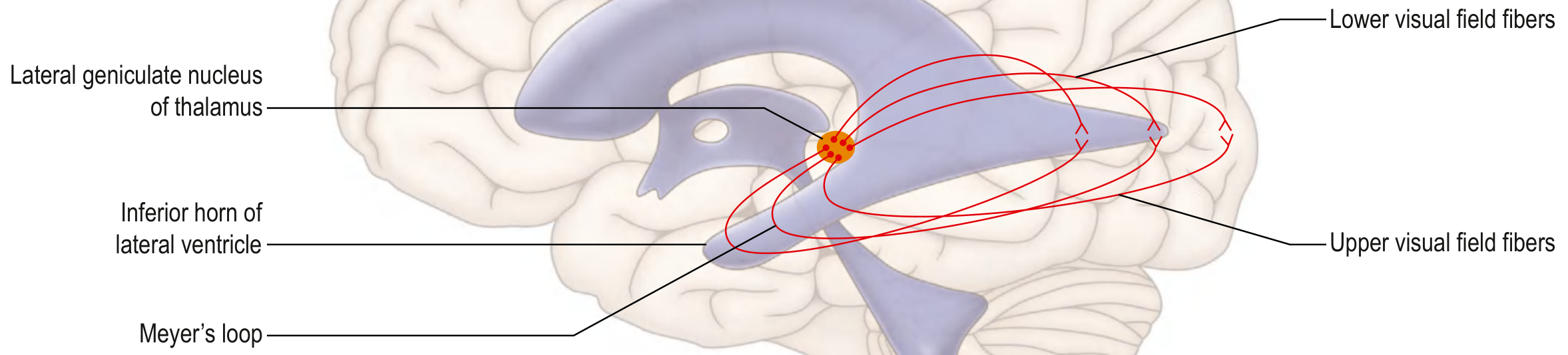
- Lower (inferior) fibers - Meyer's loop: Carry information from the superior visual field (lower retina). They sweep anteriorly around the temporal horn of the lateral ventricle before heading posteriorly to the occipital lobe. Damage here (e.g., temporal lobectomy) causes a "pie in the sky" defect - superior quadrantanopia.
- Upper (superior) fibers - Baum's loop: Carry information from the inferior visual field (upper retina). They take a more direct posterior course through the parietal lobe. Damage here causes an inferior quadrantanopia ("pie on the floor").
7. Primary Visual Cortex (V1 / Striate Cortex / Area 17)
The optic radiations terminate in the primary visual cortex (V1), located on the banks of the calcarine fissure in the medial occipital lobe (Brodmann area 17). It is also called the striate cortex because of the dense myelinated stripe (line of Gennari) visible in layer IV.
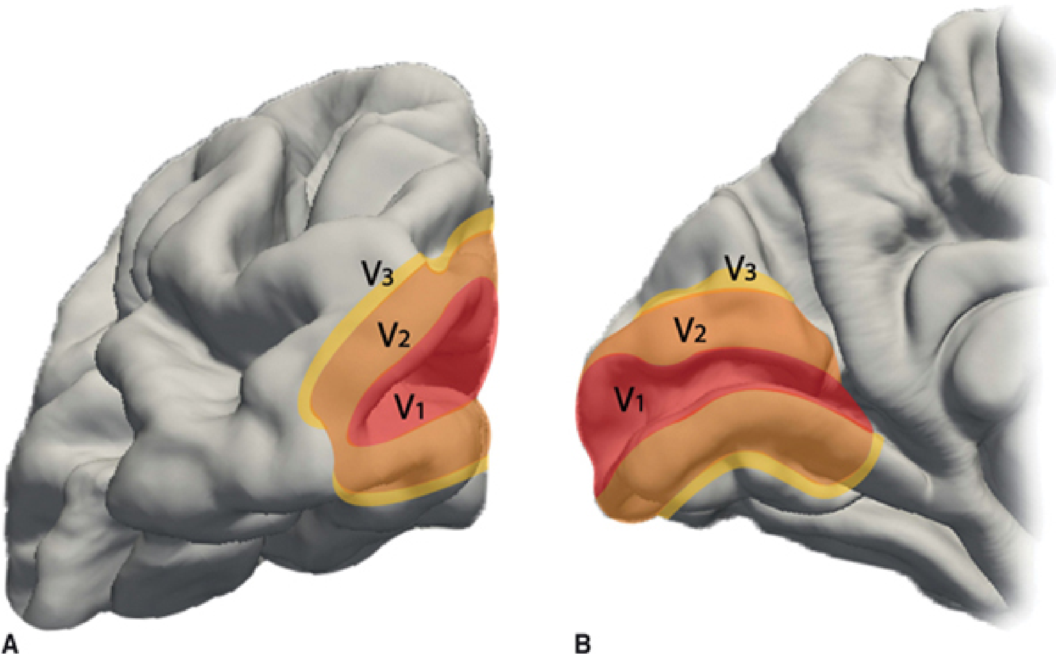
Key cortical organization:
- Superior retina (inferior visual field) → superior lip of calcarine fissure
- Inferior retina (superior visual field) → inferior lip of calcarine fissure
- Macular (central) vision → represented at the posterior pole (occipital tip), with bilateral cortical representation and spared even in large PCA strokes
- Peripheral vision → represented more anteriorly along the calcarine fissure
- Central 10-15° of vision occupies ~50-60% of the total V1 surface area (cortical magnification)
V1 then sends information to surrounding association cortex (Brodmann areas 18 and 19 = V2, V3...) via two major higher-order streams:
- Ventral "What" pathway (V1 → temporal lobe): object recognition, color, form, face recognition
- Dorsal "Where/How" pathway (V1 → parietal lobe): spatial location, motion, visuomotor guidance
Visual Field Defects and Lesion Localization
This diagram summarizes the classic visual field defects produced by lesions at different points:
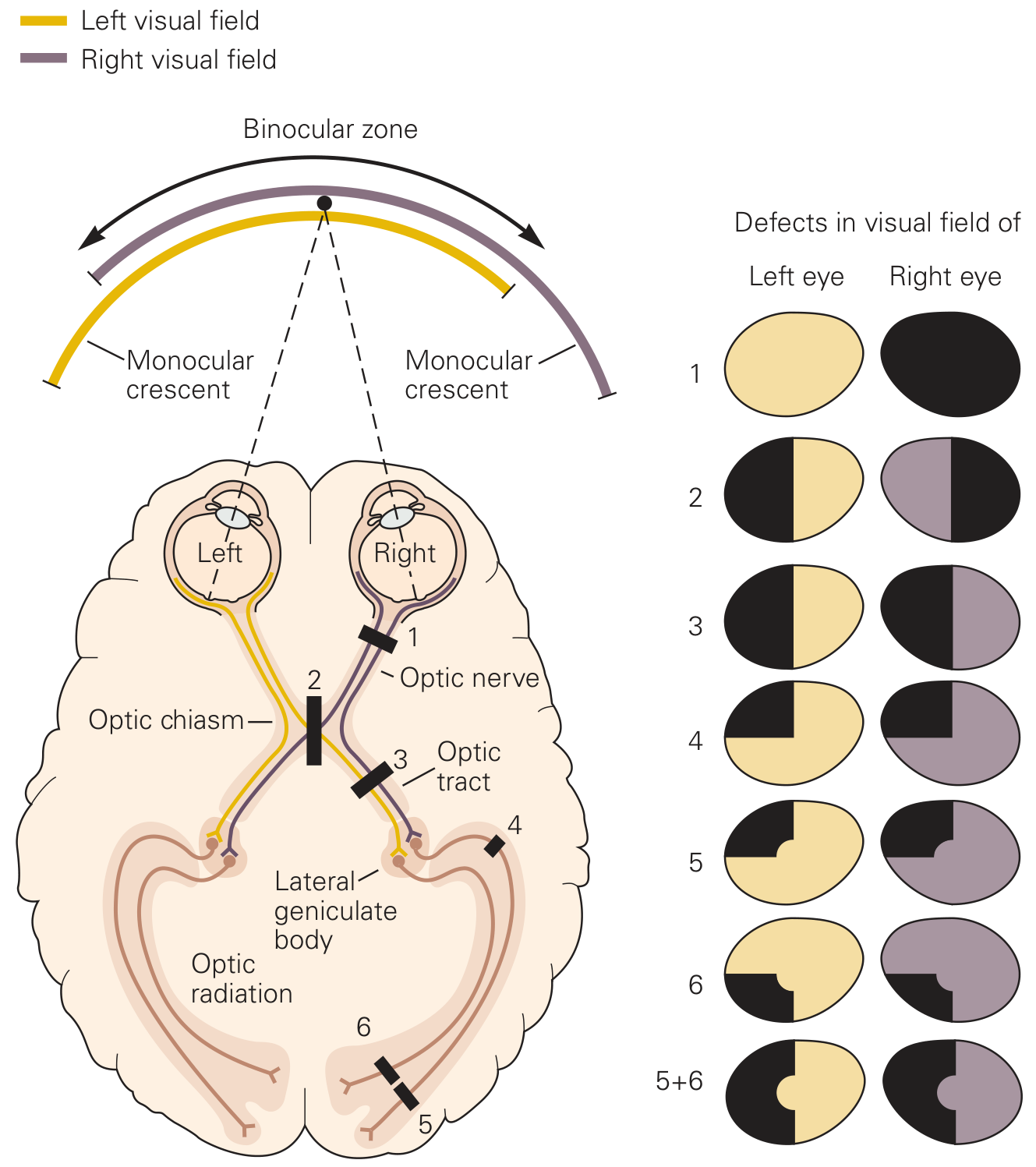
| Location | Lesion | Visual Field Defect |
|---|---|---|
| 1 - Optic nerve | Multiple sclerosis, trauma | Monocular blindness (ipsilateral eye only) |
| 2 - Optic chiasm (center) | Pituitary adenoma | Bitemporal hemianopia |
| 3 - Optic tract | Craniopharyngioma, trauma | Contralateral homonymous hemianopia (incongruous) |
| 4 - Lateral geniculate body | AntChorArt infarct | Contralateral homonymous hemianopia |
| 5 - Inferior optic radiation (Meyer's loop) | Temporal lobe lesion | Contralateral superior quadrantanopia ("pie in the sky") |
| 6 - Superior optic radiation | Parietal lobe lesion | Contralateral inferior quadrantanopia ("pie on the floor") |
| 5+6 - Optic radiation/occipital cortex | PCA stroke | Contralateral homonymous hemianopia with macular sparing |
Vascular Supply Summary
| Structure | Artery |
|---|---|
| Retina / orbital optic nerve | Ophthalmic artery (branch of ICA) |
| Anterior optic nerve | Posterior ciliary arteries |
| Intracranial optic nerve | ICA, ACA, AComArt |
| Optic chiasm | ICA, ACA, AComArt, PComArt, PCA, basilar artery |
| Optic tract | Anterior choroidal artery (AntChorArt) |
| Lateral geniculate body | AntChorArt (lateral), posterior choroidal artery (medial) |
| Optic radiation (rostral) | Middle cerebral artery (MCA) |
| Optic radiation (caudal) | Posterior cerebral artery (PCA) |
| Visual cortex | PCA (majority) |
(From Localization in Clinical Neurology, 8e)
Summary Flow
Retina (photoreceptors → bipolar cells → ganglion cells)
    ↓  [optic nerve - CN II]
Optic Chiasm  [nasal fibers cross; temporal fibers stay ipsilateral]
    ↓  [optic tract]
Lateral Geniculate Nucleus (thalamus)  [6 layers: M, P, K streams]
    ↓  [optic radiation / geniculocalcarine tract]
    ├── Meyer's loop (inferior) → lower lip of calcarine → superior visual field
    └── Baum's loop (superior) → upper lip of calcarine → inferior visual field
Primary Visual Cortex V1 (striate cortex, area 17, calcarine fissure)
    ↓
Extrastriate cortex (V2, V3, V4, V5/MT...)
    ├── Ventral stream (temporal lobe) → "What"
    └── Dorsal stream (parietal lobe) → "Where/How"
Sources: Gray's Anatomy for Students, p. 1346-1347 | Kandel Principles of Neural Science, 6th ed., p. 547-549 | Localization in Clinical Neurology, 8e, p. 327-340
This is a shared conversation. Sign in to Orris to start your own chat.