

Diagnose this case
iron deficiency anemia pale conjunctiva peripheral blood smear
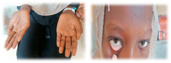
This composite clinical photograph illustrates common physical examination findings associated with severe anemia. The left panel shows the bilateral palms of a patient, demonstrating marked palmar pallor. The skin on the palms and creases appears significantly lighter and lacks the typical pinkish hue of healthy vascularized tissue, suggesting reduced hemoglobin concentration. The right panel is a close-up of the patient's face with the lower eyelid retracted to reveal the palpebral conjunctiva. Instead of the normal vibrant red or pink color, the conjunctiva exhibits conjunctival pallor, appearing whitish or very pale pink. These visual signs are critical diagnostic markers in hematology and general medicine for identifying systemic conditions such as iron-deficiency anemia or other forms of chronic blood loss. The image serves as a teaching tool for medical students and clinicians to recognize mucosal and cutaneous indicators of decreased red blood cell mass or oxygen-carrying capacity.
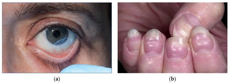
This composite of clinical photographs illustrates physical manifestations of severe anemia. Image (a) shows a close-up of a patient's eye with the lower eyelid retracted to reveal marked conjunctival pallor. The typically pink or reddish palpebral conjunctiva appears significantly pale and porcelain-white, a sign classically associated with a hemoglobin concentration below 90 g/L. Image (b) presents the fingernails of a patient demonstrating pronounced nail bed pallor. The distal nail beds lack the normal healthy pink hue. Additionally, horizontal white bands, known as Muehrcke's lines, are visible across the nail plates; these non-palpable transverse lines are associated with systemic conditions such as hypoalbuminemia and severe anemia. Together, these images demonstrate key external clinical signs used in the bedside assessment of hematological disorders and circulatory efficiency. The content is suitable for medical education regarding the physical examination findings of iron deficiency and other severe anemias.
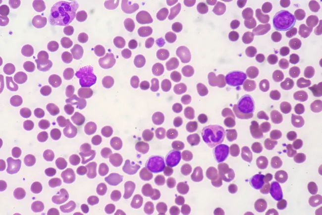
Peripheral blood smear from a patient with known chronic lymphocytic leukemia (CLL) showing autoimmune hemolytic anemia (AIHA). Brightfield microscopy of Wright-Giemsa stained smear at 1000x (oil immersion) reveals two dominant cell populations: numerous small mature lymphocytes typical of CLL and red blood cells showing spherocytic deformation. A smudge cell is evident just left of center, a classical clue for CLL. Red cells display reduced central pallor consistent with spherocytes, indicating intravascular or extravascular hemolysis driven by autoantibodies. The background shows mild polychromasia and rare nucleated RBCs, consistent with a robust reticulocyte response; the corrected reticulocyte count in this case is markedly elevated (>7%). Platelets are not significantly abnormal. These features collectively support autoimmune destruction of erythrocytes in the setting of CLL. Diagnostic significance lies in recognizing AIHA as a common autoimmune complication in CLL, with a clinical picture of sudden Hb decline and compensatory erythropoiesis. Differential considerations include drug-induced hemolysis, other causes of hemolytic anemia, or marrow infiltration by CLL. Clinical correlation includes therapy implications (steroids, rituximab) and close monitoring of hemolysis markers. This image is a valuable teaching resource for hematology morphology, AIHA in CLL, and differential diagnosis in anemia.
Diagnosis: Iron Deficiency Anemia (IDA)
Case Summary
| Feature | Finding |
|---|---|
| Age/Sex | 20-year-old female |
| Symptoms | Fatigue, exertional palpitations, breathlessness, dizziness |
| History | Irregular eating, avoids nutritious food |
| Signs | Pale conjunctiva, low BP (hypotension), fast pulse (tachycardia) |
Why Iron Deficiency Anemia?
- Young females are at particularly high risk due to menstrual blood loss combined with poor dietary iron intake
- Irregular diet + avoidance of nutritious food = inadequate iron intake (the most common cause of IDA in young women)
- Fatigue - the most common and disabling symptom; occurs even with modest reductions in iron stores
- Palpitations on exertion - the heart compensates for reduced oxygen-carrying capacity by beating faster
- Breathlessness - reduced hemoglobin means tissues are hypoxic; the respiratory rate rises to compensate
- Dizziness - reduced cerebral perfusion from anemia and low BP
- Pale conjunctiva - the hallmark bedside sign; pallor of the palpebral conjunctiva reflects reduced hemoglobin
- Low BP (hypotension) - chronic anemia reduces blood viscosity and effective circulating volume
- Fast pulse (tachycardia) - a compensatory response to maintain cardiac output despite low Hb
Pathophysiology
Investigations to Confirm
| Test | Expected Finding in IDA |
|---|---|
| CBC | Low Hb, low MCV (microcytic), low MCH (hypochromic) |
| Serum ferritin | < 15 ng/mL (most reliable test; highly specific for IDA) |
| Serum iron | Low |
| TIBC (Total Iron Binding Capacity) | Elevated (transferrin upregulated) |
| Transferrin saturation | Low (< 16%) |
| Peripheral blood smear | Microcytic, hypochromic RBCs; pencil cells |
| Reticulocyte count | Low (no reticulocytosis) |
Treatment
- Oral iron supplementation - Ferrous sulfate 325 mg (elemental iron ~65 mg) three times daily; taken on an empty stomach with vitamin C to enhance absorption
- Dietary counseling - Encourage iron-rich foods: red meat, leafy green vegetables, legumes, fortified cereals
- Identify the underlying cause - In a young female, the primary cause is dietary; however, always assess for heavy menstrual bleeding (menorrhagia)
- Monitor - Recheck CBC and reticulocyte count at 4-6 weeks; expect a reticulocytosis within 1-2 weeks of starting iron
- Treat for 3-6 months after Hb normalizes to replenish iron stores
Differential Diagnoses to Exclude
| Condition | Why Less Likely Here |
|---|---|
| Thalassemia minor | Also microcytic, but Hb A2 elevated; dietary history less relevant |
| Anemia of chronic disease | Ferritin normal or elevated; no chronic illness mentioned |
| Folate/B12 deficiency | Causes macrocytic (large RBC) anemia, not microcytic |
| Hemolytic anemia | Would show reticulocytosis and jaundice |
Physical examination performed in this case
Physical Examination — Case 5
20-year-old Female with Suspected Iron Deficiency Anemia
1. General Appearance
- Young female, appears pale and tired
- Looks thin/undernourished
- Not in acute distress at rest, but fatigued
- Anxious, slightly restless
2. Vital Signs
| Parameter | Finding | Significance |
|---|---|---|
| Blood Pressure | Low (e.g., 90/60 mmHg) | Reduced blood volume/viscosity |
| Pulse | Fast (e.g., 100-110 bpm), weak | Compensatory tachycardia |
| Respiratory Rate | Mildly elevated (18-22/min) | Compensatory for hypoxia |
| Temperature | Normal (37°C) | No infection |
| SpO₂ | Normal to mildly low | Reduced Hb but adequate ventilation |
| Weight/BMI | Underweight | Reflects poor nutrition |
3. Skin
| Finding | Detail |
|---|---|
| Pallor | Generalized pallor of skin |
| Dryness | Dry, rough skin from iron deficiency |
| Koilonychia | Spoon-shaped nails (if chronic/severe IDA) |
| Brittle nails | Nail fragility |
| Hair | Dull, thin, easily falling out (iron-deficient tissues) |
4. Eyes
| Finding | Detail |
|---|---|
| Pale palpebral conjunctiva | ✅ Key sign — hallmark of anemia (already noted in case) |
| Sclera | White (no jaundice — rules out hemolytic anemia) |
| Periorbital area | Mild puffiness possible |
5. Mouth & Tongue
| Finding | Detail |
|---|---|
| Glossitis | Smooth, red, sore tongue (atrophic papillae) |
| Angular stomatitis | Cracks/fissures at corners of mouth |
| Pallor of oral mucosa | Pale gums and buccal mucosa |
| Teeth | May reflect poor nutrition |
6. Lymph Nodes
- No lymphadenopathy (rules out malignancy, lymphoma)
7. Cardiovascular System
| Finding | Detail |
|---|---|
| Tachycardia | HR > 100 bpm |
| Hemic (flow) murmur | Soft systolic murmur at left sternal border — due to hyperdynamic circulation from low-viscosity blood |
| BP | Low — already noted |
| Peripheral pulses | Weak, rapid |
| Capillary refill | May be mildly delayed |
8. Respiratory System
- Tachypnea on exertion
- Lungs clear on auscultation
- No crackles or wheeze (no cardiac failure)
9. Abdomen
| Finding | Detail |
|---|---|
| Abdomen | Soft, non-tender |
| Spleen | May be mildly enlarged (splenomegaly) in severe/chronic IDA |
| Liver | Not enlarged |
| No masses | Rules out GI malignancy as cause |
10. Neurological
- Alert and oriented
- Mild dizziness (already noted)
- No focal deficits
- Restless legs may be reported (common in IDA)
11. Gynecological History (Relevant to Examination)
- Ask about menstrual history - heavy or prolonged periods (menorrhagia) as a contributing cause
- Menarche, cycle regularity, flow duration, clots
Summary of Key Positive Findings
| System | Positive Finding |
|---|---|
| General | Pallor, fatigue, underweight |
| Vitals | Tachycardia, low BP |
| Eyes | Pale conjunctiva |
| Mouth | Glossitis, angular stomatitis |
| Nails | Koilonychia, brittle nails |
| CVS | Flow murmur, weak rapid pulse |
| Abdomen | Possible mild splenomegaly |
Key Signs That Clinch the Diagnosis
| Sign | Mechanism |
|---|---|
| Pale conjunctiva | Low Hb = less oxyhemoglobin visible in vessels |
| Koilonychia | Iron needed for nail matrix cell growth |
| Glossitis + angular stomatitis | Iron deficiency in rapidly dividing epithelial cells |
| Flow murmur | Reduced blood viscosity → turbulent flow |
| Tachycardia + low BP | Compensatory cardiovascular response to anemia |
Generate a clinical case of this
CLINICAL CASE — Iron Deficiency Anemia
PATIENT PROFILE
| Name | Sara M. |
| Age | 20 years |
| Sex | Female |
| Occupation | University student |
| Nationality | — |
| Date of Presentation | Outpatient clinic visit |
CHIEF COMPLAINT
HISTORY OF PRESENTING ILLNESS
PAST MEDICAL HISTORY
| Chronic illnesses | None |
| Previous surgeries | None |
| Hospitalizations | None |
| Blood transfusions | None |
| Known allergies | None |
DRUG HISTORY
- No regular medications
- No NSAIDs, anticoagulants, or antacids
- No oral contraceptive use
FAMILY HISTORY
- No known family history of hematological disorders, thalassemia, or bleeding disorders
SOCIAL HISTORY
- University student, lives in a dormitory
- Irregular meal patterns; diet low in iron-rich foods
- Non-smoker, no alcohol, no recreational drugs
- Moderate physical activity (walks to class daily)
REVIEW OF SYSTEMS
| System | Findings |
|---|---|
| General | Fatigue, weakness, poor appetite |
| Cardiovascular | Palpitations on exertion, dizziness, low BP |
| Respiratory | Exertional breathlessness, no cough |
| GI | No hematemesis, no melena, no change in bowel habits |
| GU | Heavy menstrual bleeding (menorrhagia), regular cycles |
| Skin/Hair | Hair thinning, brittle nails noticed recently |
| Neurological | Dizziness on standing, no syncope, restless legs at night |
PHYSICAL EXAMINATION
Vital Signs
| BP | 90/60 mmHg (low) |
| Pulse | 108 bpm, weak and thready |
| RR | 20 breaths/min |
| Temperature | 36.8°C |
| SpO₂ | 97% on room air |
| BMI | 17.5 kg/m² (underweight) |
General
Eyes
- Pale palpebral conjunctiva bilaterally ✅ (hallmark sign)
- Sclera: white (no icterus)
Mouth
- Smooth, red tongue (glossitis)
- Cracking at corners of mouth (angular stomatitis)
- Pale oral mucosa
Nails & Skin
- Koilonychia (spoon-shaped nails) on both hands
- Dry skin, pale complexion
- Mild hair thinning
Cardiovascular
- Tachycardia (HR 108 bpm)
- Soft systolic flow murmur at left sternal border (Grade 2/6)
- Normal heart sounds, no added sounds
- Peripheral pulses: present but weak
Respiratory
- Air entry equal bilaterally
- No crackles, wheeze, or added sounds
Abdomen
- Soft, non-tender
- Mild splenomegaly (spleen just palpable, 1 cm below costal margin)
- Liver not palpable
- No masses or ascites
Neurological
- Alert and oriented x3
- No focal deficits
- Romberg's sign: negative
INVESTIGATIONS
Bedside
| Test | Result |
|---|---|
| Urine dipstick | Normal |
Hematology
| Test | Result | Normal Range |
|---|---|---|
| Hemoglobin | 7.2 g/dL ⬇ | 12–16 g/dL (female) |
| MCV | 68 fL ⬇ | 80–100 fL |
| MCH | 20 pg ⬇ | 27–33 pg |
| MCHC | 28 g/dL ⬇ | 32–36 g/dL |
| RBC | Low-normal | |
| WBC | Normal | |
| Platelets | Mildly elevated (reactive thrombocytosis) | |
| Reticulocyte count | Low |
Iron Studies
| Test | Result | Normal Range |
|---|---|---|
| Serum Ferritin | 6 ng/mL ⬇⬇ | 12–150 ng/mL |
| Serum Iron | 40 μg/dL ⬇ | 60–170 μg/dL |
| TIBC | 420 μg/dL ⬆ | 250–370 μg/dL |
| Transferrin Saturation | 9% ⬇ | 20–50% |
Peripheral Blood Smear
- Microcytic, hypochromic RBCs
- Pencil cells (elliptocytes)
- Anisocytosis and poikilocytosis
- No target cells (rules out thalassemia)
DIAGNOSIS
Iron Deficiency Anemia (IDA) — secondary to nutritional deficiency and menorrhagia
DIFFERENTIAL DIAGNOSES CONSIDERED & EXCLUDED
| Diagnosis | Reason Excluded |
|---|---|
| Thalassemia minor | No family history; no target cells; ferritin very low |
| Anemia of chronic disease | Ferritin normal/high in ACD; no chronic illness here |
| B12/Folate deficiency | Macrocytic anemia; this patient has microcytic anemia |
| Hemolytic anemia | No jaundice, no reticulocytosis, negative hemolysis markers |
MANAGEMENT
Immediate
- Oral ferrous sulfate 325 mg three times daily (take on empty stomach with vitamin C to enhance absorption)
- Avoid tea/coffee/antacids within 2 hours of iron tablet (they inhibit absorption)
Dietary Counseling
- Increase intake of: red meat, chicken, fish, leafy greens (spinach), legumes, fortified cereals
- Pair iron-rich foods with vitamin C sources (citrus, tomatoes)
- Reduce inhibitors: tea, coffee, calcium-rich foods taken with iron-rich meals
Address Underlying Cause
- Gynecology referral for menorrhagia assessment
- Consider combined oral contraceptive pill or tranexamic acid for heavy periods if confirmed
Follow-Up
| Timepoint | Action |
|---|---|
| 2 weeks | Check reticulocyte count (should rise — confirms treatment response) |
| 4–6 weeks | Repeat CBC — expect Hb to rise by ≥ 1 g/dL/week |
| 3–6 months | Continue iron after Hb normalizes to replenish stores |
PROGNOSIS
LEARNING POINTS
- Iron deficiency anemia is the most common nutritional deficiency worldwide, especially in young females
- The triad of pale conjunctiva + tachycardia + low BP strongly suggests significant anemia
- Serum ferritin < 15 ng/mL is the most specific single test for IDA
- Always look for and treat the underlying cause (diet + menorrhagia here), not just the anemia
- Classic signs - koilonychia, glossitis, angular stomatitis - appear in chronic/severe cases