
Histology of cvs topic you know am bsc anesthesia in want that topic from anatomy subject
cardiac muscle histology microscopy cross section
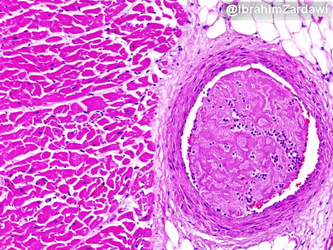
Imaging modality: Light microscopy of a hematoxylin and eosin (H&E) stained cardiac tissue section. Specimen type: paraffin-embedded myocardium with an intraluminal thrombus within a branch of a coronary artery. Imaging perspective: transverse cross-section illustrating occlusion of a coronary artery lumen by a recent thrombus and adjacent myocardial fibers. Major finding: occlusive thrombus densely eosinophilic within the arterial lumen, partially projecting into the vessel lumen and compressing flow. Vessel wall shows no striking dissection; subtle intimal thickening may reflect underlying atherosclerosis. Surrounding myocardium demonstrates preserved architectural integrity at this field of view, with no conspicuous extensive coagulative necrosis or inflammatory infiltrate evident at this early stage. The thrombus composition is suggested by a dense eosinophilic core with fibrous and platelet-rich components, typical of acute thrombosis; perivascular tissue shows mild edema. Clinical interpretation: acute coronary syndrome due to acute thrombotic occlusion of a coronary branch, likely on a preexisting atherosclerotic plaque. Diagnostic significance: provides histopathologic confirmation of thrombosis as the mechanism of ischemia; differential considerations include embolic occlusion, vasospasm with secondary thrombosis, or plaque rupture; correlate with clinical presentation and angiography. Potential educational uses: pathophysiology of myocardial ischemia, ACS, coronary thrombosis, histology of coronary arteries, and teaching images of acute myocardial infarction pathology.
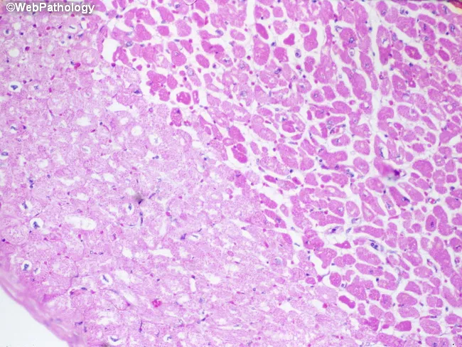
Imaging modality: Light microscopy of hematoxylin-eosin (H&E) stained cardiac tissue. Specimen is paraffin-embedded myocardial section, 5 micron thick, imaged with bright-field optics at high magnification. Anatomic region: left ventricular myocardium with an infarct in the left half of the field. The histology demonstrates pallor of necrotic cardiomyocytes (myocytolysis) produced by sarcolemmal disruption with influx of salt and water and cytoplasmic swelling. In this early post-ischemic stage, fibers lose cross-striations and display increased eosinophilia; nuclei are often absent or pyknotic. The infarct border may show waviness of fibers and mild edema; contraction bands may be present at the margin. Neutrophilic infiltration is typically minimal during the first 24 hours but may begin to appear around this window. The combination of coagulative necrosis, myocytolysis, and pallor indicates an acute myocardial infarction approximately one day old. This histologic pattern correlates with ischemia from coronary occlusion, clinical presentation of acute coronary syndrome, and aligns with diagnostic timelines (0-24 h). Clinically the image supports timing of infarct and helps distinguish acute infarction from myocarditis or chronic scar, serving as an educational reference for pathology, medical education, and research on myocardial injury dynamics. Further confirmed by clinical correlation and education.
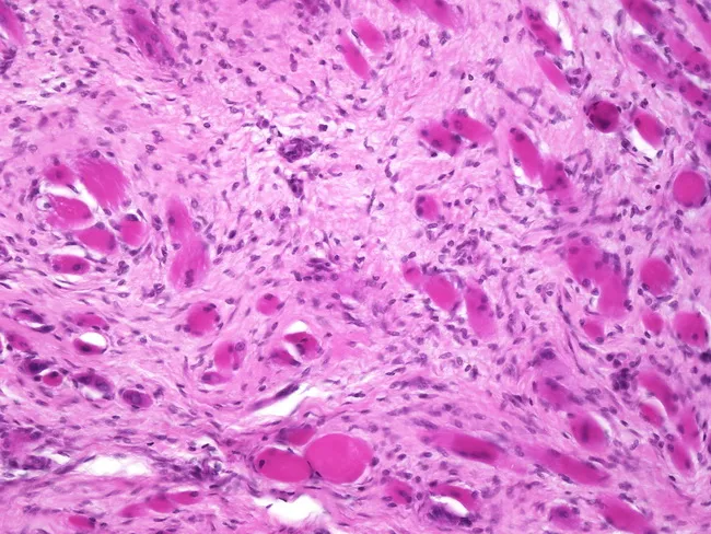
Histopathology image acquired from neonatal neck muscle (sternocleidomastoid) illustrating congenital fibromatosis of infancy (fibromatosis coli) within the lower third of the SCM. Modality: light microscopy on hematoxylin and eosin stained sections. The section shows dense, hypocellular fibrous connective tissue interposed between residual atrophic skeletal muscle fibers. The muscle fibers appear angular, fragmented, and sparsely populated by cross sectional myofibers with lost striations, while spindle-shaped fibroblasts populate the stroma. Occasional multinucleated giant cells are absent; there is minimal inflammatory infiltrate. The overall pattern is scar-like fibrosis replacing contractile parenchyma, with relative preservation of surrounding adipose tissue. The histology is characteristic of fibromatosis coli (congenital muscular torticollis), typically linked to birth trauma and obstetric deliveries (forceps, breech). Clinically, this lesion is benign and non-neoplastic, often resolving with physiotherapy or observation in mild cases; prognosis is favorable with appropriate management. Differential considerations include infantile rhabdomyosarcoma, inflammatory myopathies, and other congenital neck masses; histology helps differentiate based on absence of malignant features and presence of dense fibrous stroma with atrophic muscle. The diagnostic significance lies in correlating perinatal injury history with persistent neck tilt in newborns and guiding conservative therapy. This correlation aids targeted physiotherapy and monitoring.
Low-power brightfield light microscopy of a formalin-fixed, paraffin-embedded arterial cross-section stained with Hematoxylin and Eosin. The specimen shows a circular vessel with a well-defined, multilayered wall. The lumen is patent and lined by a narrow endothelial layer. The tunica media exhibits concentric layers of eosinophilic smooth muscle cells and occasional nuclei aligned along the wall, giving a dense, pink stippled appearance. The internal elastic lamina is not clearly resolved at this magnification. The tunica adventitia is relatively inconspicuous with sparse connective tissue. There is no overt atheromatous plaque, calcification, hemorrhage, or inflammatory infiltrate noted. Overall architecture is preserved, suggesting a normal or nonpathologic arterial segment. This image is suitable for illustrating normal vascular histology and can serve as a reference for comparison with arteriosclerotic lesions in teaching, research, and diagnostic practice. Potential clinical utility includes training in recognition of arterial layering, identifying pathologies such as atherosclerosis, vasculitis, and aneurysm formation, and correlating histology with imaging findings from ultrasound or CT angiography. The dataset supports queries for vascular histology, arterial wall ultrastructure, smooth muscle organization, and H&E staining patterns in a cross-sectional artery. The image exemplifies typical histologic cues used in vascular pathology curricula and can anchor comparative discussions of disease progression and treatment implications.
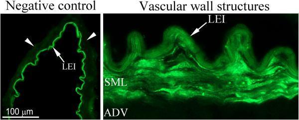
Diagnostic fluorescence microscopy images of human artery sections illustrating vascular wall histology and immunohistochemical controls. The image is divided into two panels. Left Panel (Negative Control): A cross-section of a cerebral artery where primary antibodies were omitted. It demonstrates prominent green auto-fluorescence of the lamina elastica interna (LEI), appearing as a bright, thin, undulating line. White arrowheads point to the smooth muscle cell layer (SML), which shows no specific immunoreactivity, serving as a baseline for experimental comparison. Right Panel (Vascular Wall Structures): An architectural overview of the arterial wall layers. Labeled structures include the outermost adventitial layer (ADV), the intermediate smooth muscle cell layer (SML), and the innermost lamina elastica interna (LEI). The LEI shows characteristic intense fluorescence and a folded morphology. The SML exhibits a more diffuse fluorescence pattern, representing specific staining or background across the tunica media. A 100 µm scale bar is provided for reference. This visual is intended for teaching vascular anatomy, immunohistochemical techniques, and the phenomenon of tissue auto-fluorescence.
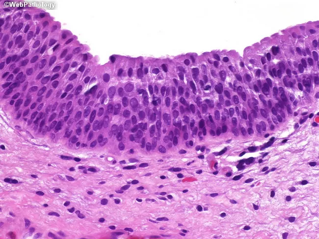
Imaging modality: light microscopy of a hematoxylin and eosin (H&E) stained histology section of bladder wall mucosa. Primary subject is the urothelium (transitional epithelium) lining the urinary bladder, with underlying lamina propria and, deeper, muscularis propria (detrusor muscle). The section depicts a cross-sectional view of the bladder mucosa in which urothelial cells form a multi-layered, stratified lining whose thickness varies with bladder distension. Superficial umbrella cells are present at the luminal surface, with rounded to slightly flattened cytoplasm and prominent nuclei; underlying intermediate and basal cells show orderly alignment and clear basement membrane orientation. The lamina propria appears as loose connective tissue containing scattered inflammatory cells and small vessels; muscularis propria is seen deeper as circular and longitudinal smooth muscle bundles. No overt cytologic atypia or invasion is evident in this normal specimen. Clinically, this histologic pattern provides a reference standard for normal urothelium, enabling recognition of pathological changes such as urothelial hyperplasia, dysplasia, carcinoma in situ, or invasive urothelial carcinoma when atypia or architectural disarray is present. Diagnostic significance includes distinguishing normal distension-related variability from disease states; differential considerations include inflammatory/reactive changes and neoplastic processes. This image is educational for medical students, residents, and researchers studying bladder histology and urothelial pathology.
blood vessel wall layers tunica intima media adventitia histology
Low-power brightfield light microscopy of a formalin-fixed, paraffin-embedded arterial cross-section stained with Hematoxylin and Eosin. The specimen shows a circular vessel with a well-defined, multilayered wall. The lumen is patent and lined by a narrow endothelial layer. The tunica media exhibits concentric layers of eosinophilic smooth muscle cells and occasional nuclei aligned along the wall, giving a dense, pink stippled appearance. The internal elastic lamina is not clearly resolved at this magnification. The tunica adventitia is relatively inconspicuous with sparse connective tissue. There is no overt atheromatous plaque, calcification, hemorrhage, or inflammatory infiltrate noted. Overall architecture is preserved, suggesting a normal or nonpathologic arterial segment. This image is suitable for illustrating normal vascular histology and can serve as a reference for comparison with arteriosclerotic lesions in teaching, research, and diagnostic practice. Potential clinical utility includes training in recognition of arterial layering, identifying pathologies such as atherosclerosis, vasculitis, and aneurysm formation, and correlating histology with imaging findings from ultrasound or CT angiography. The dataset supports queries for vascular histology, arterial wall ultrastructure, smooth muscle organization, and H&E staining patterns in a cross-sectional artery. The image exemplifies typical histologic cues used in vascular pathology curricula and can anchor comparative discussions of disease progression and treatment implications.
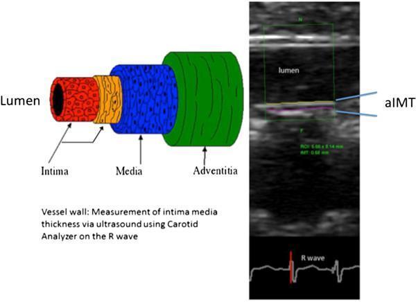
This composite educational graphic illustrates the assessment of aortic intima-media thickness (aIMT) in pediatric clinical research. On the left, an anatomical diagram of a blood vessel cross-section details the vascular wall architecture: the lumen (center), the Tunica Intima (red), the Tunica Media (blue), and the Tunica Adventitia (green). On the right, a longitudinal B-mode ultrasound image of an infant aorta demonstrates the clinical application of this model. The ultrasound features a green region of interest (ROI) where semi-automated edge-detection software identifies the far-wall interfaces of the intima and media. The specific 'aIMT' measurement is highlighted between these layers. At the bottom of the ultrasound panel, a synchronized electrocardiogram (ECG) trace with a highlighted R-wave indicates that measurements are gated to end-diastole for consistency. This material is designed to teach diagnostic imaging techniques used for evaluating early vascular changes and cardiovascular risk markers in infant populations.
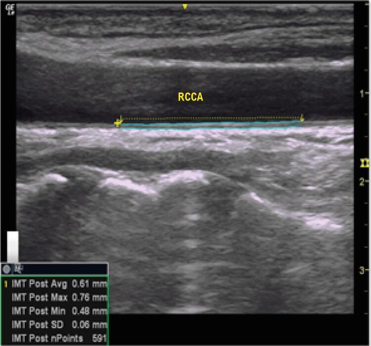
This diagnostic image is a longitudinal B-mode ultrasound of the Right Common Carotid Artery (labeled RCCA), demonstrating the assessment of Carotid Intima-Media Thickness (CIMT). The vessel lumen is shown as an anechoic (dark) space, while the posterior arterial wall exhibits a characteristic double-line pattern representing the interface between the blood-intima and the media-adventitia. A cyan measurement tool is overlaid on a plaque-free segment of the posterior wall, tracing the tunica intima and tunica media layers. A data box in the lower left corner provides quantitative metrics: an average IMT of 0.61 mm, a maximum of 0.76 mm, a minimum of 0.48 mm, and a standard deviation of 0.06 mm across 591 measurement points. This imaging modality is primarily used in cardiovascular risk stratification and the screening of subclinical atherosclerosis, providing a non-invasive surrogate marker for systemic vascular health and future cardiovascular events.
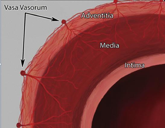
This anatomical diagram provides a cross-sectional view of a large systemic artery wall, illustrating the structural layers and the microvascular supply. The diagram identifies three distinct histological layers: the tunica intima (innermost lining), the tunica media (thick muscular middle layer), and the tunica adventitia (outermost connective tissue layer). A primary focus of the illustration is the 'vasa vasorum'—the 'vessels of the vessels.' The vasa vasorum are depicted as a branching network of small capillaries originating from larger nutrient vessels on the external surface of the adventitia. These microvessels penetrate through the adventitia and extend into the outer portions of the tunica media, highlighting the physiological mechanism for delivering oxygen and nutrients to thick-walled vessels where simple diffusion from the lumen is insufficient. The image serves as a pedagogical tool for understanding vascular histology and the pathophysiology of arterial wall maintenance in conditions such as atherosclerosis or aortic disease.
heart wall layers pericardium myocardium endocardium histology
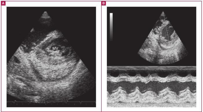
Diagnostic ultrasound images demonstrating classic echocardiographic features of advanced endomyocardial fibrosis (EMF). Panel A presents a short-axis transthoracic echocardiogram view showing the characteristic 'layering' effect of the posterior cardiac wall. This layering consists of three distinct, thickened zones: the hyperechoic endocardium, the myocardium, and the thickened pericardium. The posterior mitral valve (MV) leaflet is clearly visible and tethered to the underlying fibrotic endocardium, a hallmark of endocardial restriction. Panel B displays an M-mode echocardiogram across the posterior wall, further highlighting the anatomical separation of the three layers. The M-mode confirms significant endocardial thickening and a fibrosed, thickened pericardium associated with small peripheral pericardial effusions. These images illustrate the progressive fibrotic transformation of the heart's internal and external layers, which leads to restrictive physiology and valvular dysfunction. This material is suitable for advanced cardiology education focusing on restrictive cardiomyopathies and tropical cardiology.
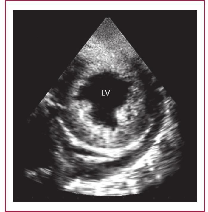
This diagnostic ultrasound image presents a short-axis echocardiographic view of the left ventricle (LV) in a patient with endomyocardiopericardial fibrosis (EMPF). The image demonstrates a characteristic three-layered 'layering' effect of the posterior cardiac wall. The innermost layer, the endocardium, appears as a dense, intensely hyperechoic (bright) band that is significantly thickened, engulfing the posterior papillary muscle. Adjacent to this is the myocardium, which maintains a distinct, less echogenic texture. The outermost layer shows a densely fibrosed and calcified pericardium, characterized by high-intensity echoes. An associated pericardial effusion is visible as an anechoic (dark) space between the myocardial and pericardial layers. This combination of endocardial thickening, myocardial preservation, and pericardial involvement is pathognomonic for advanced stages of endomyocardial fibrosis (EMF), often leading to restrictive or constrictive cardiac physiology.
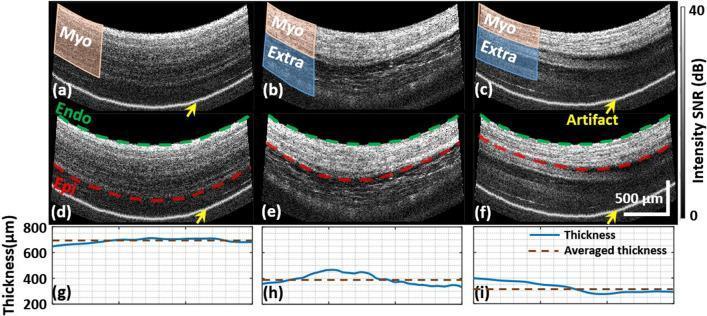
Educational figure illustrating the segmentation and thickness measurement of the myocardial wall using Polarized Sensitive Optical Coherence Tomography (PSOCT). Panels (a-c) present grayscale structural cross-sectional images of the cardiac wall. The myocardium (Myo) is highlighted in orange, while extra-myocardial tissues (Extra), such as adipose or pericardium, are highlighted in blue. Panels (d-f) demonstrate the corresponding automated or manual segmentation of the wall layers, with dashed green lines identifying the endocardium (Endo) and dashed red lines identifying the epicardium (Epi). A scale bar indicates 500 µm. Yellow arrows point to imaging artifacts originating from the probe. Panels (g-i) provide quantitative line graphs showing the calculated wall thickness in microns (µm). The solid blue line tracks real-time thickness variations across the scanned area, while the dashed brown line indicates the mean averaged thickness. This figure serves to teach the methodology for distinguishing myocardial boundaries from adjacent anatomical structures to ensure accurate clinical measurement of cardiac wall dimensions.
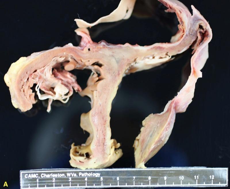
This clinical photograph displays a gross pathological cross-section of a human heart from an autopsy, illustrating a large left ventricular pseudoaneurysm (LVPA). The specimen shows a clear distinction between the true muscular wall of the left ventricle and the pseudoaneurysmal sac. The primary left ventricular myocardium is thick, measuring approximately 1 cm, with a solid muscular texture and reddish-tan coloration. In contrast, the pseudoaneurysmal walls, positioned in the upper right, are markedly thinned and lack myocardial layers. These pathological walls exhibit a more fibrous, less dense texture with yellowish-tan and pinkish-white hues, consistent with a sac composed of fibrin and organized thrombus rather than true endocardium and myocardium. The internal architecture reveals an irregular ventricular chamber shape and a fistulous tract or communication point leading to the pseudoaneurysm. This specimen demonstrates cardiac remodeling and morphological changes typical of a late-stage complication of infective endocarditis or myocardial injury, illustrating the high risk of rupture or heart failure associated with such large cardiac pseudoaneurysms.
Purkinje fibers conducting system heart histology
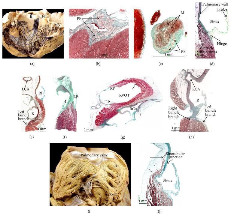
This composite educational resource details the cardiac conduction system and ventricular anatomy through gross clinical photographs and histological sections. Figure (a) displays a bovine heart with Indian ink highlighting the left bundle branch and dense Purkinje network branching around the aortic sinuses (R, N) and mitral valve (MV). Figures (b) and (c) are Masson's trichrome-stained sections showing Purkinje fibers (PF) as distinct light-colored structures within the subendocardial matrix and false tendons. Figures (d), (g), and (j) illustrate the histology of the pulmonary valve, highlighting the hinge attachment to the ventricular myocardium and fiber orientation within the right ventricular outflow tract (RVOT). Sagittal sections (e, f, h) demonstrate the anatomical relationships between the right atrium (RA), aortic root, and conduction tissue, specifically the right and left bundle branches near the right coronary artery (RCA). Figure (i) provides an endocardial view of the RVOT, showing the septomarginal trabeculation (SMT) and supraventricular crest (SC). These materials illustrate the structural basis for electrical propagation and the anatomical proximity of conduction pathways to valvar structures.
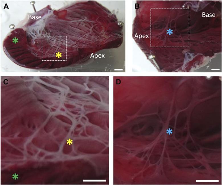
This set of clinical photographs demonstrates the cardiac Purkinje fiber network in a rabbit heart model following chemical ablation. Images A and C show the left ventricle (LV) endocardial surface after exposure to Lugol solution, stained with Triphenyl Tetrazolium Chloride (TTC). In these images, the Purkinje fiber network appears as a prominent white, mesh-like structure (yellow asterisk), indicating widespread cellular necrosis. The underlying bulk myocardium remains largely red (green asterisk), suggesting it is unaffected by the superficial chemical treatment. In contrast, images B and D display the untreated right ventricle (RV) of the same heart. Here, the Purkinje fibers (blue asterisk) stain red/pink, consistent with healthy, viable tissue. The comparison illustrates the specific targeting of the subendocardial conduction system by the chemical agent. The magnified views in C and D highlight the morphological differences between the necrotic, opacified fibers in the treated LV versus the intact, translucent fibers in the control RV. Key anatomical landmarks including the base and apex are labeled for orientation.
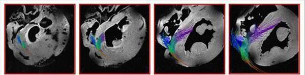
This diagnostic imaging series consists of four sequential high-resolution MR microscopy frames demonstrating cardiac fiber tracking of the conduction system. The images provide a 3D visualization of the specialized conduction pathways from the base of the heart toward the midwall. The tracking originates behind the non-coronary leaflet at the cardiac base. Color-coded diffusion tensor imaging (DTI) or similar fiber tractography techniques are utilized to differentiate pathways: blue fibers represent the bundle proceeding down the right ventricular subendocardium, while purple fibers illustrate the branch traversing the interventricular septum before descending into the left ventricular subendocardium. As the sequence progresses from frame A to D, the left ventricular branch expands into a fan-like structure, ultimately terminating in free-running Purkinje fibers within the ventricular cavity. This visual highlights the micro-anatomical orientation of the His-Purkinje system, essential for understanding ventricular depolarization and electrical signaling in cardiac physiology.
Histology of the Cardiovascular System (CVS)
1. WALL OF THE HEART
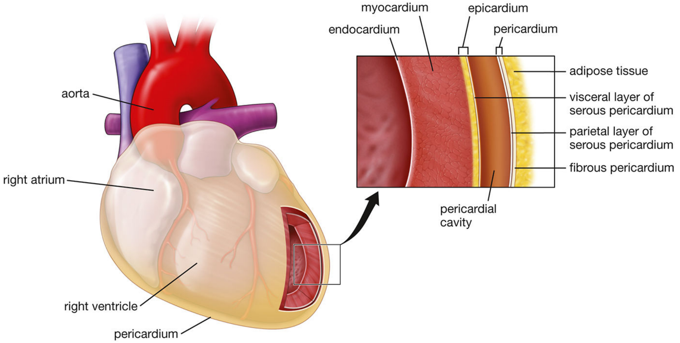
A. Epicardium (Visceral Pericardium)
- The outermost layer of the heart wall
- Consists of a single layer of mesothelial cells resting on underlying loose connective tissue and adipose tissue
- Contains coronary arteries, cardiac veins, and nerves embedded in the adipose tissue
- Is reflected back at the great vessels to become the parietal layer of serous pericardium
- Between visceral and parietal layers = pericardial cavity (15-50 mL serous fluid)
- Clinical note: accumulation of excess fluid in this cavity = cardiac tamponade
B. Myocardium
- The thickest and most important layer - composed of cardiac muscle
- Atrial myocardium is thinner than ventricular myocardium
- Ventricles must generate greater pressure to eject blood into systemic/pulmonary circulations
- Left ventricular wall is thicker than right (higher systemic resistance)
C. Endocardium
- The innermost layer, lining the cardiac chambers
- Consists of:
- Endothelium (simple squamous) on the luminal surface
- Subendothelial layer of connective tissue
- Subendocardial layer containing Purkinje fibers (modified cardiac myocytes of the conducting system)
- Continuous with the tunica intima of blood vessels at the great vessel orifices
2. CARDIAC MUSCLE HISTOLOGY
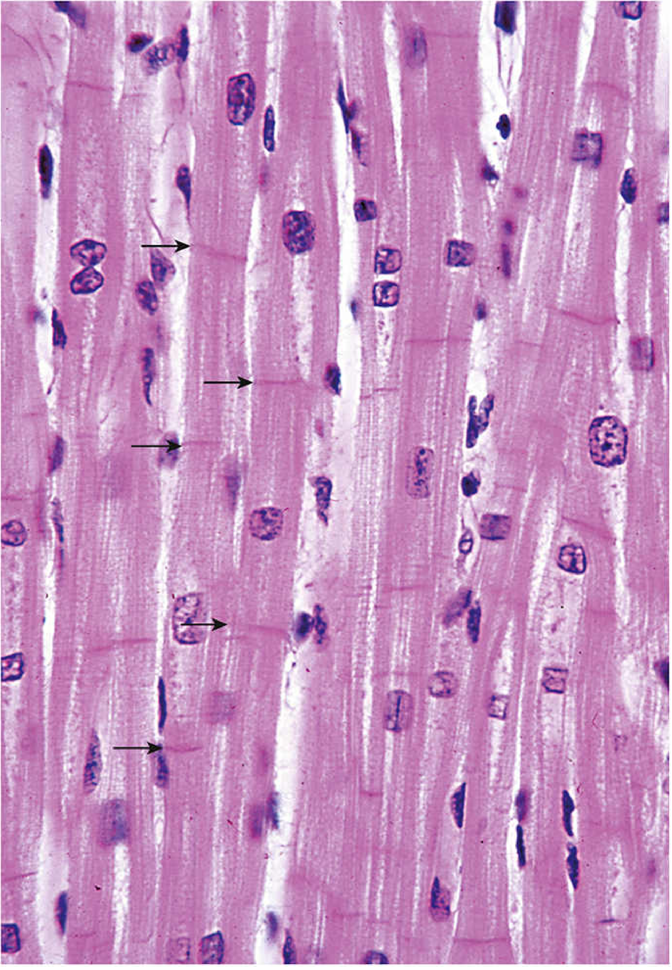
Key Features Under Light Microscope:
| Feature | Cardiac Muscle | Skeletal Muscle |
|---|---|---|
| Cell shape | Cylindrical, branched | Long, unbranched cylinders |
| Nuclei | 1-2, centrally placed | Multiple, peripherally placed |
| Cross-striations | Present | Present |
| Intercalated discs | Present (unique feature) | Absent |
| Diameter | 15-30 µm | Variable |
| Length | 85-120 µm | Very long |
| Endomysium | Present (rich capillary network) | Present |
Cell Organelles (Ultrastructure):
- Mitochondria: Occupy up to 40% of cell volume - higher than any other muscle type. Large, with densely packed cristae. Located between myofibrils and around the nucleus
- Glycogen granules: Energy reserve, located adjacent to myofibrils
- Lipofuscin pigment granules: Perinuclear, "wear and tear" pigment - age-related
- T-tubules: Located at the Z-line level (cf. skeletal muscle - at A-I junction); larger diameter than skeletal muscle T-tubules
- Sarcoplasmic reticulum: Less developed than in skeletal muscle; forms a "diad" (not triad) with T-tubules
- Atrial granules: In atrial cardiomyocytes only - contain Atrial Natriuretic Factor (ANF) and Brain Natriuretic Factor (BNF) - both are diuretics that inhibit renin/aldosterone and cause vasodilation. BNF levels rise in congestive heart failure
Intercalated Discs:
-
Transverse component (perpendicular to myofibrils):
- Fascia adherens - the major constituent; attachment site for thin (actin) filaments
- Macula adherens (desmosomes) - reinforce adhesion between cells
-
Lateral component (parallel to myofibrils):
- Gap junctions - provide ionic continuity between cells; act as electrical synapses allowing rapid impulse conduction → cells contract as a functional syncytium
3. CONDUCTING SYSTEM
| Component | Location | Histology |
|---|---|---|
| SA node | Right atrial wall, near SVC opening | Small, pale-staining cells with few myofibrils |
| AV node | Floor of right atrium, near tricuspid valve | Similar to SA node |
| Bundle of His | Interventricular septum | Transitional cells |
| Purkinje fibers | Subendocardial layer | Large, pale cells with few peripheral myofibrils and abundant glycogen |
4. HEART VALVES
| Layer | AV Valves | Semilunar Valves | Composition |
|---|---|---|---|
| Towards atrium/outflow | Atrialis | Ventricularis | Elastic fibers + endothelium |
| Middle | Spongiosa | Spongiosa | Loose connective tissue, proteoglycans |
| Towards ventricle/vessel | Fibrosa | Fibrosa | Dense collagen (gives structural strength) |
5. BLOOD VESSELS - GENERAL PLAN
Tunica Intima (Innermost)
- Endothelium - simple squamous epithelium on luminal surface
- Subendothelial connective tissue
- Internal elastic membrane (IEM) - prominent in muscular arteries (appears as a wavy, refractile line on H&E)
- Functions of endothelium:
- Selective permeability barrier
- Secretes anticoagulants (prevents clotting)
- Secretes vasoconstrictors and vasodilators (e.g., NO, endothelin)
- Regulates immune responses
Tunica Media (Middle)
- Circumferentially arranged vascular smooth muscle cells
- Elastic lamellae interspersed between smooth muscle layers
- Bounded by internal and external elastic membranes
- Thickest in arteries; thinner in veins
Tunica Adventitia (Outermost)
- Collagen fibers (predominantly Type I) with few elastic fibers
- Contains vasa vasorum (tiny vessels supplying the vessel wall)
- Contains nervi vasorum (autonomic nerve network regulating vessel tone)
- Thickest layer in veins
6. CLASSIFICATION OF ARTERIES
A. Large / Elastic Arteries (e.g., Aorta, pulmonary trunk, brachiocephalic)
- Tunica media: multiple concentric elastic lamellae with smooth muscle between them
- No fibroblasts in the media
- Function: Windkessel effect - store energy during systole, release during diastole
- On H&E: alternating pink (smooth muscle) and wavy (elastic) layers
B. Medium / Muscular Arteries (e.g., coronary, femoral, radial)
- Tunica media: many layers of smooth muscle with fewer elastic lamellae
- Prominent internal elastic membrane (well-defined wavy line in intima)
- Less prominent external elastic membrane
- Function: regulate blood distribution
C. Small Arteries and Arterioles
- Small arteries: 2-20 smooth muscle layers in media
- Arterioles: 1-2 smooth muscle layers in media
- Primary regulators of vascular resistance - control blood flow to capillary beds
- Precapillary sphincters on metarterioles regulate flow into capillaries
7. CAPILLARIES
| Type | Wall | Basal Lamina | Location |
|---|---|---|---|
| Continuous | Uninterrupted endothelium | Continuous | Muscle, lung, CNS, skin |
| Fenestrated | Endothelium with pores (fenestrae) | Continuous | Kidney glomerulus, intestinal villi, endocrine glands |
| Discontinuous / Sinusoidal | Large gaps between cells | Discontinuous / absent | Liver, spleen, bone marrow |
8. VEINS
| Type | Diameter | Key Features |
|---|---|---|
| Postcapillary venules | <0.1 mm | Pericytes present; high endothelial venules in lymphoid tissue |
| Small veins | <1 mm | Thin media, prominent adventitia |
| Medium veins | <10 mm | Valves present (especially in limb veins) |
| Large veins (e.g., vena cava, portal vein) | >10 mm | May have myocardial sleeves in tunica adventitia |
- Artery: round lumen, thick media, narrow adventitia
- Vein: collapsed / irregular lumen, thin media, adventitia is the thickest layer, valves present
9. KEY ANESTHESIA RELEVANCE
- Cardiac tamponade - fluid in pericardial cavity compresses heart, needs emergency pericardiocentesis
- Endothelium - target of many anesthetic/vasoactive drugs; produces NO (vasodilation) and endothelin (vasoconstriction)
- Intercalated discs (gap junctions) - explain why cardiac arrhythmias can propagate through the entire myocardium
- BNP/BNF levels - used clinically to diagnose and monitor heart failure
- Purkinje fibers - basis for conduction blocks and arrhythmias relevant in perioperative management
- Arterioles and precapillary sphincters - primary site of vascular resistance regulation; target of vasopressors/vasodilators used in anesthesia
Can you give rralated questions and answers from this topic
Q&A: Histology of the Cardiovascular System
SECTION A: Short Answer / One-Liners
- Epicardium (visceral pericardium)
- Myocardium (cardiac muscle)
- Endocardium (endothelium + connective tissue)
- Fascia adherens - major component; anchors actin (thin) filaments
- Macula adherens (desmosomes) - reinforce cell-to-cell adhesion
- Tunica intima - innermost (endothelium + subendothelial CT + internal elastic membrane)
- Tunica media - middle (smooth muscle + elastic lamellae)
- Tunica adventitia - outermost (collagen + vasa vasorum + nervi vasorum)
- Artery: Tunica media is thickest
- Vein: Tunica adventitia is thickest
- Continuous - uninterrupted endothelium (muscle, CNS, lung)
- Fenestrated - pores/fenestrae present (kidney, intestine, endocrine glands)
- Discontinuous/Sinusoidal - large gaps, discontinuous basal lamina (liver, spleen, bone marrow)
SECTION B: Structured / Applied Questions
- Shape: Short, cylindrical, branched cells arranged end to end
- Nucleus: 1-2, centrally placed, euchromatic (pale-staining)
- Cross-striations: Present (due to sarcomere arrangement of actin and myosin)
- Intercalated discs: Darkly staining, irregular step-like bands crossing the fiber - represent junctions between adjacent cells
- Endomysium: Thin connective tissue sheath with a rich capillary network around each fiber
- Branching: Fibers branch and interconnect, allowing force to be distributed across the myocardium
- Size: ~15-30 µm diameter, 85-120 µm length
| Feature | Artery | Vein |
|---|---|---|
| Lumen shape | Round, regular | Collapsed/irregular |
| Wall thickness | Thick | Thin |
| Tunica media | Thick, prominent | Thin |
| Thickest layer | Tunica media | Tunica adventitia |
| Internal elastic membrane | Well-defined | Poorly defined or absent |
| Valves | Absent | Present (especially limbs) |
| Lumen: wall ratio | Smaller lumen relative to wall | Larger lumen relative to wall |
| Type | Example | Tunica Media |
|---|---|---|
| Elastic (large) artery | Aorta, pulmonary trunk | Multiple elastic lamellae alternating with smooth muscle; no fibroblasts |
| Muscular (medium) artery | Coronary, femoral, radial | Many layers of smooth muscle, fewer elastic lamellae; prominent internal elastic membrane |
| Small artery / Arteriole | Pre-capillary vessels | 1-2 smooth muscle layers; arterioles are the main regulators of vascular resistance |
| Layer position | AV Valves (mitral/tricuspid) | Semilunar Valves (aortic/pulmonary) | Composition |
|---|---|---|---|
| Atrial/outflow side | Atrialis | Ventricularis | Elastic fibers |
| Middle | Spongiosa | Spongiosa | Loose CT, proteoglycans (cushioning) |
| Ventricular/vessel side | Fibrosa | Fibrosa | Dense collagen (structural strength) |
- Found in atrial cardiomyocytes (not ventricular), in the juxtanuclear cytoplasm
- Size: 0.3-0.4 µm
- Contain two hormones:
- Atrial Natriuretic Factor (ANF) - also called ANP
- Brain Natriuretic Factor (BNF) - also called BNP
- Both hormones:
- Are diuretics (increase urinary sodium excretion)
- Inhibit renin and aldosterone secretion
- Inhibit vascular smooth muscle contraction (vasodilation)
- Clinical significance: BNP levels rise in congestive heart failure and are used as a diagnostic/monitoring biomarker in clinical practice
- Loose connective tissue
- Purkinje fibers (modified cardiac myocytes of the conduction system - bundle of His branches and terminal Purkinje network)
- This zone is the most vulnerable to ischemia during low cardiac output states (e.g., hypotension under anesthesia) because it is farthest from coronary blood supply
- Conduction abnormalities (bundle branch blocks, arrhythmias) seen intraoperatively originate from disruption here
- Monitoring for ST-segment depression (subendocardial ischemia) on ECG is standard during anesthesia
SECTION C: MCQ-Style
SECTION D: Diagram-Based Questions
- Pericardial cavity (with mesothelial lining)
- Fibrous pericardium (outermost)
- Parietal serous pericardium
- Visceral serous pericardium = Epicardium (mesothelium + adipose tissue + coronary vessels)
- Myocardium (cardiac muscle)
- Endocardium (endothelium + subendothelial CT + subendocardial layer with Purkinje fibers)
- Transverse component (perpendicular to myofibrils):
- Fascia adherens
- Macula adherens (desmosomes)
- Lateral component (parallel to myofibrils):
- Gap junctions
- T-tubule at Z-line level
QUICK REVISION TABLE
| Topic | Key Point to Remember |
|---|---|
| Cardiac muscle nucleus | Central (1-2 per cell) |
| Intercalated disc - electrical link | Gap junctions (lateral component) |
| Intercalated disc - mechanical link | Fascia adherens + desmosomes (transverse) |
| T-tubule position | Z-line level |
| Mitochondria % in cardiac cell | 40% of cell volume |
| ANF/BNF found in | Atrial granules of atrial cardiomyocytes |
| Thickest artery layer | Tunica media |
| Thickest vein layer | Tunica adventitia |
| Purkinje fibers location | Subendocardial layer |
| Valves - strongest layer | Fibrosa (dense collagen) |
| Capillary in liver | Sinusoidal (discontinuous) |
| Capillary in kidney | Fenestrated |
| Capillary in CNS | Continuous (blood-brain barrier) |
| Arteriole function | Primary regulator of vascular resistance |
| Pericardial fluid volume (normal) | 15-50 mL |