
fracture dislocations of shoulder and management
fracture dislocation shoulder glenohumeral
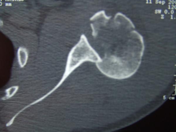
This axial CT scan slice of the shoulder demonstrates a posterior glenohumeral dislocation with associated complex fractures of the proximal humerus. The humeral head is displaced posteriorly relative to the glenoid fossa. A prominent 'reverse Hill-Sachs lesion' (trough line) is visible as an impaction fracture on the anterior aspect of the humeral head where it has abutted the posterior glenoid rim. Additionally, the image shows comminuted fractures involving the greater and lesser tuberosities, characterized by sharp cortical disruption and displaced bone fragments. The normal congruent relationship between the rounded humeral head and the concave glenoid is lost, illustrating a severe musculoskeletal injury pattern often resulting from high-energy trauma or forced internal rotation. This diagnostic image serves as a key educational example of posterior instability complications and the utility of computerized tomography in evaluating complex periarticular shoulder fractures.
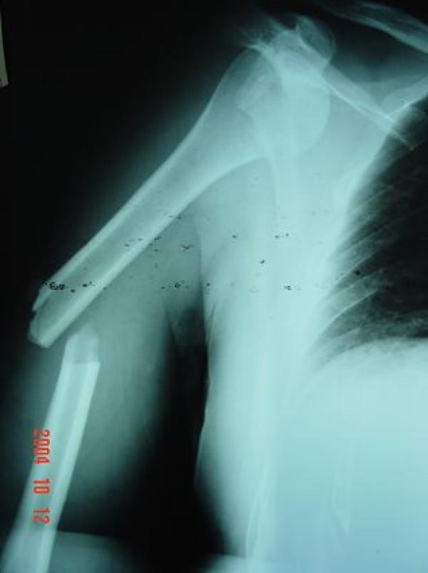
Anteroposterior (AP) radiograph of the right shoulder and humerus demonstrating a concomitant proximal injury and midshaft fracture. The glenohumeral joint shows an anterior dislocation, characterized by the displacement of the humeral head inferior and anterior to the glenoid fossa (subcoracoid position). Distal to the shoulder joint, there is a complete, transverse fracture through the middle third of the humeral shaft. The fracture shows significant lateral displacement and overriding (shortening) of the distal fragment relative to the proximal fragment. The clavicle and visible thoracic cage appear intact. This combination of injuries is clinically significant as the humeral shaft fracture complicates the standard leverage maneuvers used for closed reduction of the shoulder dislocation. Educational focus includes orthopedic trauma assessment, recognition of multi-level upper extremity injuries, and surgical planning for fracture-dislocation management.
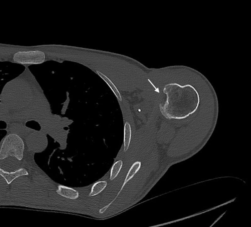
This diagnostic axial CT image of the shoulder demonstrates a posterior glenohumeral dislocation or subluxation in a 39-year-old patient. The humeral head is displaced posteroinferiorly relative to the glenoid fossa. A prominent impaction fracture is visible on the anteromedial aspect of the humeral head (indicated by the white arrow), characteristic of a reverse Hill-Sachs lesion. This type of bony defect typically occurs when the anterior humeral head impacts against the posterior glenoid rim during a posterior dislocation event. Additionally, small radiopaque hyperdensities are visible in the anterior periarticular soft tissues, representing metallic suture anchors from a prior McLaughlin-type remplissage surgical procedure. The posterior glenoid rim appears relatively well-preserved, without evidence of a significant bony Bankart lesion. This image serves as a clinical example of chronic posterior shoulder instability and the associated characteristic secondary bony changes often seen in patients with repetitive trauma or seizure-related dislocations.
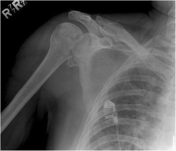
Anteroposterior (AP) radiograph of the right shoulder demonstrating a posterior glenohumeral dislocation. The humeral head is laterally displaced and lacks its normal overlap with the glenoid fossa, manifesting the 'light bulb sign' due to fixed internal rotation. A prominent cortical impaction fracture is visible on the anteromedial aspect of the humeral head, representing a reverse Hill-Sachs lesion (McLaughlin lesion), which typically occurs when the humeral head impacts against the posterior glenoid rim. There is a widened glenohumeral joint space, often referred to as the 'rim sign.' The anatomical landmarks of the clavicle, acromion, and proximal humeral shaft are otherwise intact. This diagnostic image is representative of orthopedic trauma imaging for shoulder instability and is used to identify associated osseous injuries that may require surgical intervention, such as rotational osteotomy.
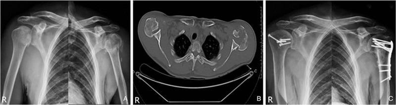
This composite educational graphic depicts the diagnostic imaging and postoperative management of bilateral posterior shoulder fracture-dislocations. (A) Anteroposterior (AP) radiographs of both shoulders demonstrate bilateral proximal humeral fractures. The humeral heads show a loss of normal overlap with the glenoid fossa, manifesting as the 'light bulb sign,' which is characteristic of internal rotation in posterior dislocations. (B) An axial CT scan through the glenohumeral joints provides definitive visualization of the pathology: a posterior 2-part fracture-dislocation on the right side and a complex posterior 4-part fracture-dislocation on the left side, where the humeral head is displaced posterior to the glenoid. (C) Postoperative AP radiographs illustrate the results of open reduction and internal fixation (ORIF). The right proximal humerus is stabilized with multiple cancellous bone screws, while the left humerus is reconstructed using an anatomical locked plating system. The images demonstrate successful restoration of glenohumeral alignment and stable internal fixation of the fractures.
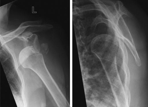
This diagnostic image consists of two radiographic views of the left shoulder (anteroposterior and Y-view) demonstrating a traumatic anterior glenohumeral dislocation. The humeral head is significantly displaced anteriorly and inferiorly relative to the glenoid fossa. Associated with the dislocation is a comminuted avulsion fracture of the greater tuberosity. A distinct, irregular bony fragment is visible, having been pulled from its anatomical position on the proximal humerus and displaced superiorly and laterally. The scapula, including the glenoid rim and acromion, appears otherwise intact, though normal joint alignment is lost. This clinical image is characteristic of acute shoulder trauma and is used to illustrate the typical presentation of a combined glenohumeral dislocation and rotator cuff-related bony avulsion, common in adult patients.
"shoulder" AND "fracture dislocation" AND "management"
Fracture-Dislocations of the Shoulder: Classification and Management
Overview
Anatomy and Four "Parts" (Codman-Neer Foundation)
- Articular head fragment
- Greater tuberosity (attached to supraspinatus, infraspinatus, teres minor - pulls posteromedially)
- Lesser tuberosity (attached to subscapularis - pulls anteromedially)
- Humeral shaft (pulled anteromedially by pectoralis major)
- Rockwood and Green's Fractures in Adults, 10th ed. 2025, p. 1335-1336
- Miller's Review of Orthopaedics, 9th ed., p. 901
Neer Classification
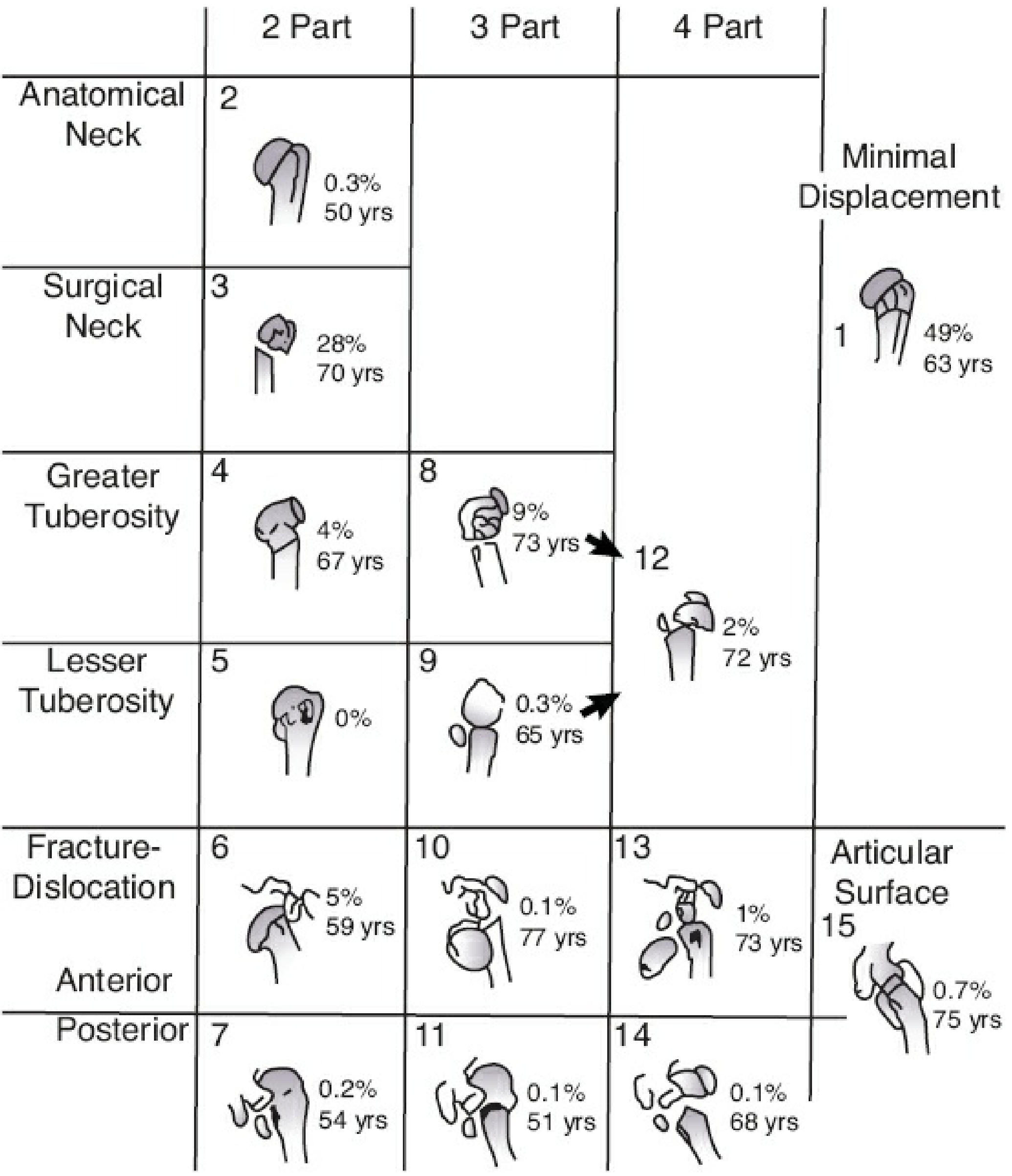
| Category | Description |
|---|---|
| 1-part | Nondisplaced / minimally displaced (any number of fracture lines); 49% of cases |
| 2-part | One segment displaced; e.g., surgical neck, anatomic neck, or isolated tuberosity |
| 3-part | Two segments displaced - always involves surgical neck + either GT or LT |
| 4-part | All four segments displaced; highest risk of AVN |
| Articular surface (head-splitting) | Split through the articular surface; almost always requires replacement |
- 2-part fracture-dislocation (anterior or posterior): 5% and 0.2% respectively
- 3-part fracture-dislocation: 0.1% each direction
- 4-part fracture-dislocation: 1% anterior, 0.1% posterior
Types of Fracture-Dislocation by Direction
Anterior Fracture-Dislocations
- Greater tuberosity avulsion (most common fracture with anterior dislocation) - pulled by supraspinatus/infraspinatus
- Surgical neck fracture - combined with dislocation in severe trauma
- Hill-Sachs lesion (posterior impaction defect on humeral head)
- Bankart lesion (anterior-inferior glenoid rim fracture)
Posterior Fracture-Dislocations
- "Light bulb sign" on AP x-ray - fixed internal rotation gives a rounded humeral head
- "Rim sign" - widened glenohumeral joint space >6 mm
- "Trough line" - reverse Hill-Sachs lesion (impaction fracture on anterior humeral head)
- CT scan or axillary view is mandatory to confirm
Predictors of Avascular Necrosis (Hertel Criteria)
- Fracture through the anatomic neck
- Loss of medial periosteal hinge (disruption of medial calcar soft tissue)
- Medial metadiaphyseal extension <8 mm (calcar <8 mm)
- Bailey and Love's Short Practice of Surgery, 28th ed., p. 457
- Miller's Review of Orthopaedics, 9th ed., p. 902
Management Principles
1. One-Part Fracture-Dislocation (Minimal Displacement)
- Closed reduction of the dislocation
- Standard reduction techniques (Cunningham, Stimson, Kocher - with care)
- Post-reduction: sling immobilization 3-4 weeks, then physiotherapy
- Most can be managed non-operatively
2. Two-Part Fracture-Dislocation
- Closed reduction often successful; the GT fracture frequently reduces with the dislocation
- If GT displaced >5 mm post-reduction, surgical fixation is required (suture anchors, screws, or plate) to restore rotator cuff function
- Displacement >5 mm leads to impingement and supraspinatus dysfunction
- Usually requires open reduction as the fracture prevents lever-arm mechanics for closed reduction
- Fixation options: locking plate (ORIF), intramedullary nail, percutaneous Kirschner wires
- Pye's Surgical Handicraft notes: "The fracture often makes reduction of the shoulder impossible, and operative reduction may be necessary."
- If reverse Hill-Sachs lesion <20% of articular surface: closed reduction under anaesthesia acceptable
- Reverse Hill-Sachs 20-40%: McLaughlin procedure - transfer of the subscapularis tendon (with or without lesser tuberosity) into the defect, or remplissage-equivalent
- Reverse Hill-Sachs >40%: arthroplasty (hemi or reverse total shoulder)
3. Three-Part Fracture-Dislocation
- Generally operative for most patients
- ORIF with locking proximal humeral plate is the standard for younger patients with good bone stock
- The intact tuberosity fragment is used for rotational control intraoperatively
- Elderly/low-demand patients with osteoporosis: reverse total shoulder arthroplasty (rTSA) is increasingly preferred, as it does not rely on tuberosity healing
- In three-part greater tuberosity fracture-dislocation: head segment is internally rotated by subscapularis (unopposed pull)
- In three-part lesser tuberosity fracture-dislocation: head segment is externally rotated/abducted by posterosuperior rotator cuff
4. Four-Part Fracture-Dislocation
- Young patients (<50 years): Aggressive attempt at ORIF, accepting higher AVN risk rather than performing arthroplasty in a young patient
- Elderly/osteoporotic patients: Primary reverse total shoulder arthroplasty (rTSA) is now widely preferred
- Does NOT depend on tuberosity healing - functions on deltoid power
- Better functional outcomes than hemiarthroplasty in this group
- Hemiarthroplasty remains an option but outcomes depend critically on tuberosity healing and rotator cuff integrity
5. Head-Splitting Fracture-Dislocation
- The articular surface is split/comminuted
- Almost invariably requires arthroplasty (hemiarthroplasty or reverse TSA depending on age/rotator cuff status)
Surgical Techniques
| Technique | Best Indication |
|---|---|
| Closed reduction + percutaneous pinning (CRPP) | 2-part surgical neck in young patients with good bone |
| ORIF - locking proximal humeral plate | 3- and 4-part fractures in young/active patients with reasonable bone stock |
| Intramedullary nail | 2-part surgical neck fractures |
| Hemiarthroplasty | Selected 3/4-part fractures when head is viable but tuberosities repairable |
| Reverse total shoulder arthroplasty (rTSA) | 4-part fracture-dislocations in elderly, osteoporosis, unreconstructable head, or failed fixation |
| McLaughlin procedure | Posterior FD with reverse Hill-Sachs 20-40% |
Associated Neurovascular Injuries
-
Axillary nerve - most commonly injured (courses 5 cm below the acromion through the quadrilateral space)
-
Brachial plexus - infraclavicular injury pattern; axillary artery injury is rare but possible in elderly patients with atherosclerosis
-
Neurotmesis of axillary nerve in only ~5% of cases; most are neurapraxia/axonotmesis with good prognosis for spontaneous recovery
-
Always assess deltoid sensation and function (axillary nerve) and distal neurovascular status before and after reduction
-
Rockwood and Green's Fractures in Adults, 10th ed. 2025, p. 788
Imaging
| Modality | Role |
|---|---|
| AP shoulder + scapular Y + axillary view (trauma series) | Primary assessment |
| CT scan | Mandatory for complex fractures, posterior dislocations, preoperative planning |
| MRI | Rotator cuff integrity, soft tissue assessment when surgery is planned |
Clinical Images
Complications
| Complication | Notes |
|---|---|
| Avascular necrosis | Most feared; highest in 4-part fracture-dislocations; Hertel criteria predict risk |
| Malunion | Particularly tuberosity malunion - causes impingement and rotator cuff dysfunction |
| Nonunion | Most common after 2-part surgical neck; nonunion of GT post-arthroplasty causes loss of elevation |
| Stiffness | Common; rehabilitation may take months; permanent above-head limitation in elderly |
| Screw cutout | Most common hardware complication after locking plate fixation |
| Axillary nerve injury | Lateral pins in CRPP place it most at risk |
| Infection | More common in open procedures |
Summary Algorithm
Fracture-Dislocation of Shoulder
        |
        ├── 1-part/minimally displaced ──> Closed reduction + sling + physio
        |
        ├── 2-part
        │    ├── GT + anterior dislocation ──> Closed reduction; fix GT if >5mm displaced
        │    ├── Surgical neck + dislocation ──> Often needs ORIF (CRPP, locking plate, IMN)
        │    └── LT + posterior (McLaughlin) ──> Based on defect size: reduction / McLaughlin procedure / arthroplasty
        |
        ├── 3-part ──> Young/good bone: ORIF locking plate
        │              Elderly/osteoporosis: rTSA
        |
        ├── 4-part ──> Young: ORIF (accept AVN risk)
        │              Elderly: Primary rTSA (preferred)
        |
        └── Head-splitting ──> Arthroplasty (hemi or rTSA)
- Rockwood and Green's Fractures in Adults, 10th ed. 2025 - detailed classification, pathoanatomy, fracture-dislocation specifics
- Miller's Review of Orthopaedics, 9th ed. - Neer classification, treatment indications, Hertel AVN criteria
- Bailey and Love's Short Practice of Surgery, 28th ed. - vascularity predictors, fixation methods, arthroplasty selection
- Pye's Surgical Handicraft, 22nd ed. - operative indications, closed vs. open reduction