
discuss the new defintion for PPH management of PPH including modern and conservative surgical management from williams with flow chart proper explanation diagrams, for 10 mark answer m.s main exam
Reading File
Now let me search the medical library for PPH content from Williams Obstetrics in parallel:
Finding Sources
Finding Sources
I need to find Williams Obstetrics specifically. Let me search for it:
Finding Sources
Finding Sources
Finding Sources
Finding Sources
Williams Obstetrics is not in the library. Let me now pull from Creasy & Resnik (the best available OB text) and supplement with web search for the Williams-specific content:
Reading File
Finding Sources
Searching the Web
Williams Obstetrics PPH postpartum hemorrhage new definition 1000ml 500ml management 2022 2024
Excellent! Now let me get more detailed content on surgical management of PPH from Creasy & Resnik and also search for B-Lynch, uterine artery ligation content:
Reading File
Finding Sources
Searching the Web
Williams Obstetrics 26th edition PPH surgical management B-Lynch compression suture uterine artery ligation hysterectomy
I found direct Williams Obstetrics content. Let me extract more from the Williams PDF and also get Schwartz's surgical management content:
Reading Web Page
https://repository.stikesrspadgs.ac.id/44/1/Williams%20Obste…
Reading File
I have rich content now. Let me fetch the B-Lynch suture image from the Williams PDF content and also get ACOG/tranexamic acid info:
Loading Image
Searching the Web
Williams Obstetrics PPH 4T causes uterine atony tone trauma tissue thrombin tranexamic acid management steps 2022
I now have comprehensive information. Let me compile the complete 10-mark MS exam answer:
Postpartum Hemorrhage (PPH): Definition, Management, and Surgical Options
(As per Williams Obstetrics - 10 Mark MS Main Exam Answer)
1. DEFINITION (New/Revised)
Classical Definition (WHO 2006 / Traditional):
- Vaginal delivery: Blood loss > 500 mL within 24 hours of delivery
- Caesarean delivery: Blood loss > 1000 mL within 24 hours
New ACOG Definition (2017 - Now Standard in Williams Obstetrics):
PPH = Blood loss ≥ 1000 mL, OR any blood loss accompanied by signs/symptoms of hypovolemia within 24 hours of delivery, regardless of mode of delivery.
This unified definition replaced the dual threshold because:
- Estimated blood loss (EBL) by visual inspection underestimates actual loss by ~50%
- Hypovolemia symptoms reliably appear at ~1000 mL loss
- Quantitative blood loss (QBL) measurement is now mandated (calibrated drapes/weighing)
Classification of PPH:
| Type | Timing | Blood Loss |
|---|---|---|
| Primary PPH | Within 24 hours of delivery | ≥ 1000 mL or hypovolemia signs |
| Secondary PPH | 24 hours to 12 weeks postpartum | > 500 mL after 24 hours |
| Minor (RCOG) | - | 500-1000 mL |
| Major (RCOG) | - | > 1000 mL |
| Severe | - | > 2000 mL or hypovolemic shock |
2. ETIOLOGY - THE "4 T's" (Williams Obstetrics)
┌─────────────────────────────────────────────────────────────┐
│                    CAUSES OF PPH - 4 T's                    │
├──────────┬─────────────────────────────────────────────────-┤
│  TONE    │ Uterine atony (MOST COMMON - 70-80%)             │
│          │ Over-distended uterus, prolonged labour, MgSO₄   │
├──────────┼─────────────────────────────────────────────────-┤
│  TRAUMA  │ Lacerations (cervix, vagina, perineum), uterine  │
│          │ rupture, uterine inversion, extensions of LSCS   │
├──────────┼─────────────────────────────────────────────────-┤
│  TISSUE  │ Retained placenta, retained cotyledons,          │
│          │ placenta accreta spectrum, abnormal placentation  │
├──────────┼─────────────────────────────────────────────────-┤
│  THROMBIN│ Coagulopathy - DIC, pre-existing bleeding        │
│          │ disorders, anticoagulant use, HELLP syndrome      │
└──────────┴─────────────────────────────────────────────────-┘
3. MANAGEMENT FLOWCHART
                    POSTPARTUM HEMORRHAGE
                    Blood loss ≥ 1000 mL OR signs of hypovolemia
                              │
                              ▼
               ┌──────────────────────────┐
               │    IMMEDIATE RESUSCITATION│
               │  • Call for help / MICU   │
               │  • 2 large bore IV lines  │
               │  • Oxygen supplementation │
               │  • CBC, coags, crossmatch │
               │  • Foley catheter         │
               │  • Quantify blood loss    │
               └───────────┬──────────────┘
                           │
                           ▼
               ┌──────────────────────────┐
               │  IDENTIFY THE CAUSE (4T's)│
               │  • Uterine examination    │
               │  • Inspect birth canal    │
               │  • Check placenta         │
               │  • Check coagulation      │
               └───────────┬──────────────┘
                           │
              ┌────────────┴────────────┐
              ▼                         ▼
         UTERINE ATONY           TRAUMA/TISSUE/THROMBIN
         (TONE)                  • Repair lacerations
              │                  • Remove retained tissue
              ▼                  • Correct coagulopathy
    ┌─────────────────┐
    │  MEDICAL MANAGEMENT│
    │  (Step 1)           │
    └─────────┬───────────┘
              │
              ▼
    ┌─────────────────────────────────┐
    │         UTEROTONICS             │
    │ 1. Oxytocin 20-40 U in 1L RL   │
    │    infuse @100mU/min            │
    │ 2. Methylergonovine 0.2mg IM    │
    │    (avoid IV; avoid in HTN)     │
    │ 3. Carboprost 250 μg IM q15 min │
    │    (max 8 doses; CI: asthma)    │
    │ 4. Misoprostol 800-1000 μg      │
    │    sublingual/rectal            │
    │ 5. Tranexamic acid 1g IV        │
    │    (within 3 hrs of birth)      │
    └─────────┬───────────────────────┘
              │ FAILS
              ▼
    ┌────────────────────────────────────┐
    │  CONSERVATIVE SURGICAL MANAGEMENT  │
    │         (Step 2)                   │
    └────────────────────────────────────┘
              │
    ┌─────────┴────────────┐
    ▼                      ▼
UTERINE TAMPONADE    COMPRESSION
• Bakri balloon      SUTURES
• Foley catheter     • B-Lynch
• Gauze packing      • Hayman
• SOS Bakri          • Cho square suture
              │
              ▼
       PELVIC VESSEL LIGATION
       • O'Leary Uterine artery ligation
       • Internal Iliac (Hypogastric) artery ligation
              │
              ▼
       ANGIOGRAPHIC EMBOLIZATION
       (where facilities available)
              │ STILL FAILS
              ▼
    ┌────────────────────────┐
    │  PERIPARTUM HYSTERECTOMY│
    │  (Definitive - Step 3)  │
    └────────────────────────┘
4. MEDICAL MANAGEMENT IN DETAIL
A. Oxytocin (First-line)
- 20-40 units in 1000 mL Ringer's Lactate IV infusion
- Rate: do not exceed 100 mU/min (antidiuretic risk above this)
- Never give IV bolus - causes hypotension, especially with hypovolemia
- Mechanism: direct uterine smooth muscle contraction
B. Methylergonovine / Ergometrine
- 0.2 mg IM only (NOT IV - risk of hypertension, CVA)
- Contraindicated in: hypertension, pre-eclampsia, Raynaud's
- Duration of action: 3 hours IM
C. Carboprost (15-methyl PGF₂α)
- 250 μg IM, repeat every 15 min; maximum 8 doses (2000 μg)
- Contraindicated in asthma (bronchoconstriction)
- Caution in cardiovascular disease
D. Misoprostol (PGE₁ analogue)
- 800-1000 μg sublingually or rectally
- Used when oxytocin fails; safe, no refrigeration needed
- Role in resource-limited settings
E. Tranexamic Acid (TXA) - WOMAN Trial Evidence
- 1 g IV over 10 minutes; repeat 1 g if bleeding continues after 30 min
- Must be given within 3 hours of delivery
- Antifibrinolytic: prevents plasminogen → plasmin conversion
- Reduces PPH-related mortality; does not increase thromboembolism
- Recommended by WHO/FIGO 2022 as part of standard PPH package
5. CONSERVATIVE SURGICAL MANAGEMENT
A. Uterine Tamponade
Bakri Balloon (Intrauterine Balloon Tamponade):
- Silicone balloon inserted into uterine cavity
- Inflated with 250-500 mL saline
- Left in place 24-36 hours
- "Tamponade test" - if balloon controls bleeding, avoids laparotomy
- Also: Foley catheter (for low implantation site), SOS Bakri, Sengstaken-Blakemore tube
Uterine Packing:
- Sterile gauze packing of uterine cavity
- Less preferred, but effective in selected cases (Creasy & Resnik)
B. Uterine Compression Sutures (Williams Obstetrics, Chapter 41)
B-Lynch Suture (1997 - "Brace Suture / Suspenders")
- Introduced by Christopher B-Lynch in 1997
- Purpose: compresses anterior and posterior uterine walls together
- Uses No. 2 chromic catgut suture
Technique (from Williams Obstetrics Fig. 41-34):
Step 1: Needle enters below LSCS incision,
        pierces lower uterine segment into cavity
Step 2: Brought up and over the fundus anteriorly
Step 3: Passes to posterior uterine wall
Step 4: Enters posterior lower uterine segment
Step 5: Crosses to contralateral side
Step 6: Reversed - suture brought around fundus
        posterior to anterior
Step 7: Exits through contralateral side of
        hysterotomy incision
Step 8: Both ends tied in midline, compressing the uterus
Appearance: Like "suspenders" holding the uterus compressed
Outcomes (Williams):
- B-Lynch (2005): 948 cases, only 7 failures (99.3% success)
- Kayem et al (2011): 211 women, 25% failure rate (varies by indication)
Complications:
- Uterine ischemic necrosis + peritonitis (rare)
- Uterine cavity synechiae (subsequent infertility)
- Total uterine necrosis (if combined with bilateral artery ligations)
- Wall defects in subsequent pregnancy
Modifications:
- Hayman suture - simpler, does not require opening the uterus
- Cho square sutures - for localized bleeding areas
- Pereira suture - horizontal and vertical sutures combined
- Nelson modification
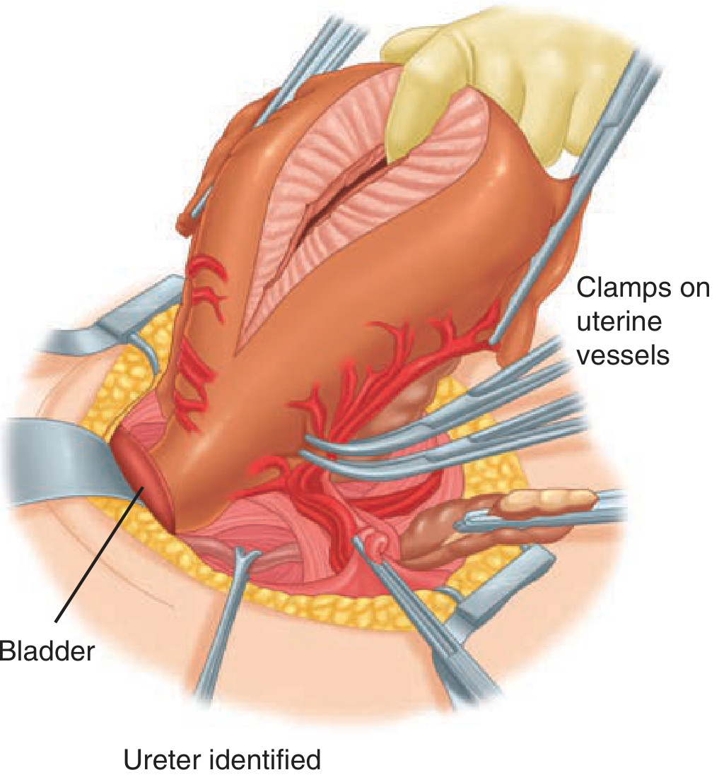
Here is the surgical image from Schwartz's/Williams showing clamps on uterine vessels during peripartum hysterectomy:
C. Pelvic Vessel Ligation
1. O'Leary Uterine Artery Ligation
- Described by O'Leary and O'Leary
- Series of sutures placed around branches of uterine artery + through myometrium
- Results in vessel compression against uterine wall
- Performed when major uterine laceration is not found at laparotomy
- First step in laparotomy for PPH (before hypogastric ligation)
2. Internal Iliac (Hypogastric) Artery Ligation (Burchell, 1964)
- If uterine artery ligation fails
- Mechanism (Burchell): Transient decrease in blood pressure and flow in regional pelvic vessels → hemostasis through clot formation
- Isolated 3 cm distal to bifurcation with external iliac artery
- Preserves fertility - collateral circulation is ample; women have delivered normally in subsequent pregnancies
- Success rate: Only ~50% (technically difficult, per Williams)
- Drawbacks: Technically demanding; risk to ureter; only 50% success
- Transient complications: mild bladder dysfunction, buttock pain (self-resolving)
Vascular Supply Anatomy:
Aorta
  └── Common Iliac
          ├── External Iliac → Femoral
          └── Internal Iliac (Hypogastric)
                  ├── Anterior Division
                  │       └── Uterine artery ← LIGATION POINT
                  │       └── Vaginal artery
                  │       └── Inferior vesical
                  └── Posterior Division
                          (Preserved - supplies gluteal region)
3. Stepwise Devascularisation (Abdrabbo's Steps):
- Unilateral uterine artery ligation
- Bilateral uterine artery ligation
- Utero-ovarian ligament ligation
- Bilateral internal iliac ligation
D. Angiographic Embolization
- Uterine artery embolization (UAE) via interventional radiology
- Balloon/embolization catheters in anterior divisions of internal iliac arteries
- Placed pre-operatively (in placenta accreta) or intraoperatively
- Requires facility + trained personnel
- Advantage: Fertility-preserving
- Used in haemodynamically stable patients
6. DEFINITIVE SURGICAL MANAGEMENT
Peripartum Hysterectomy (Last Resort)
Indications:
- Failure of all conservative measures
- Uterine rupture
- Placenta accreta spectrum (percreta, increta)
- Atony unresponsive to all treatments
- Patient with no further desire for fertility
Approach:
- Patient in semilithotomy position (allows simultaneous vaginal inspection)
- Sterile drapes permit vaginal/cervical exam by assistant to confirm cessation of bleeding
- Same steps as non-pregnant hysterectomy but DIFFERENT due to:
- Engorged, friable vessels
- Pliable, thinned tissues
- Distorted anatomy
Types:
- Total hysterectomy - preferred (removes cervix + body)
- Subtotal (supracervical) hysterectomy - faster, less morbidity; acceptable if bleeding is controlled
Key Steps:
- Identify and protect ureter (crucial - distorted anatomy)
- Bladder reflection downward
- Clamp uterine vessels bilaterally (see image above)
- Ligate uterosacral and cardinal ligaments
- Close vaginal vault (total) or leave cervix (subtotal)
Monitoring: CVP line / Swan-Ganz catheter for haemodynamic monitoring in massive PPH (Creasy & Resnik)
7. BLOOD PRODUCT RESUSCITATION (Massive Transfusion Protocol)
| Component | Ratio | Rationale |
|---|---|---|
| pRBC | 1 | Oxygen carrying capacity |
| FFP | 1 | Clotting factors |
| Platelets | 1 | 1:1:1 ratio (damage control resuscitation) |
| Cryoprecipitate | As needed | Fibrinogen < 2 g/L |
- Target Hb: > 8 g/dL in active PPH
- Target fibrinogen: > 2 g/L
- Avoid: Normal saline (hyperchloraemic acidosis); use Lactated Ringer's
- Point-of-care testing: ROTEM/TEG for coagulopathy guidance
8. COMPLETE MANAGEMENT ALGORITHM (Summary Table)
STEP 1 - MEDICAL (All cases of PPH)
├── Uterine massage (bimanual compression)
├── Oxytocin 20-40 U IV infusion
├── Methylergonovine 0.2 mg IM
├── Carboprost 250 μg IM (if no asthma)
├── Misoprostol 800 μg sublingual/rectal
├── Tranexamic acid 1 g IV (within 3 hrs)
└── IV fluids + blood products

STEP 2 - CONSERVATIVE SURGICAL (If Step 1 fails)
├── A. Uterine Tamponade
│     └── Bakri balloon / Foley / Gauze packing
├── B. Angiographic embolization (if stable + facilities)
└── (If above fail → Laparotomy)
    ├── C. Uterine compression sutures
    │     └── B-Lynch / Hayman / Cho
    ├── D. O'Leary uterine artery ligation
    └── E. Internal iliac artery ligation

STEP 3 - DEFINITIVE SURGICAL (Last resort)
└── Peripartum Hysterectomy
      ├── Total (preferred)
      └── Subtotal (faster, emergency)
Key High-Yield Points for MS Exam:
| Point | Detail |
|---|---|
| New PPH definition | ≥ 1000 mL OR hypovolemia signs (ACOG 2017) |
| Most common cause | Uterine atony (70-80%) |
| 4 T's | Tone, Trauma, Tissue, Thrombin |
| First uterotonic | Oxytocin (IV infusion, never IV bolus) |
| Carboprost CI | Asthma |
| Methylergonovine CI | Hypertension |
| TXA timing | Within 3 hours, 1g IV (WOMAN trial) |
| B-Lynch introduced | B-Lynch et al. 1997 |
| B-Lynch mechanism | Mechanical compression of uterine walls |
| B-Lynch suture material | No. 2 chromic catgut |
| Hayman modification | Simpler, no need to open uterus |
| Internal iliac ligation success | Only ~50% (technically difficult) |
| Hypogastric ligation mechanism | Burchell - reduces pulse pressure → thrombosis |
| Fertility after hypogastric ligation | Preserved (collateral circulation) |
| Last resort | Peripartum hysterectomy |
| Semilithotomy position | Allows vaginal exam during laparotomy |
Sources: Williams Obstetrics Ch. 41 (Obstetrical Hemorrhage); Creasy & Resnik's Maternal-Fetal Medicine; Schwartz's Principles of Surgery; ACOG Practice Bulletin 183 (2017); FIGO Recommendations 2022; WOMAN Trial (tranexamic acid)
This is a shared conversation. Sign in to Orris to start your own chat.