


Make me understnad this in a very bery easy way as per the order of book
🧠 8th Cranial Nerve - The Hearing Pathway (Super Easy)
What is this nerve?
- Hearing part (cochlear)
- Balance part (vestibular)
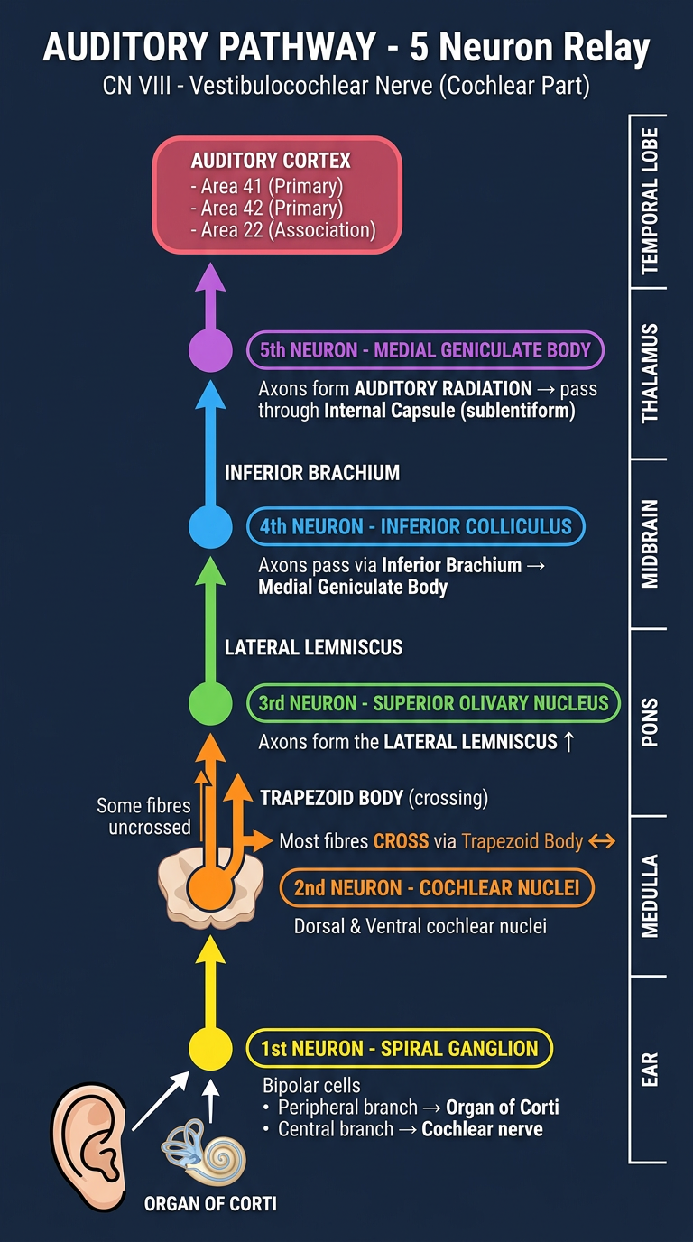
The Journey of Sound - 5 Relay Stations (Neurons)
🥇 NEURON 1 - In the Ear (Spiral Ganglion)
- These cells are bipolar (they have two branches - one going out, one going in)
- The outer branch → goes to the Organ of Corti (the actual hearing organ inside the cochlea - like a microphone)
- The inner branch → forms the cochlear nerve which travels to the brainstem
- The cochlear nerve ends at the dorsal and ventral cochlear nuclei (in the medulla)
🎤 Think of it as: the ear picks up sound → sends signal to the brainstem
🥈 NEURON 2 - In the Brainstem (Cochlear Nuclei, Medulla)
- Most axons cross to the opposite side (through the trapezoid body)
- They end in the superior olivary nucleus
- Some fibres also end in the nucleus of trapezoid body or lateral lemniscus
- A few fibres stay on the same side (uncrossed)
↔️ Think of it as: most sound signals CROSS to the other side of the brain here
🥉 NEURON 3 - Superior Olivary Nucleus (Pons)
- These neurons send their axons upward forming the lateral lemniscus
- The lateral lemniscus travels up and reaches the inferior colliculus (in the midbrain)
🛣️ Think of it as: signals travel up a highway called the lateral lemniscus
4️⃣ NEURON 4 - Inferior Colliculus (Midbrain)
- Axons pass through the inferior brachium (a small arm-like connection)
- They reach the medial geniculate body (in the thalamus)
- Note: Some lateral lemniscus fibres skip the inferior colliculus and go directly to the medial geniculate body
🔁 Think of it as: a processing/relay station in the midbrain before reaching the thalamus
5️⃣ NEURON 5 - Medial Geniculate Body (Thalamus)
- Axons form the auditory radiation
- They pass through the sublentiform part of the internal capsule
- They reach the auditory cortex in the temporal lobe (Areas 41 and 42)
- Area 41 = primary auditory cortex (actually hears/processes the sound)
- Area 42 = also primary auditory
- Area 22 = association auditory cortex (understands the meaning of sound)
🎧 Think of it as: the final destination - your brain consciously hears and understands the sound!
Quick Summary Table
| Neuron | Location | What it does |
|---|---|---|
| 1st | Spiral ganglion (ear) | Picks up sound from Organ of Corti |
| 2nd | Cochlear nuclei (medulla) | Crosses to opposite side via trapezoid body |
| 3rd | Superior olivary nucleus (pons) | Sends signals up via lateral lemniscus |
| 4th | Inferior colliculus (midbrain) | Relays to thalamus via inferior brachium |
| 5th | Medial geniculate body (thalamus) | Sends to auditory cortex via auditory radiation |
The Final Destination - Auditory Cortex (Temporal Lobe)
- High frequency sounds → processed at one end
- Low frequency sounds → processed at another end
- Located deep in the lateral sulcus of the temporal lobe
- Area 41 & 42 = hear the sound
- Area 22 = understand/associate the sound (like understanding speech)
- Sound crosses mainly at the 2nd neuron (trapezoid body) - so each ear sends signals to BOTH sides of the brain
- The pathway goes: Ear → Medulla → Pons → Midbrain → Thalamus → Temporal Cortex
- There are 5 neurons total in the pathway
Create a visual summary diagram of all 5 neurons in the auditory pathway
A clean, detailed medical education diagram showing the 5-neuron auditory pathway from the ear to the brain. The diagram should be vertical, flowing from bottom to top, with a dark navy blue background and bright colors for clarity. Layout from bottom to top: 1. At the bottom: Draw an EAR icon on the left. Label "ORGAN OF CORTI" with a small cochlea spiral. Arrow pointing up. 2. NEURON 1 (yellow circle): Label "1st NEURON - SPIRAL GANGLION" in a yellow rounded box. Text below: "Bipolar cells • Peripheral branch → Organ of Corti • Central branch → Cochlear nerve". Located at level: Inner Ear / Cochlea. 3. Arrow going up to a small structure labeled "MEDULLA" with level indicator on the right side. 4. NEURON 2 (orange circle): Label "2nd NEURON - COCHLEAR NUCLEI" in an orange rounded box. Text: "Dorsal & Ventral cochlear nuclei". Show a CROSSING arrow going to the right (labeled "Most fibres CROSS via Trapezoid Body ↔") and a small straight arrow (labeled "Some fibres uncrossed"). Level: Medulla. 5. Arrow going up labeled "TRAPEZOID BODY (crossing)" to level PONS. 6. NEURON 3 (green circle): Label "3rd NEURON - SUPERIOR OLIVARY NUCLEUS" in a green rounded box. Text: "Axons form the LATERAL LEMNISCUS ↑". Level: Pons. 7. Arrow going up labeled "LATERAL LEMNISCUS" to level MIDBRAIN. 8. NEURON 4 (blue circle): Label "4th NEURON - INFERIOR COLLICULUS" in a blue rounded box. Text: "Axons pass via Inferior Brachium → Medial Geniculate Body". Level: Midbrain. 9. Arrow going up labeled "INFERIOR BRACHIUM" to THALAMUS level. 10. NEURON 5 (purple circle): Label "5th NEURON - MEDIAL GENICULATE BODY" in a purple rounded box. Text: "Axons form AUDITORY RADIATION → pass through Internal Capsule (sublentiform)". Level: Thalamus. 11. Arrow going up to TEMPORAL LOBE (pink/red box at top): "AUDITORY CORTEX - Area 41 (Primary) - Area 42 (Primary) - Area 22 (Association)". On the RIGHT side of the diagram, show vertical level labels: EAR → MEDULLA → PONS → MIDBRAIN → THALAMUS → TEMPORAL LOBE. Title at the top in bold white text: "AUDITORY PATHWAY - 5 Neuron Relay" Subtitle: "CN VIII - Vestibulocochlear Nerve (Cochlear Part)" Style: Clean medical textbook illustration, color-coded neurons, clear arrows, easy to read font, professional anatomy diagram
| Color | Neuron | Location |
|---|---|---|
| 🟡 Yellow | 1st Neuron | Spiral Ganglion (Ear) |
| 🟠 Orange | 2nd Neuron | Cochlear Nuclei (Medulla) |
| 🟢 Green | 3rd Neuron | Superior Olivary Nucleus (Pons) |
| 🔵 Blue | 4th Neuron | Inferior Colliculus (Midbrain) |
| 🟣 Purple | 5th Neuron | Medial Geniculate Body (Thalamus) |
| 🔴 Pink/Red | Final destination | Auditory Cortex - Areas 41, 42, 22 (Temporal Lobe) |

Make me understand this in simple way as per the order of book
📖 BD Chaurasia - Page 77 (Simple Explanation)
PART 1: VESTIBULAR PATHWAY (Balance Pathway)
What are the receptors? (What picks up the signal?)
| Receptor | Location | What it detects |
|---|---|---|
| Maculae (of saccule & utricle) | Inner ear | Static balance - like knowing you're standing still or tilting |
| Crista ampullaris (of semicircular ducts) | Inner ear | Kinetic balance - movement, rotation |
How do the nerve fibres travel?
- Bundle 1: Fibres from the crista of anterior & lateral semicircular canals + some maculae fibres → travel through the superior vestibular area of the internal acoustic meatus
- Bundle 2: Fibres from crista of posterior semicircular canal → travel through foramen singulare
- Bundle 3: Most fibres from maculae of utricle & saccule → travel through the inferior vestibular area
Neuron 1 - Vestibular Ganglion
- Cell bodies are in the vestibular ganglion (inside internal acoustic meatus)
- Their central processes form the vestibular nerve
- The vestibular nerve ends in the vestibular nuclei (in the brainstem)
Neuron 2 - Vestibular Nuclei (in brainstem)
- Via the inferior cerebellar peduncle (vestibulocerebellar tract)
- For coordination and smooth movement
- Through the medial longitudinal bundle (MLF)
- To motor nuclei of CN III, IV, VI (eye movement) and CN XI (neck)
- This is why your eyes reflexively move when your head turns! (vestibulo-ocular reflex)
- Through the vestibulospinal tract
- To anterior horn cells of spinal cord
- Controls trunk and limb muscles for balance
- For conscious awareness of balance
🎯 The big picture: Through this pathway, the labyrinth (inner ear) can control movements of the eyes, head, neck and trunk - basically your entire posture and balance!
PART 2: CLINICAL ANATOMY (Diseases - Very Important for Exams!)
👂 DEAFNESS - 3 Types
- Sound waves fail to reach the cochlea
- Problem is in the outer/middle ear (like wax blockage, perforated eardrum, fluid in middle ear)
- The nerve itself is fine
- Sound reaches the cochlea, but there is failure to produce or transmit action potential
- Problem can be in:
- The cochlea itself
- The cochlear nerve
- The cochlear nerve's central connections
- A bilateral or dominant posterior temporal lobe lesion
- The ear and nerve work perfectly - the person CAN hear sounds
- But they CANNOT understand spoken language (like hearing noise but not making sense of words)
🌀 VERTIGO
- What it is: An illusion of rotatory movement - the patient feels the environment is spinning around them
- Cause: Disturbed orientation of the body in space
- Due to: Disease of the vestibular nerve
Easy way to remember: Vertigo = "the room is spinning" - it's NOT dizziness, it's a spinning illusion
🔔 TINNITUS
- What it is: A sensation of buzzing, ringing, hissing or singing in the ear
- Can be:
- Unilateral (one ear) or bilateral (both ears)
- High pitch or low pitch
- Continuous or intermittent (comes and goes)
🌊 MENIERE'S SYNDROME
- Classic triad: Tinnitus + Vertigo + Hearing loss (all three together, recurrent attacks)
- Plus: Sensitivity to noises
- Who gets it: Middle-aged or older persons
- Cause: Increase in volume of endolymph (the fluid inside the inner ear increases, causing pressure)
Memory trick: Meniere's = More endolymph = Middle-aged
🧠 ACOUSTIC NEUROMA
- What it is: A slow growing benign tumor of neurolemmal (Schwann) cells
- Grows on the vestibulocochlear nerve (CN VIII)
- First symptom: Early loss of hearing (because hearing fibres are affected first)
Quick Summary Table
| Condition | Key Feature | Remember as |
|---|---|---|
| Conductive deafness | Sound can't reach cochlea | Outer/middle ear problem |
| Sensorineural deafness | Nerve/cochlea problem | Inner ear/nerve |
| Cortical deafness | Hears but can't understand | Brain problem |
| Vertigo | Room spinning illusion | Vestibular nerve disease |
| Tinnitus | Ringing/buzzing in ear | Any pitch, any side |
| Meniere's syndrome | Tinnitus + Vertigo + Deafness | Excess endolymph |
| Acoustic neuroma | Benign Schwann cell tumor | Early hearing loss |
Tell the vestibular pathway in short
Vestibular Pathway (Short & Simple)
Neuron 1 - In the Inner Ear
- Maculae (saccule & utricle) → static balance
- Crista ampullaris (semicircular ducts) → kinetic balance (movement)
Neuron 2 - Vestibular Nuclei → sends fibres to 4 places:
| Destination | Pathway | Effect |
|---|---|---|
| Cerebellum | Inferior cerebellar peduncle (vestibulocerebellar tract) | Coordination |
| Eye/neck muscles (CN III, IV, VI, XI) | Medial Longitudinal Bundle (MLF) | Eyes move when head turns |
| Spinal cord (anterior horn) | Vestibulospinal tract | Balance of trunk & limbs |
| Thalamus → Frontal cortex | - | Conscious awareness of balance |
One Line Summary:
Inner ear receptors → Vestibular ganglion (N1) → Vestibular nuclei (N2) → Cerebellum / Eye muscles / Spinal cord / Cortex


Make me understand this in a bery very simple way as per the order of book
🧠 9th Cranial Nerve - GLOSSOPHARYNGEAL NERVE
(BD Chaurasia - Pages 77 to 80)
STEP 1: What is this nerve? (Introduction)
- It is the 9th cranial nerve
- It is the nerve of the 3rd branchial arch (means it developed from the 3rd gill arch in the embryo)
What does it DO? (Functions in one shot)
| Function | Where |
|---|---|
| Motor | Stylopharyngeus muscle (only one muscle!) |
| Secretomotor | Parotid gland (makes saliva) |
| Taste (Gustatory) | Posterior 1/3 of tongue + circumvallate papillae |
| Sensory | Pharynx, tonsil, soft palate, posterior 1/3 of tongue, carotid body, carotid sinus |
Easy way to remember: "Gloss-o-Pharyngeal" = Glosso (tongue) + Pharyngeal (throat) - it's the tongue + throat nerve!
STEP 2: Functional Components (5 types of fibres)
1. SVE - Special Visceral Efferent (Motor fibre)
- Arises from nucleus ambiguus (in medulla)
- Supplies the stylopharyngeus muscle (a muscle that lifts the pharynx during swallowing)
2. GVE - General Visceral Efferent (Secretomotor/Parasympathetic fibre)
- Arises from inferior salivatory nucleus (in medulla)
- Preganglionic fibres travel to the otic ganglion
- From otic ganglion, postganglionic fibres join the auriculotemporal nerve → reach the parotid gland
- This makes the parotid gland secrete saliva
3. GVA - General Visceral Afferent (General sensory fibre)
- Cell bodies in inferior ganglion of the nerve
- Carries sensations from: pharynx, palate, posterior 1/3 of tongue, tonsil, carotid body, carotid sinus
- Central processes go to lower part of nucleus of solitary tract
4. SVA - Special Visceral Afferent (Taste fibre)
- Cell bodies in inferior ganglion
- Carries taste from posterior 1/3 of tongue + circumvallate papillae
- Central processes go to nucleus of solitary tract
5. GSA - General Somatic Afferent (Somatic sensory fibre)
- Cell bodies in inferior ganglion
- Carries general sensations from middle ear, proprioception from stylopharyngeus
- Central processes go to nucleus of spinal tract of trigeminal
STEP 3: The Three Nuclei (in upper part of medulla)
| Nucleus | Type | Function |
|---|---|---|
| Nucleus ambiguus | Branchiomotor (SVE) | Motor to stylopharyngeus |
| Inferior salivatory nucleus | Parasympathetic (GVE) | Secretomotor to parotid |
| Nucleus of tractus solitarius | Sensory (GVA + SVA) | Receives taste + visceral sensations |
STEP 4: Course and Relations (How the nerve travels)
Inside the skull (Intracranial course):
- Fibres pass forwards and laterally between the olivary nucleus and inferior cerebellar peduncle, through the reticular formation of the medulla
- At the base of the brain, the nerve is attached by 3-4 filaments to the posterolateral sulcus of the medulla (just above the rootlets of vagus nerve)
- These filaments unite to form a single trunk which passes forward and laterally toward the jugular foramen, crossing and grooving the jugular tubercle of the occipital bone
Leaving the skull:
- The nerve leaves the skull through the jugular foramen (middle part), anterior to vagus and accessory nerves. It has its own separate sheath of dura mater
- In the jugular foramen, it is lodged in a deep groove leading to the cochlear canaliculus, and is separated from vagus and accessory nerves by the inferior petrosal sinus
Outside the skull (Extracranial course):
STEP 5: The Two Ganglia
| Ganglion | Size | Features |
|---|---|---|
| Superior ganglion | Small | Detached part of inferior ganglion - gives NO branches |
| Inferior ganglion | Larger | Sits in notch on lower border of petrous temporal bone - gives communicating and tympanic branches |
STEP 6: Branches and Distribution (6 branches)
1. 🦻 Tympanic Nerve (most important branch!)
- Branch of the inferior ganglion
- Enters the middle ear through tympanic canaliculus
- Forms the tympanic plexus in the middle ear
- Distributes to: middle ear, auditory tube, mastoid antrum and air cells
- One branch of this plexus = Lesser petrosal nerve
- Contains preganglionic secretomotor fibres for parotid gland
- Relays in otic ganglion
- Post-ganglionic fibres join auriculotemporal nerve → reach parotid gland
2. 🩸 Carotid Branch
- Descends on the internal carotid artery
- Supplies carotid sinus (pressure receptor) and carotid body (oxygen sensor)
3. 🗣️ Pharyngeal Branches
- Join with vagus and sympathetic fibres to form the pharyngeal plexus
- Supplies mucous membrane of pharynx and palate
4. 💪 Muscular Branch
- Supplies the stylopharyngeus muscle (the ONLY muscle CN IX supplies!)
5. 🍒 Tonsillar Branches
- Supply the tonsil
- Join the lesser palatine nerves to form a plexus
- Fibres go to soft palate and palatoglossal arches
6. 👅 Lingual Branches
- Carry taste AND general sensations from the posterior 1/3 of tongue including circumvallate papillae
Quick Summary - CN IX in One Picture
GLOSSOPHARYNGEAL NERVE (CN IX)
|
Jugular Foramen
|
Inferior Ganglion
/ \
Tympanic nerve Carotid branch
(Middle ear → (Carotid sinus
Otic ganglion → & carotid body)
Parotid gland)
|
Around Stylopharyngeus
|
Pharyngeal branches → Pharynx & palate
Muscular branch → Stylopharyngeus
|
Submandibular region
|
Tonsillar branches → Tonsil, soft palate
Lingual branches → Post 1/3 tongue (taste + sensation)
Memory Tricks
| Thing to remember | Trick |
|---|---|
| Only muscle CN IX supplies | "Style" - Stylopharyngeus |
| Taste area | Posterior 1/3 of tongue (CN VII does anterior 2/3!) |
| Leaves skull via | Jugular foramen (along with CN X and XI) |
| Parotid gland secretion | Inferior salivatory → Otic ganglion → Auriculotemporal → Parotid |
| Carotid body/sinus | CN IX monitors blood pressure (sinus) and oxygen (body) |
Tell the pathway in short
CN IX - Glossopharyngeal Nerve: Course (Short)
Intracranial (Inside skull)
At Jugular Foramen
Extracranial (Outside skull) - 3 steps
One Line Summary
Medulla → Jugular foramen → Between IJV & ICA → Around stylopharyngeus → Between carotids → Pharynx → Deep to hyoglossus → Tongue & tonsil