
Act as a peditric radiologist with specialisation in temporal bone (CT and MRI). Explain pediatric temporal bone anatomy, pathologies, their imaging appearances, congenital anomalies, cochlear implant basics in a structured format. Use professional terms and explain in depth
temporal bone CT axial cochlea vestibule semicircular canal anatomy
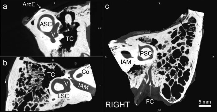
This diagnostic image consists of three high-resolution micro-CT scans (planes a, b, and c) of the right human temporal bone, focusing on the inner ear anatomy. Plane (a), known as the Pöschl plane, demonstrates the anterior semicircular canal (ASC) in its circular orientation, the vestibule (V), and the overlying arcuate eminence (ArcE), with the tympanic cavity (TC) located laterally. Plane (b) is an axial view tilted parallel to the lateral semicircular canal (LSC), showing its characteristic 'signet ring' appearance, the vestibule (V), the internal acoustic meatus (IAM), the cochlea (Co), and Bill’s bar (asterisk). Plane (c) displays the posterior semicircular canal (PSC) plane, revealing the PSC and common crus in relation to the IAM and the facial canal (FC). These planes are used for precise anatomical mapping and surgical planning for procedures like stapedotomy. The images illustrate the complex spatial relationships between the vestibular system, the facial nerve, and the middle ear space.
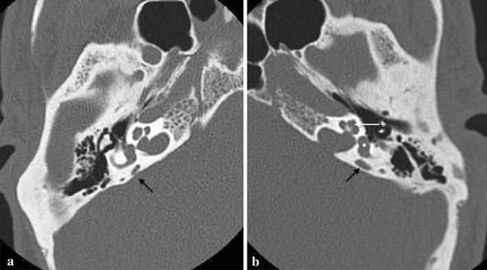
This diagnostic image consists of two high-resolution axial CT scans (labeled a and b) of the temporal bone at the level of the internal auditory canal (IAC), demonstrating the classic radiological features of Enlarged Vestibular Aqueduct (EVA) syndrome. In both images, black arrows point to a significantly dilated vestibular aqueduct, which appears as a broad, fluid-filled bony canal extending posteriorly and laterally from the vestibule toward the posterior cranial fossa. For anatomical reference in image b, the cochlea is marked with a white arrow and the vestibule with an asterisk. Despite the prominent aqueduct enlargement, the cochlea displays normal turns and internal architecture, and the vestibule and semicircular canals appear morphologically preserved. This bilateral finding is a frequent cause of congenital or progressive sensorineural hearing loss in pediatric patients. The images contrast the pathological widening of the aqueduct with the otherwise normal anatomy of the otic capsule.
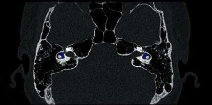
This diagnostic axial CT scan displays the skull base and temporal bones with a focus on the inner ear anatomy. The image demonstrates high-attenuation cortical bone surrounding the pneumatized spaces of the mastoid air cells and the petrous portion of the temporal bone. Bilaterally, the vestibule of the inner ear is highlighted with a blue overlay, illustrating its central position within the bony labyrinth. The highlighted vestibule appears as a low-attenuation, fluid-filled ovoid space situated between the anteriorly located cochlea and the posteriorly oriented semicircular canals. Other visible landmarks include the sphenoid sinus and the internal auditory canal. The image serves as a clinical reference for segmenting small, high-precision anatomical structures of the ear, which is vital for diagnosing vestibulocochlear disorders and planning microsurgical interventions. The high contrast between the dense otic capsule and the delicate membranous labyrinth components is characteristic of high-resolution temporal bone imaging.
cholesteatoma CT temporal bone erosion mastoid
Mondini malformation incomplete partition cochlea congenital inner ear
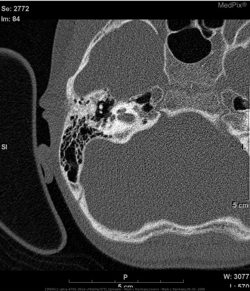
cochlear implant post-operative CT electrode array
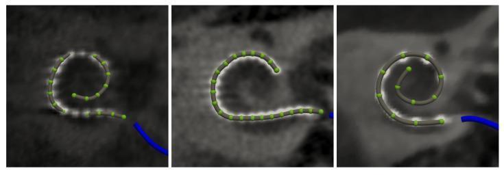
This clinical diagnostic image features three side-by-side panels demonstrating automated electrode array detection from cochlear imaging (CT/CBCT). Each panel displays a grey, elongated cochlear implant electrode array superimposed on radiological cross-sections of the temporal bone. The arrays follow the natural spiral morphology of the cochlea, with individual electrodes represented by equidistant green spherical markers. The left image shows an Oticon Medical EVO electrode array with a tight spiral configuration. The middle image depicts a Cochlear Nucleus CI622 with a more open 'C-shaped' curvature. The right image illustrates a MED-EL FLEX24 array with a moderate spiral turn. A blue curvilinear element at the base of each array denotes the extracochlear connection point. This visual serves to validate the performance of automated segmentation software (Nautilus) across different commercial implant designs, focusing on tonotopic mapping, insertion depth, and accurate spatial localization of electrodes within the scala tympani or scala vestibuli for post-operative clinical assessment.
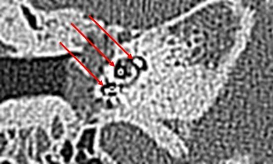
This diagnostic image is a Multi-Slice Computed Tomography (MSCT) scan of the temporal bone, focusing on the cochlear anatomy. The image demonstrates a translocated cochlear implant electrode. Three red arrows point to hyperdense metallic artifacts representing the electrode array within the basal and first turns of the cochlea. The visualization highlights a scalar translocation, where the electrode has deviated from its intended position (typically the scala tympani) into adjacent cochlear compartments such as the scala vestibuli. The image shows the characteristic diminishing signal and metallic blooming artifact associated with medical implants in CT imaging. This visual serves as a clinical example of post-operative assessment for cochlear implant positioning and the identification of insertion trauma or electrode misplacement. The high-resolution cross-sectional view allows for the evaluation of the electrode's relationship to the basilar membrane and osseous spiral lamina.
congenital aural atresia external auditory canal CT
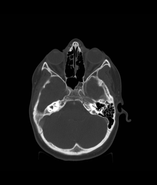
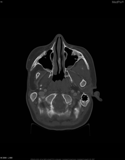
mastoiditis coalescent CT scan temporal bone pediatric complication
Pediatric Temporal Bone: A Comprehensive Radiological Guide
SECTION 1: OVERVIEW OF IMAGING MODALITIES
1.1 High-Resolution Computed Tomography (HRCT)
- Slice thickness: 0.5-0.625 mm with multiplanar reconstruction in axial and coronal planes
- Bone algorithm (kernel): Sharp/ultra-sharp kernel to resolve fine bony detail
- Field of view: Small, centered on each temporal bone individually
- Radiation dose: Low-dose protocols with iterative reconstruction are mandatory in pediatric patients (ALARA principle)
- No contrast is typically required for most indications
- External auditory canal (EAC) atresia/stenosis
- Middle ear ossicular chain integrity
- Mastoid pneumatization and air cell architecture
- Otic capsule bony detail (cochlea, vestibule, semicircular canals)
- Tegmen and lateral wall of the epitympanum (scutum)
- Facial nerve canal (tympanic and mastoid segments)
- Internal auditory canal (IAC) bony dimensions
1.2 MRI of the Temporal Bone
- T2 SPACE/CISS/FIESTA (heavily T2-weighted 3D sequences): Gold standard for visualizing the fluid-filled membranous labyrinth, cochlear nerve within the IAC, and perilymph/endolymph spaces. The term "seven-up and coke down" is a useful mnemonic - the facial nerve is superior-anterior, cochlear nerve inferior-anterior, superior vestibular nerve superior-posterior, and inferior vestibular nerve inferior-posterior within the IAC on sagittal-oblique views.
- T1 pre/post-gadolinium: Labyrinthine enhancement indicates labyrinthitis or nerve enhancement (geniculate ganglion enhancement on T1 post-Gd can be normal due to perineural vessels; labyrinthine segment enhancement is always abnormal)
- Non-EPI Diffusion-Weighted Imaging (non-EPI DWI): Critical for cholesteatoma detection. Non-echo-planar DWI is preferred over standard EPI-DWI because it produces far fewer susceptibility artifacts at the bone-soft tissue interface at the skull base.
- MRI for cochlear implant candidates: Currently the modality of choice - it demonstrates cochlear nerve caliber, cochlear patency (fibrosis/ossification), and IAC anatomy. - Cummings Otolaryngology, p. 3710
SECTION 2: PEDIATRIC TEMPORAL BONE ANATOMY
2.1 Macroscopic Osseous Anatomy
| Part | Features | Pediatric Notes |
|---|---|---|
| Squamous | Largest part; forms lateral temporal fossa; zygomatic process anteriorly; suprameatal (Macewen's) triangle | Pterion - junction with greater wing of sphenoid and parietal bone - overlies the middle meningeal artery |
| Petromastoid | Petrous pyramid; contains inner ear, IAC, and carotid canal; mastoid portion fills with air cells | Petrous part is full adult size at birth; mastoid process absent at birth |
| Tympanic | C-shaped ring forming most of the EAC wall and floor of the bony canal | Incomplete ring at birth (Santorini's fissures persist briefly) |
| Styloid | Styloid process; stylomastoid foramen | At birth the stylomastoid foramen is near the lateral skull surface - the extracranial facial nerve is extremely superficial and vulnerable |
2.2 External Auditory Canal (EAC)
- The bony EAC is formed primarily by the tympanic part (posterior, inferior, anterior walls) and squamous/petromastoid portions (superior/posterior-superior wall)
- Length: approximately 2.5 cm in adults; significantly shorter in neonates
- Normal adult caliber: 5-9 mm
- The anterior wall also forms the posterior wall of the temporomandibular joint (TMJ) - TMJ effusion or condylar pathology can be appreciated on temporal bone CT
2.3 Middle Ear (Tympanic Cavity)
| Compartment | Location | Key Structures |
|---|---|---|
| Epitympanum (Attic) | Superior to tympanic membrane (TM) | Head of malleus, body and short process of incus; Prussak's space (lateral epitympanic recess) |
| Mesotympanum | Level of TM | Manubrium of malleus, long process of incus, stapes, oval window, round window niche, cochlear promontory, tensor tympani |
| Hypotympanum | Below TM | Jugular bulb (variable height); aberrant internal carotid artery |
| Protympanum | Anterior; Eustachian tube orifice | Carotid canal posterolateral wall |
- Malleus: Head (in attic), neck, manubrium (handle in mesotympanum)
- Incus: Body articulates with malleus head; long process (medial to TM) terminates in lenticular process; short process points posteriorly (useful landmark)
- Stapes: Superstructure (anterior and posterior crura + capitulum) and footplate (fixed in oval window by annular ligament)
- Ice cream cone sign: Normal malleoincudal articulation on coronal CT - the head of malleus forms the "cone" and the body of incus forms the "scoop"
- Scutum (lateral attic wall): The sharp bony spur separating the EAC from the epitympanum. Blunting or erosion of the scutum is an early and sensitive sign of pars flaccida cholesteatoma.
- Tegmen tympani: Thin bony roof separating middle ear from middle cranial fossa
- Facial nerve tympanic segment: Runs in the facial nerve canal on the medial wall of the middle ear, just superior to the oval window niche - at risk in cholesteatoma surgery
2.4 Inner Ear (Otic Capsule / Bony Labyrinth)
- 2.5-2.75 turns in a normal adult cochlea
- Three scalae: scala vestibuli (superior), scala media/cochlear duct (contains organ of Corti and endolymph), scala tympani (inferior)
- Basal turn of cochlea is visible on axial CT as a round/oval structure anterior to the IAC
- Modiolus: central bony axis of the cochlea - a normal modiolus is visible on HRCT
- Tonotopic organization: high frequencies (up to 20 kHz) in the basal turn; low frequencies (20 Hz) at the apex
- Central chamber of the bony labyrinth
- Contains utricle and saccule (static labyrinth - detects gravity/linear acceleration)
- Normal dimensions: anteroposterior ~4-5 mm
- Three orthogonal rings: superior (anterior), lateral (horizontal), posterior
- Lateral SCC is the most commonly visible on standard axial HRCT and serves as a key surgical landmark
- Each SCC has an ampullated end containing the crista ampullaris
- Superior and lateral SCCs share the common crus
- Normal width: 4-8 mm (variation is significant)
- Contains: facial nerve (VII, anterior-superior), cochlear nerve (VIII, anterior-inferior), superior vestibular nerve (posterior-superior), inferior vestibular nerve (posterior-inferior)
- Bill's bar: Vertical bony crest within the lateral IAC separating the facial from superior vestibular nerve
- Transverse crest (falciform crest): Horizontal bony crest separating superior (facial + superior vestibular) from inferior (cochlear + inferior vestibular) nerves
- Labyrinthine segment - shortest, from IAC fundus to geniculate ganglion
- Tympanic (horizontal) segment - runs medial to the middle ear, superior to oval window
- Mastoid (vertical) segment - descends to stylomastoid foramen
SECTION 3: IMAGING ANATOMY ON STANDARD CT PLANES
Key Axial CT Landmarks (superior to inferior):
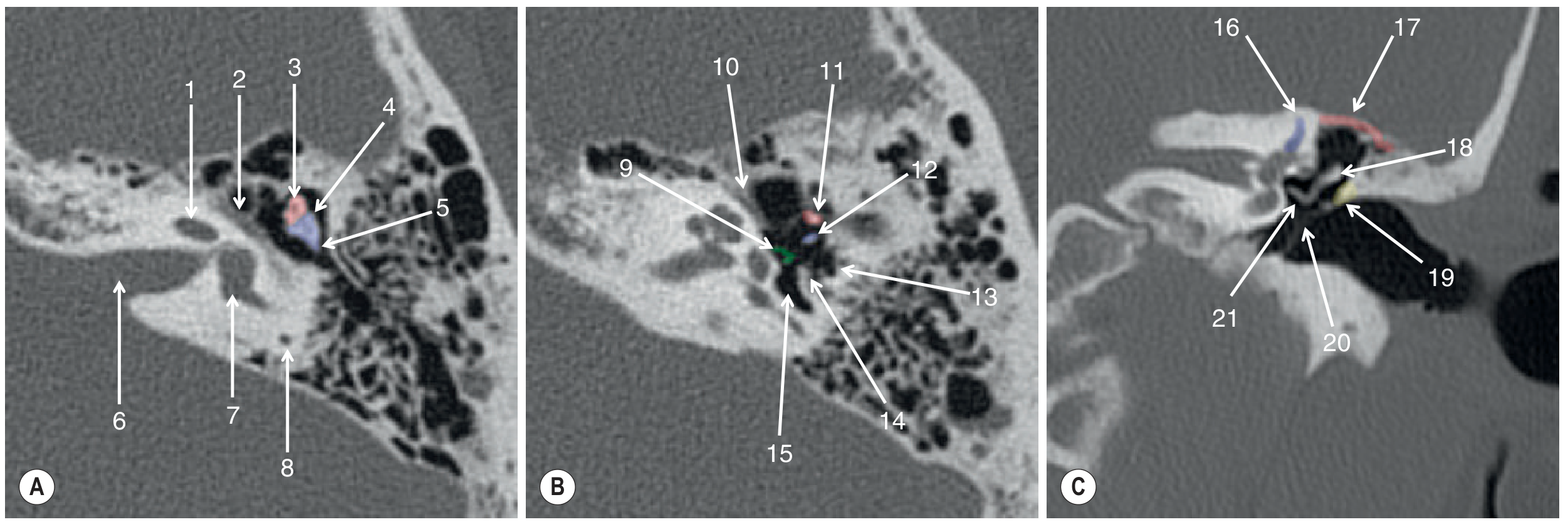
- Superior cuts: Superior SCC (arcuate eminence), tegmen
- Mid-cuts: IAC, cochlea, vestibule, lateral SCC (signet ring appearance), malleus head, incus body
- Lower cuts: Basal turn of cochlea, round window niche, hypotympanum, EAC
- Anterior: Cochlea, tensor tympani, carotid canal
- Mid: Scutum, malleus manubrium, oval window, facial nerve tympanic segment
- Posterior: Posterior SCC, sigmoid sinus, mastoid air cells
SECTION 4: CONGENITAL ANOMALIES
4.1 Classification of Inner Ear Malformations (Sennaroglu Classification)
| Grade | Anomaly | CT/MRI Features |
|---|---|---|
| Michel deformity | Complete aplasia of inner ear | No cochlea, no vestibule, no SCCs; dense petrous bone; CI contraindicated |
| Cochlear Aplasia | Absent cochlea with present vestibule/SCCs | Flat anterior otic capsule; present but malformed vestibule |
| Common Cavity | Cochlea and vestibule merged into single ovoid/round cyst | Single featureless ovoid cavity without internal structure; no modiolus; CI possible but outcomes variable |
| Incomplete Partition Type I (IP-I, "Cystic cochlea") | No modiolus, no inter-scalar septa; cochlear cyst-like appearance | Cochlea appears round/cystic on axial CT with no internal architecture; enlarged vestibule; often associated with CSF gusher during CI surgery |
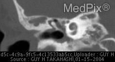
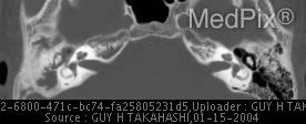
| Incomplete Partition Type II (IP-II, "Mondini malformation") | Deficient inter-scalar partitioning; only basal turn normally formed; dilated vestibule; large vestibular aqueduct | On axial CT: basal turn normal, middle and apical turns fused/cystic; modiolus partially present; enlarged vestibular aqueduct (>1.5mm at midpoint, >2mm at operculum) |
| Incomplete Partition Type III (IP-III) | X-linked deafness; absent modiolus; "bolt-hole" appearance | IAC opens directly into cochlea; high perilymph gusher risk; dilated IAC fundus |
| Cochlear Hypoplasia | Small but formed cochlea | Cochlear diameter <4 mm; fewer turns; preserved internal architecture |
4.2 Enlarged Vestibular Aqueduct (EVA) / Large Vestibular Aqueduct Syndrome (LVAS)
- Vestibular aqueduct midpoint diameter >1.5 mm (Cincinnati criteria) or >2 mm at the operculum (Valvassori criteria)
- On MRI T2: enlarged endolymphatic duct and sac posterior to the posterior SCC
- SNHL is often progressive and may be precipitated by minor head trauma or Valsalva maneuver (classic history in a child with progressive hearing loss after a fall)
- Associated with Pendred syndrome (EVA + hypothyroid goiter; SLC26A4 gene mutation)
- May coexist with Mondini malformation (IP-II)
- Both MRI and CT are recommended: MRI to demonstrate enlarged endolymphatic duct and sac; CT to confirm bony aqueduct dilation
4.3 External and Middle Ear Congenital Anomalies
- EAC absent (atresia) or severely stenotic
- Most unilateral; associated with microtia
- Jahrsdoerfer Grading Scale on HRCT guides surgical candidacy (10-point scale):
- Stapes present (2 pts), oval window open (1 pt), middle ear space (1 pt), facial nerve (1 pt), malleoincudal complex (1 pt), mastoid pneumatization (1 pt), incudostapedial connection (1 pt), round window (1 pt), appearance of external ear (1 pt)
- Score ≥7/10 = good surgical candidate for canaloplasty/tympanoplasty
- Atretic plate replacing the EAC (bony atresia most common; fibrous atresia less common)
- Ossicular chain anomalies: fused malleus-incus mass fixed to atretic plate or medial wall (very common); hypoplastic stapes; absent stapes
- Small or absent middle ear space
- Anteriorly displaced tympanic segment of facial nerve (critical surgical risk)
- Oval window stenosis/atresia (affects surgical planning)
- Normal inner ear in most cases (key point: cochlear function preserved; bone conduction thresholds should be tested)
- Congenital stapes fixation (most common cause of isolated congenital conductive hearing loss)
- Malleus-incus fusion or fixation to the lateral attic wall
- Absent long process of incus
- These are often identified on HRCT as thickened or fused ossicular segments
4.4 Branchial Apparatus Anomalies
- First branchial cleft cysts/sinuses: May present as a preauricular pit or mass; Type I is near the EAC (superficial to parotid), Type II extends into the EAC or even into the middle ear
- CT shows a cystic structure +/- tract in relationship to the EAC; intimate relationship to the facial nerve mandates pre-operative MRI/CT to identify nerve course
SECTION 5: ACQUIRED PATHOLOGIES
5.1 Otitis Media (OM) and Mastoiditis
- CT is not routinely indicated for uncomplicated AOM
- When imaged: opacification of the middle ear cleft and mastoid air cells (bilateral opacification of mastoid air cells in a young child is frequently a normal or incidental finding)
- CT indication: Suspected complication (see below)
- CT findings: Opacification of mastoid air cells WITH loss/destruction of bony septa between air cells (coalescence); periosteal reaction/subperiosteal abscess (lateral to mastoid cortex); erosion of mastoid cortex
- Subperiosteal abscess appears as a rim-enhancing collection on CT with contrast, displacing the pinna anterolaterally (the ear sticks out - classic clinical sign)
| Complication | CT Finding | MRI Finding |
|---|---|---|
| Subperiosteal abscess | Lateral cortex erosion; soft tissue collection | Rim-enhancing collection with restricted diffusion |
| Bezold's abscess | Erosion of mastoid tip; collection tracking into neck (under SCM) | Collection along SCM |
| Sigmoid sinus thrombosis | Absence of normal flow void in sigmoid sinus | "Delta sign" on MRV; T1 hyperintense thrombus |
| Epidural empyema | Lenticular epidural collection | Restricts on DWI; rim enhancement |
| Labyrinthitis | Normal CT | Labyrinthine enhancement on T1 Gd |
| Meningitis | CT may be normal or show meningeal enhancement | Leptomeningeal enhancement |
| Petrous apicitis (Gradenigo's) | Opacification + destruction of petrous apex | Petrous apex fluid + enhancement |
5.2 Cholesteatoma
-
Congenital Cholesteatoma (CC): "An expanding cystic mass with keratinizing squamous epithelium located medially to the intact tympanic membrane, assumed to be present at birth but usually diagnosed during infancy or early childhood in patients with no prior history of otorrhea, perforation, or previous ear surgery" - Scott-Brown's Vol. 2, p. 4511. The most accepted etiology is persistence of epidermoid cell rests in the anterior epitympanum.
-
Acquired Cholesteatoma: Much more common. Pars flaccida type (Prussak's space, lateral attic) is most common; pars tensa type occurs in posterior mesotympanum.
- Stage I: Single quadrant, no ossicular or mastoid involvement
- Stage II: Multiple quadrants, no ossicular or mastoid involvement
- Stage III: Ossicular involvement/erosion; no mastoid
- Stage IV: Mastoid extension
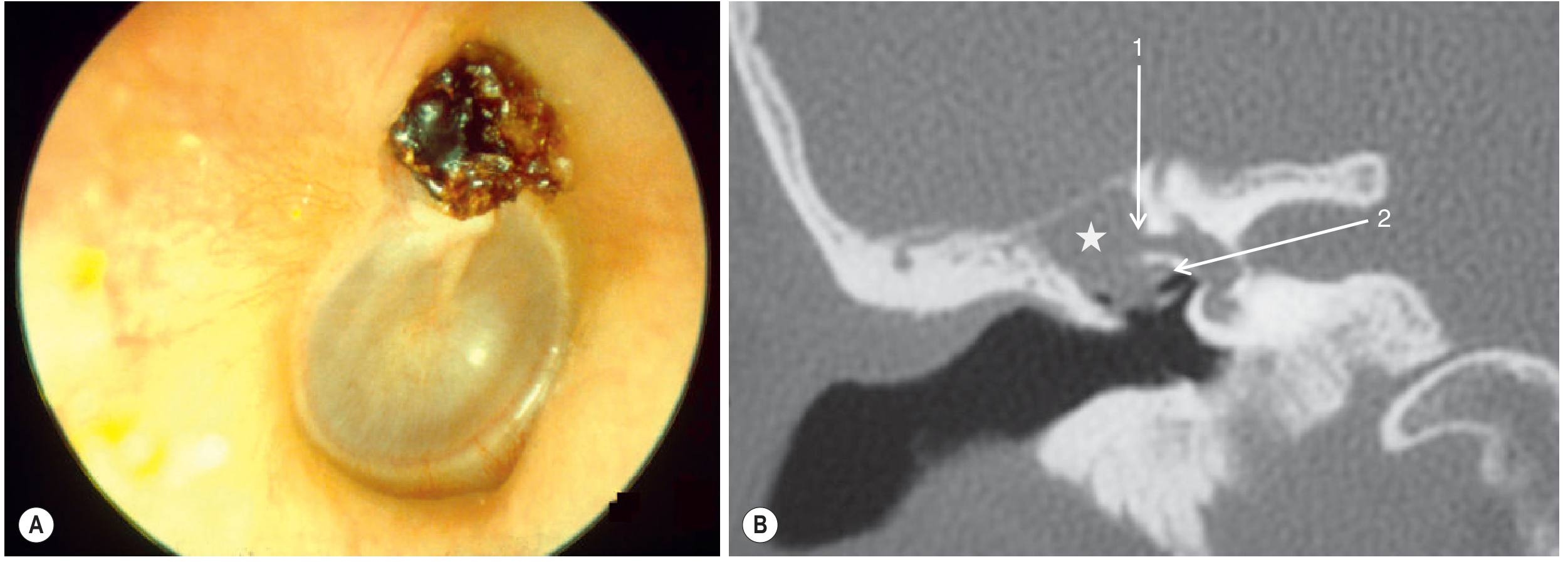
- Soft tissue opacification in the affected compartment (attic most commonly for pars flaccida type)
- Scutum erosion: Key early sign on coronal CT - blunting or frank erosion of the lateral attic wall
- Ossicular erosion: Long process of incus eroded most commonly; stapes superstructure; lenticular process
- Lateral SCC fistula: Erosion of the otic capsule over the lateral SCC - associated with vestibular symptoms; requires careful identification pre-operatively
- Tegmen erosion: Extension into middle cranial fossa
- Facial nerve canal erosion: Tympanic segment most vulnerable
- Sigmoid sinus plate erosion: Risk of venous sinus thrombosis
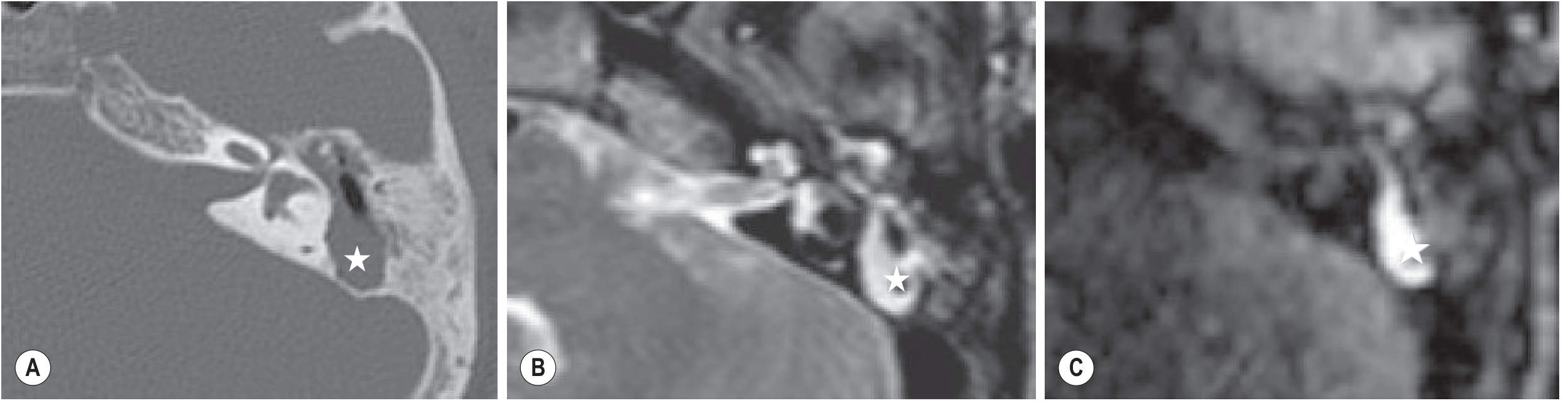
- Non-EPI DWI (HASTE-based or PROPELLER/BLADE DWI): Cholesteatoma = markedly restricted diffusion (bright on b1000, dark on ADC). This is the key differentiating feature from granulation tissue, scar, fluid, or mucosal disease which do NOT restrict
- Preferred for surveillance after canal-wall-up (CWU) surgery - detects residual/recurrent disease (minimum lesion size detectability ~3-5 mm with newer sequences)
- T1: Isointense to brain
- T2: Mildly hyperintense to brain, but non-specific
- Which compartments are involved (attic, antrum, mesotympanum, petrous apex)?
- Ossicular chain integrity - what is eroded?
- Is the tegmen intact?
- Is the SCC intact (lateral SCC fistula)?
- Is the facial nerve canal intact (tympanic/mastoid segment)?
- Is the sinus plate intact?
- Mastoid pneumatization (hypo-pneumatized mastoids are almost invariably present in cholesteatoma patients)
- IAC integrity
5.3 Otosclerosis
- Typically presents in the third decade but can present in adolescence in pediatric practice
- Pathophysiology: Normal dense endochondral bone of otic capsule replaced by foci of vascular spongy ("otospongiosis") bone
- 85% bilateral on CT; family history common
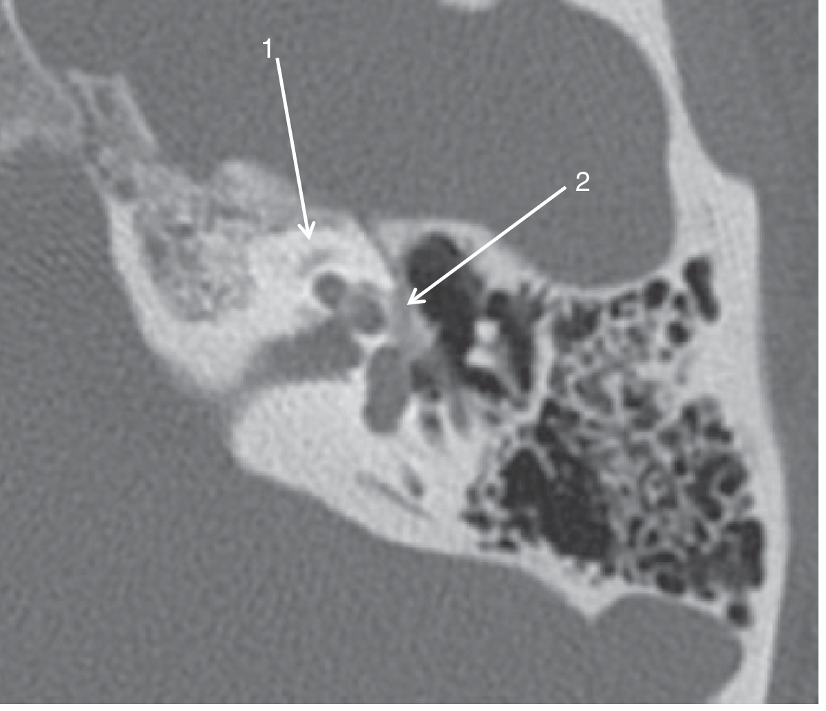
- Fenestral otosclerosis: Halo of lucency (less dense bone) anterior to the oval window niche at the fissula ante fenestram - the most common focus; associated with pure conductive hearing loss from stapes fixation
- Retrofenestral (pericochlear) otosclerosis: Lucent halo surrounding the basal turn of the cochlea ("double ring sign" or "halo sign") - associated with mixed or sensorineural hearing loss; less common
- Both types can coexist
5.4 Tympanosclerosis
- Sequela of chronic OM
- CT finding: Foci of dense calcification - punctate or web-like - within the middle ear cleft or tympanic membrane
- Calcification may surround ossicles causing fixation and conductive hearing loss
- When only the TM is calcified: myringosclerosis
- Suspensory ligaments and tensor tympani tendon may also calcify
5.5 Temporal Bone Fractures
- Longitudinal fractures (80%): Secondary to temporoparietal trauma; fracture extends along the long axis of petrous pyramid; usually involves EAC; spares inner ear; presents with conductive hearing loss and hemotympanum; facial nerve palsy in 20% but often incomplete and recovers
- Transverse fractures (20%): Secondary to frontal/occipital trauma; crosses petrous axis; traverses inner ear structures; causes irreversible SNHL and vestibular dysfunction; facial nerve palsy in 50% (often complete); may produce pneumolabyrinth (air within the labyrinth on CT - pathognomonic)
- Otic capsule-sparing (OCS) fractures (90%): Fracture does not violate the bony labyrinth; better prognosis; SNHL uncommon
- Otic capsule-violating (OCV) fractures (10%): Transgresses cochlea, vestibule or SCCs; high association with SNHL (>90%), facial nerve injury, CSF leakage (otorrhoea or rhinorrhoea via the Eustachian tube)
- Fracture line through petrous bone (may be subtle - review multiplanar reformats carefully)
- Hemotympanum (opacified middle ear without air-fluid level - blood appears denser than fluid acutely)
- Pneumolabyrinth (air in cochlea/vestibule - pathognomonic of labyrinthine transgression)
- Pneumocephalus
- Ossicular disruption: incudostapedial (IS) joint separation is most common, followed by malleoincudal (MI) dislocation; malleus and stapes are more firmly anchored
- Carotid canal involvement (risk of traumatic ICA dissection/pseudoaneurysm - report specifically, recommend CTA)
- Facial nerve canal involvement (localize segment - guides surgical approach)
5.6 Paragangliomas (Glomus Tumors)
- Most common tumors causing pulsatile tinnitus in adults; can present in adolescents
- Glomus tympanicum: Arises on the cochlear promontory on the medial wall of the mesotympanum; presents early with pulsatile tinnitus; seen as a vascular mass behind the TM on otoscopy
5.7 Vestibular Schwannoma
- Most common CPA tumor; most common cause of asymmetrical SNHL
- Bilateral vestibular schwannoma = Neurofibromatosis type 2 (NF-2) - ALWAYS screen for this in pediatric bilateral cases
- MRI is the gold standard (CT can miss small intracanalicular lesions)
- T2: Hypointense to CSF within the IAC
- T1 post-Gd: Avid homogeneous enhancement (large tumors may be heterogeneous due to cystic change)
- Shape: "Ice cream on a cone" for tumors with an intracanalicular and CPA component
- Angle with posterior petrous wall: Acute angle (vs. obtuse angle for meningioma)
- CPA cistern involvement with displacement of structures
5.8 Labyrinthitis and Labyrinthitis Ossificans
- Inflammation of the membranous labyrinth, usually from bacterial meningitis (most feared cause) or extension from otitis media
- MRI post-Gd: Pathological enhancement of the membranous labyrinth (particularly the cochlea and vestibule)
- CT: Normal in acute stage
- Progressive fibrosis followed by new bone formation within the cochlear lumen
- Most common cause: bacterial meningitis (especially Streptococcus pneumoniae)
- CT: New bone (increased density/sclerosis) within the cochlear turns - basal turn first and most severely affected
- MRI T2: Loss of normal high T2 signal within the cochlear lumen (dark turns = fibrosis/ossification)
- Critical implication for cochlear implantation: obliterated cochlea makes electrode insertion technically challenging or impossible; requires drill-out techniques; outcomes less predictable
- Urgent pre-CI MRI is indicated in post-meningitis children - ossification can begin within weeks after meningitis; early CI before complete ossification is recommended
SECTION 6: COCHLEAR IMPLANTATION (CI) - RADIOLOGICAL PERSPECTIVE
6.1 Pre-Operative Imaging Assessment
- MRI temporal bone (T2 CISS/FIESTA + T1 post-Gd) as primary modality - Cummings Otolaryngology, p. 3710
- HRCT temporal bone as complementary modality
- Both together provide maximum information for surgical planning
- Cochlear nerve (CN) caliber - Critical. Cochlear nerve aplasia or severe hypoplasia on parasagittal oblique T2 through the IAC is an absolute contraindication to CI (the device has nothing to stimulate). The CN should be equal to or larger than the adjacent vestibular nerve.
- IAC dimensions - Narrow IAC (< 2 mm) on CT suggests CN deficiency; direct CN visualization on MRI is superior and required
- Cochlear patency - T2 signal within the cochlea; loss indicates fibrosis/ossification (labyrinthitis ossificans)
- Inner ear malformation - As classified above; most malformations can receive CI with modified techniques; common cavity and IP-I carry gusher risk
- Cochlear anatomy and any ossification/sclerosis
- Facial nerve course - identify any dehiscence or aberrant course (tympanic segment position relative to oval window; mastoid course)
- Mastoid pneumatization (guides surgical approach)
- Position and patency of round window (standard cochleostomy site)
- Jugular bulb dehiscence or high-riding jugular bulb (may interfere with cochleostomy)
- Aberrant ICA (rare but catastrophic if unrecognized during surgery)
- IAC dimensions - to correlate with MRI CN assessment
- Tegmen height - relevant to avoid middle fossa approach complications
6.2 Cochlear Implant Components
- External component: Microphone, speech processor, transmitter coil
- Internal component (surgically implanted):
- Receiver-stimulator: Placed in a bony well in the squamous part of the temporal bone
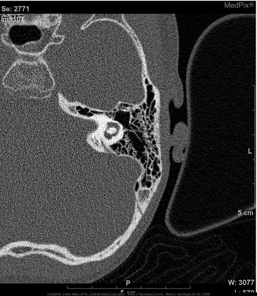
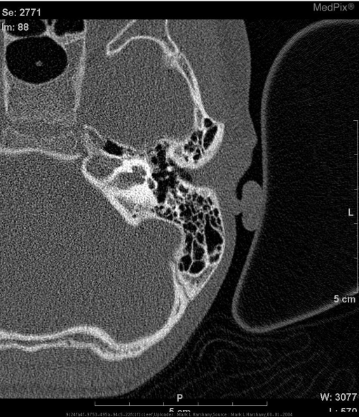
- Electrode array: Inserted through the cochleostomy (or round window approach) into the scala tympani of the basal turn; aims to stimulate the spiral ganglion cells along the full cochlear length
6.3 Post-Operative Imaging
- Electrode migration or malposition suspected clinically
- Device malfunction
- Facial nerve stimulation (may indicate translocation into scala vestibuli near facial nerve)
- Post-operative infection/complication evaluation
- Identifies electrode array position within the cochlea
- Detects scalar translocation (electrode leaving scala tympani into scala vestibuli, especially at the first turn) - associated with worse hearing outcomes
- Insertion depth assessment (360° in basal turn = approximately covering frequencies up to ~500 Hz)
- Identifies tip fold-over (kinking of the electrode at the apex)
- MRI post-CI: Requires careful review of device MRI-compatibility documentation. Most modern CI systems are MRI-conditional at 1.5 T (and some at 3 T) but the internal magnet must be considered; scanning protocols require specific precautions

6.4 Contraindications to Cochlear Implantation
| Absolute | Relative/Modified Approach |
|---|---|
| Cochlear nerve aplasia on MRI | Cochlear nerve hypoplasia (consider CI ± ABI) |
| Michel deformity (total aplasia) | Common cavity, IP-I, IP-II (modified technique, gusher management) |
| Completely obliterated cochlea (post-ossification) | Partial obliteration (drill-out technique, shorter array) |
| Active untreated middle ear infection | Chronic suppurative OM (staged approach) |
| Retrocochlear pathology (central deafness) | Auditory neuropathy spectrum disorder (ABI may be preferred) |
SECTION 7: SPECIAL PEDIATRIC CONSIDERATIONS
7.1 Mastoid Pneumatization
- Mastoid air cells begin as a single air cell (the mastoid antrum, which is present at birth and communicates with the epitympanum via the aditus ad antrum)
- Progressive pneumatization occurs from birth to puberty
- Pneumatization is impaired by chronic otitis media - hence hypopneumatized mastoids are virtually universal in cholesteatoma patients (both congenital and acquired)
- On CT: Count and assess the extent of air cells; measure mastoid size
7.2 Congenital CMV and Hearing Loss
- Congenital cytomegalovirus (cCMV) is the leading non-genetic cause of congenital SNHL
- MRI brain (not temporal bone CT) is the key imaging study:
- Periventricular calcifications (CT: punctate calcifications adjacent to the lateral ventricles)
- Cortical malformations (lissencephaly, pachygyria)
- Cerebellar hypoplasia
- White matter signal abnormalities (T2 hyperintensity)
- Temporal bone MRI: may show cochlear enhancement in active labyrinthitis; CT usually normal
7.3 Post-Meningitis Hearing Loss
- Urgent audiological assessment and imaging required
- T2 MRI showing loss of cochlear fluid signal and T1 Gd showing labyrinthine enhancement precedes CT-detectable ossification
- Window period for CI before complete ossification may be as short as 4-6 weeks in severe cases
- Bilateral imaging even if hearing loss is unilateral, as ossification can be asymmetric
7.4 Pendred Syndrome
- SLC26A4 mutation (autosomal recessive)
- Classic triad: SNHL + enlarged vestibular aqueduct + thyroid goiter (euthyroid/hypothyroid)
- Imaging: bilateral EVA + IP-II (Mondini cochlea) in most cases
- Hearing loss is usually progressive and can be precipitated by minor trauma
SECTION 8: SYSTEMATIC REPORTING APPROACH
External Ear
- EAC present/stenotic/atretic
- Soft tissue density within the EAC
- Atretic plate morphology and thickness (if CAA)
Middle Ear
- Middle ear cleft opacification/aeration
- Ossicular chain integrity (malleus, incus, stapes individually)
- Scutum morphology
- Tegmen integrity
- Facial nerve canal (tympanic segment)
- Oval window and round window
- Hypotympanum (jugular bulb height, ICA)
- Eustachian tube patency
Inner Ear
- Cochlea: number of turns, modiolus, partitioning (IP classification if abnormal)
- Vestibule: size, morphology
- Semicircular canals: present/absent/dysplastic; lateral SCC integrity in cholesteatoma
- Vestibular aqueduct: measure at midpoint and operculum
- IAC: dimensions bilaterally; asymmetry
Mastoid
- Degree of pneumatization
- Air cell opacification
- Bony septal integrity (coalescence)
- Cortical integrity
- Sigmoid sinus plate
Adjacent Structures
- Tegmen (middle cranial fossa)
- Posterior fossa plate
- Carotid canal integrity
- Jugular foramen
- Stylomastoid foramen region
Key References
- Scott-Brown's Otorhinolaryngology Head & Neck Surgery, Vol 2 - Chapter on Temporal Bone Anatomy and Pathology
- Cummings Otolaryngology Head and Neck Surgery - Chapters on Cochlear Implantation (congenital deafness evaluation) and Pediatric Cholesteatoma
- Grainger & Allison's Diagnostic Radiology - Chapter 61: Temporal Bone Imaging (comprehensive imaging review)
- Shambaugh: Surgery of the Ear - Diagnostic Techniques section