
Everything i need to know from multiple myeloma
Reading File
Finding Sources
Finding Sources
Reading File
Reading File
Reading File
Reading File
Reading File
Reading File
Reading File
Reading File
Reading File
Reading File
Reading File
Reading File
Finding Sources
Searching PubMed
"multiple myeloma"[MeSH Terms] AND treatment
Searching the Web
multiple myeloma 2025 2026 treatment updates new FDA approvals
Multiple Myeloma: A Complete Overview
What It Is
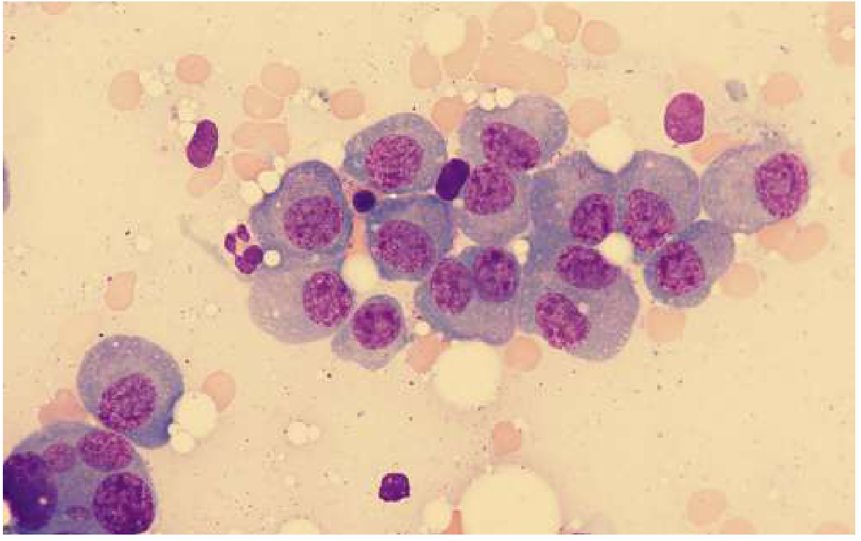
Multiple myeloma is a malignancy of terminally differentiated B cells (plasma cells) characterized by clonal plasma cell proliferation in the bone marrow, production of a monoclonal immunoglobulin (M protein), and end-organ damage - classically bone destruction, anemia, hypercalcemia, and renal failure (Goldman-Cecil Medicine, p. 1976-1982). It is the second most common hematologic malignancy, accounting for roughly 1% of all cancers and about 10% of blood cancers, with ~30,000 new US cases/year (Robbins & Kumar Basic Pathology, p. 2869). Median age at diagnosis is about 65-70 years; it's more common in men and nearly twice as common in Black patients as in White patients.
The Disease Spectrum
Multiple myeloma sits on a continuum that starts almost universally from a precursor state:
- MGUS (monoclonal gammopathy of undetermined significance): serum M protein <3 g/dL, clonal bone marrow plasma cells <10%, no myeloma-defining events. Progresses to myeloma at ~1%/year.
- Smoldering multiple myeloma: M protein ≥3 g/dL and/or 10-60% clonal bone marrow plasma cells, but no end-organ damage. Progression risk is much higher (~10%/year for the first 5 years).
- Symptomatic multiple myeloma: ≥10% clonal bone marrow plasma cells (or biopsy-proven plasmacytoma) PLUS at least one myeloma-defining event.
Diagnostic Criteria - "Myeloma-Defining Events"
Per International Myeloma Working Group criteria, diagnosis requires clonal bone marrow plasma cells ≥10% (or biopsy-proven plasmacytoma) plus one or more of the classic CRAB features or newer biomarkers:
- Calcium elevation (>11 mg/dL)
- Renal insufficiency (creatinine >2 mg/dL or CrCl <40 mL/min)
- Anemia (Hb <10 g/dL or >2 g/dL below normal)
- Bone lesions (one or more lytic lesions on imaging)
- Additional biomarkers of malignancy: clonal bone marrow plasma cells ≥60%, serum free light chain ratio ≥100, or >1 focal lesion on MRI
Pathophysiology
Nearly all myeloma evolves from MGUS via a "two-hit" genetic model (risk of progression is fixed at ~1%/year regardless of MGUS duration). Key mechanisms (Robbins Pathology, p. 2869-2883; Goldman-Cecil Medicine):
- Chromosomal translocations fusing the IgH locus (14q32) to oncogenes (cyclin D1/D3, MAF, FGFR3/MMSET); RAS mutations and MYC abnormalities drive progression from MGUS. Deletions of 17p and 1p, and amplification of 1q, are associated with adverse prognosis.
- IL-6, produced by bone marrow stromal cells, is the major growth factor supporting plasma cell proliferation.
- Bone destruction: myeloma cells upregulate RANKL on stromal cells/osteoblasts while reducing osteoprotegerin (OPG), activating osteoclasts; simultaneously, DKK1 and other factors suppress osteoblasts. This produces pure osteolytic lesions with hypercalcemia and pathologic fractures.
- Renal damage: light-chain (Bence Jones) cast nephropathy in the distal tubules ("myeloma kidney"), light chain deposition disease, AL amyloidosis (~10% of patients), hypercalcemia-induced injury, and Fanconi syndrome.
- Immune dysfunction: despite elevated total immunoglobulin, functional antibody production is suppressed, causing recurrent bacterial infections.
Clinical Presentation
- Bone pain (back/chest most common) is present at diagnosis in >2/3 of patients; height loss from vertebral collapse
- Fatigue/weakness from anemia (present in ~75% at diagnosis, nearly universal eventually)
- Recurrent infections from functional hypogammaglobulinemia
- Renal insufficiency (~20% at diagnosis)
- Hypercalcemia (15-20% at diagnosis) - anorexia, nausea, polyuria, confusion
- Increased risk of deep vein thrombosis (compounded by immunomodulatory drug therapy)
- Radiculopathy/spinal cord compression from vertebral collapse

Workup
- Labs: CBC (normocytic anemia), serum protein electrophoresis + immunofixation, urine electrophoresis + immunofixation, serum free light chain assay, beta-2-microglobulin, albumin, LDH, calcium, creatinine. Combined serum/urine studies detect an M protein in ~97% of patients (IgG 52%, IgA 21%, light-chain only 16%). ~3% are "nonsecretory."
- Bone marrow biopsy/aspirate: clonal plasma cells, flow cytometry (CD38+/CD138+, CD19-/CD45-), FISH cytogenetics for risk stratification (t(4;14), t(14;16), t(11;14), del17p, 1q amplification)
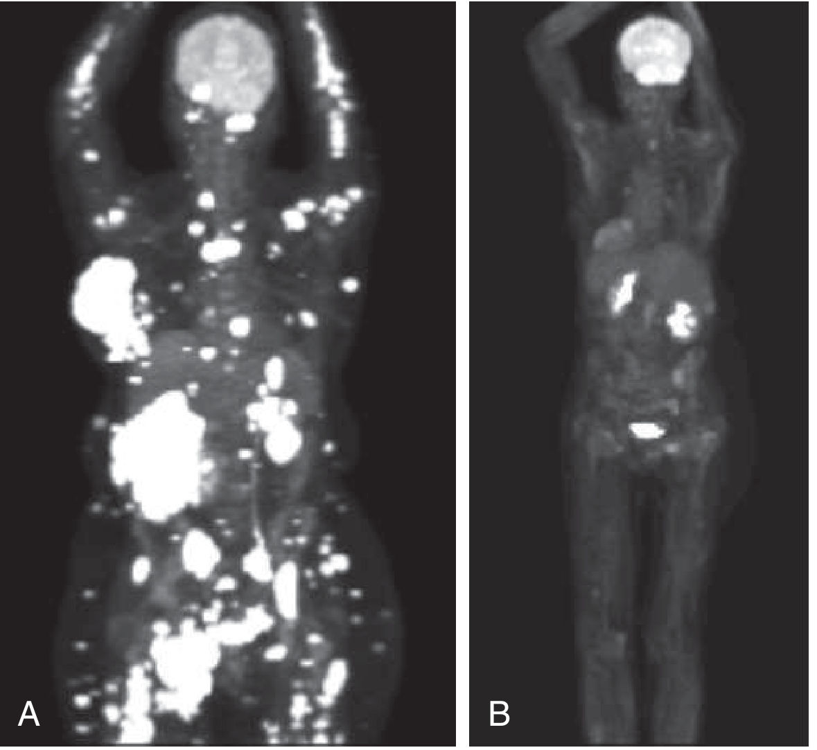
- Imaging: low-dose whole-body CT or PET-CT are now preferred over plain films (conventional radiographs miss disease and are being phased out); MRI for spinal involvement/cord compromise
Staging (Revised International Staging System, R-ISS)
| Stage | Criteria |
|---|---|
| I | Albumin >3.5 g/dL, β2-microglobulin <3.5 mg/L, no high-risk cytogenetics, normal LDH |
| II | Neither I nor III |
| III | β2-microglobulin >5.5 mg/L AND (high-risk cytogenetics [t(4;14), t(14;16), del(17p)] OR elevated LDH) |
High-risk cytogenetics: t(14;16), t(14;20), t(4;14), del(17p), 1q amplification, p53 mutation.
Standard-risk: t(11;14), t(6;14), trisomies.
Treatment
Smoldering myeloma
Risk-stratified by M protein >2 g/dL, free light chain ratio >20, and bone marrow plasma cells >20%. Low/intermediate risk: observe every 3-4 months. High-risk: lenalidomide ± low-dose dexamethasone for ~2 years reduces progression risk.
Newly diagnosed, transplant-eligible (~50% of patients)
Induction with a triplet (e.g., bortezomib-lenalidomide-dexamethasone [VRd] or daratumumab-based quadruplets) → autologous stem cell transplant → lenalidomide maintenance (bortezomib added for high-risk disease).
Newly diagnosed, transplant-ineligible
- VRd (bortezomib/lenalidomide/dexamethasone) for ~9 months then lenalidomide maintenance, or
- DRd (daratumumab/lenalidomide/dexamethasone) until progression
- As of January 2026, the FDA approved Darzalex Faspro (daratumumab/hyaluronidase) plus VRd specifically for transplant-ineligible newly diagnosed patients.
Relapsed/refractory disease
Almost all patients eventually relapse. General principle: use ≥2 drugs the patient hasn't received or is not refractory to.
- Immunomodulatory drugs: thalidomide, lenalidomide, pomalidomide (all carry VTE risk, need prophylaxis)
- Proteasome inhibitors: bortezomib, carfilzomib, ixazomib
- Monoclonal antibodies: daratumumab/isatuximab (anti-CD38), elotuzumab (anti-SLAMF7)
- Bispecific antibodies (target BCMA or GPRC5D): teclistamab, elranatamab, talquetamab - newer options; teclistamab + daratumumab combo was FDA-approved March 2026
- CAR-T cell therapy: idecabtagene vicleucel (ide-cel), ciltacabtagene autoleucel (cilta-cel) - target BCMA; main toxicities are cytokine release syndrome and neurotoxicity
- Other: venetoclax (only for t(11;14) subtype), selinexor, bendamustine, belantamab mafodotin (BCMA-ADC, re-approved with bortezomib/dex Oct 2025)
Managing Complications
- Hypercalcemia: hydration, steroids, bisphosphonates (zoledronic acid/pamidronate)
- Renal failure/cast nephropathy: aggressive hydration, treat underlying myeloma, plasmapheresis if severe light-chain burden, dialysis if needed
- Bone disease: zoledronic acid or pamidronate monthly (reduces fractures, may improve survival; watch for osteonecrosis of jaw with prolonged use), denosumab as alternative, kyphoplasty for vertebral fractures
- Infection prevention: pneumococcal/influenza/COVID vaccination, antiviral prophylaxis (acyclovir) with bortezomib, IVIG for recurrent severe infections
Prognosis
Currently considered incurable, but survival has improved substantially. Median survival is approximately 6-7 years in patients >65, and approximately 12 years in transplant-eligible patients <65, varying widely by stage and cytogenetic risk. A rare aggressive terminal phase with pancytopenia and rapid tumor growth carries survival of only months.
Variant/Related Entities
- Plasma cell leukemia: >5% circulating plasma cells or absolute count ≥500/μL; aggressive
- Solitary plasmacytoma of bone: localized disease without systemic myeloma-defining events
- AL amyloidosis: light-chain deposition as amyloid, occurring in ~10% of myeloma patients
Note on Recent Evidence
Recent systematic reviews (2024-2025) continue to refine outcomes data in non-transplant-eligible patients and treatment-outcome heterogeneity in developing countries (PMIDs: 41027476, 41078459, 39368634) - these support but don't contradict the textbook-based approach above. The field is moving fast: several new FDA approvals occurred in late 2025/early-mid 2026 (Darzalex Faspro + VRd for transplant-ineligible NDMM, teclistamab + daratumumab for R/R disease, subcutaneous isatuximab, belantamab mafodotin combinations), reflecting a general trend toward daratumumab/isatuximab-based quadruplet regimens and increasing reliance on minimal residual disease (MRD) negativity as a trial endpoint given very high response rates with modern therapy.
If you want, I can go deeper into any single piece - diagnostic workup algorithm, a specific drug's dosing/side effects, CAR-T eligibility, or how a specific complication (e.g., renal failure) is managed step by step.
Multiple myeloma pathophysiology and complications
I already have this covered from our previous discussion. Here's the focused breakdown on pathophysiology and complications:
Pathophysiology
Origin and genetic evolution
Nearly all multiple myeloma evolves from a premalignant precursor state (MGUS), following what's essentially a "two-hit" genetic model - the risk of progression from MGUS is fixed at roughly 1%/year regardless of how long the MGUS has been present.
- Primary genetic event: chromosomal translocations fusing the immunoglobulin heavy chain (IgH) locus on chromosome 14q32 to oncogenes - most commonly cyclin D1 or cyclin D3, but also MAF and FGFR3/MMSET in other subtypes. This causes dysregulation of D-cyclins, driving increased plasma cell proliferation.
- Secondary events driving MGUS → myeloma progression: RAS mutations and MYC abnormalities are the two main drivers. Later in disease, deletions of chromosome 17p and 1p, and amplification of 1q, emerge and correlate with more aggressive, treatment-resistant disease. MYC translocations appear late and are associated with aggressive biology.
- Microenvironment: interleukin-6 (IL-6), secreted mainly by bone marrow stromal cells and macrophages, is the principal growth factor sustaining plasma cell proliferation. Angiogenesis and abnormal cytokine paracrine loops within the marrow niche further support tumor growth.
Mechanism of bone destruction
This is the pathophysiologic hallmark of myeloma:
- Myeloma cells upregulate RANKL (receptor activator of NF-κB ligand) expression on osteoblasts and stromal cells, while simultaneously reducing osteoprotegerin (OPG), RANKL's decoy receptor.
- The resulting increase in the RANKL/OPG ratio drives osteoclast activation and bone resorption.
- Other osteoclast-activating factors include MIP-1α, SDF-1α, IL-3, IL-1β, and IL-6.
- Simultaneously, osteoblast differentiation is suppressed by IL-3, IL-7, and DKK1 (dickkopf-1), a Wnt-pathway inhibitor.
- Net effect: pure osteolytic lesions (no compensatory bone formation), leading to hypercalcemia, osteopenia, and pathologic fractures.
Immune dysfunction
Myeloma cells impair normal B-cell function through poorly understood mechanisms. Paradoxically, although total serum immunoglobulin is elevated (due to the monoclonal M protein), production of functional, diverse antibodies is suppressed - creating a state of functional hypogammaglobulinemia despite hyperglobulinemia. This underlies the marked susceptibility to bacterial infection.
Renal injury mechanisms (multifactorial)
- Light-chain cast nephropathy ("myeloma kidney"): free light chains (Bence Jones protein) precipitate with Tamm-Horsfall protein to form large, waxy, laminated casts obstructing the distal tubules and collecting ducts. Severity correlates directly with the quantity of urinary free light chain.
- Hypercalcemia: from bone resorption, causes nephrogenic diabetes insipidus, dehydration, and vasoconstriction, worsening renal perfusion.
- AL amyloidosis: light chains misfold and deposit as amyloid in the glomeruli/interstitium, occurring in ~10% of myeloma patients, producing nephrotic-range proteinuria.
- Light chain deposition disease: non-amyloid, linear deposition of light chains in glomerular/tubular basement membranes.
- Acquired Fanconi syndrome: proximal tubular dysfunction from light chain toxicity, causing glycosuria, phosphaturia, aminoaciduria.
- Contributing factors: dehydration, hyperuricemia, and increased susceptibility to pyelonephritis (from hypogammaglobulinemia).
Complications and Their Management
1. Bone disease / skeletal complications
- Lytic lesions, osteopenia, pathologic fractures (vertebrae, skull, ribs, pelvis, proximal humerus/femur most common sites); vertebral collapse can cause height loss and radiculopathy or cord compression.
- Management: zoledronic acid or pamidronate monthly (reduce fracture risk, bone pain, cord compression risk; may improve survival). After 1-2 years, dosing interval can be extended to reduce osteonecrosis of the jaw risk. Denosumab is an alternative. Kyphoplasty for vertebral fractures. Radiation therapy for localized, refractory painful lesions or cord compression.
2. Hypercalcemia
- Present in 15-20% at diagnosis; presents with anorexia, nausea, vomiting, polyuria, polydipsia, constipation, weakness, confusion, stupor.
- Management: aggressive hydration, corticosteroids, bisphosphonates (zoledronic acid/pamidronate) - corrects hypercalcemia in nearly all patients.
3. Renal insufficiency
- Present in ~20% at diagnosis (creatinine >2 mg/dL); most often from light-chain cast nephropathy and/or hypercalcemia.
- Management: aggressive IV hydration + furosemide to maintain high urine flow if not oliguric; allopurinol if hyperuricemic; plasmapheresis (daily x5 days) if cast nephropathy suspected with very high free light chains (>150 mg/dL); hemodialysis if symptomatic azotemia; rapid initiation of anti-myeloma therapy (e.g., bortezomib-based regimens) is critical since reducing the light-chain burden is the definitive treatment.
4. Infection
- Due to functional hypogammaglobulinemia, neutropenia (from marrow infiltration/chemo), and immunosuppressive therapy.
- Management: prompt antibiotic therapy for bacterial infections; prophylactic trimethoprim-sulfamethoxazole with high-dose steroids; acyclovir prophylaxis with bortezomib (reactivation of herpes zoster); IVIG for recurrent severe infections with hypogammaglobulinemia; pneumococcal, influenza, and COVID vaccination for all patients.
5. Anemia
- Normocytic, normochromic; present in ~75% at diagnosis, eventually in nearly all patients. From marrow infiltration and disease burden.
- Management: treat underlying myeloma; erythropoietin for anemia persisting despite treatment.
6. Neurologic complications
- Radiculopathy (most common - thoracic/lumbosacral, from vertebral collapse or nerve compression)
- Spinal cord compression (up to 10% of patients) - from vertebral collapse or extramedullary plasmacytoma; presents with severe back pain, leg weakness/paresthesia, bowel/bladder dysfunction - treated with dexamethasone and/or urgent radiation, surgical decompression if deficits progress
- Peripheral neuropathy - uncommon from myeloma itself, usually from associated amyloidosis (or treatment-related, e.g., bortezomib/thalidomide)
7. Hyperviscosity
- Less common than in Waldenström macroglobulinemia but can occur with high M protein burden.
8. Thrombosis
- Increased baseline risk of DVT/PE, compounded by immunomodulatory drugs (lenalidomide, thalidomide, pomalidomide) - requires routine antithrombotic prophylaxis during treatment.
9. Progression to aggressive/terminal phase
-
A subset develop an acute terminal phase: rapid tumor growth, pancytopenia, subcutaneous soft-tissue plasmacytomas, paradoxically decreased M protein, fever - survival limited to months.
-
Related aggressive variant: plasma cell leukemia (>5% circulating plasma cells or absolute count ≥500/μL in peripheral blood).
-
Goldman-Cecil Medicine International Edition, p. 1976-1982
-
Robbins & Kumar Basic Pathology, p. 2869-2883
Let me know if you'd like me to expand on any single mechanism (e.g., RANKL/OPG pathway in more depth, or the molecular genetics of high-risk cytogenetics) or pull supporting images (bone marrow histology, PET/CT findings, skull lytic lesions).
This is a shared conversation. Sign in to Orris to start your own chat.