
Hybridoma Technology
I'll start with a quick overview while I search the medical library for detailed content.
Hybridoma technology is a method for producing monoclonal antibodies (MAbs) by fusing an antibody-producing B lymphocyte with a myeloma (immortal cancer) cell to create a "hybridoma" cell line. The resulting hybrid cells combine the antibody specificity of the B cell with the indefinite proliferative capacity of the myeloma cell. Developed by Kohler and Milstein in 1975, it revolutionized diagnostics and therapeutics.
Searching sources...
Reading File
Finding Sources
Finding Sources
Good results from multiple authoritative sources. Now let me read the most relevant sections in parallel.
Reading File
Reading File
Reading File
Excellent content. Let me now fetch the hybridoma process diagram and read the detailed steps from Janeway's + the Cellular & Molecular Immunology text.
Loading Image
Reading File
Finding Sources
Now I have all the content I need. Let me also fetch the Janeway's process diagram and read the Goodman & Gilman section on HAT selection.
Loading Image
Reading File
I now have comprehensive, well-sourced content from multiple authoritative textbooks along with two excellent diagrams. Here is the complete answer:
Hybridoma Technology
1. Historical Background
Hybridoma technology was developed by Georges Köhler and César Milstein in 1975, for which they received the Nobel Prize in Physiology or Medicine in 1984. Their key insight was fusing an antibody-producing spleen cell (B lymphocyte) with an immortal myeloma cell to create a permanently dividing hybrid cell line that secretes a single, defined antibody.
Prior to this, the only source of homogeneous antibody was the serum of patients with multiple myeloma - but the antigen specificity of those myeloma proteins was unknown, limiting their usefulness. - Janeway's Immunobiology 10e
2. Principle
A hybridoma combines two essential properties in one cell:
| Property | Source |
|---|---|
| Specific antibody production | Immunized B cell / plasma cell |
| Immortality (indefinite proliferation) | Myeloma (tumor) cell |
The resulting cell line secretes a monoclonal antibody (MAb) - a single, homogeneous antibody of one defined specificity and isotype.
3. Step-by-Step Process
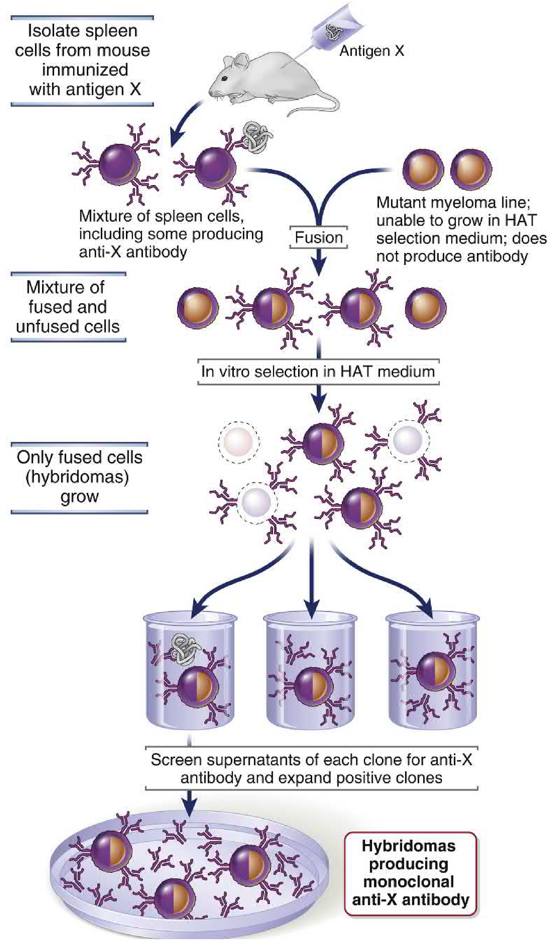
Here is the full workflow, illustrated by the diagram from Cellular & Molecular Immunology (Abbas):
And from Janeway's Immunobiology 10e:
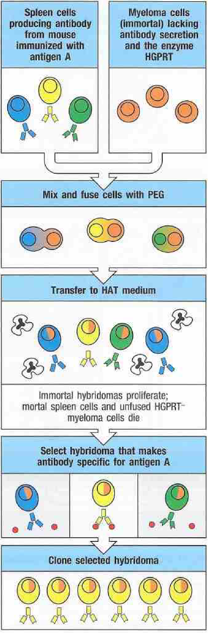
Step 1 - Immunization
A mouse is immunized with the desired antigen (e.g., antigen X or A). To maximize spleen B cell activation, an intravenous booster injection is given 3 days before harvesting the spleen. The spleen is then removed and a suspension of B lymphocytes (including plasma cells secreting anti-X antibody) is prepared.
Step 2 - Selection of Myeloma Partner
A mutant myeloma cell line is used that:
- Cannot produce its own antibody (ensuring the only antibody produced by the hybridoma comes from the B cell partner)
- Lacks the enzyme HGPRT (hypoxanthine-guanine phosphoribosyltransferase) - this makes it unable to survive in HAT selection medium
Step 3 - Cell Fusion
Spleen B cells and myeloma cells are mixed and fused together using polyethylene glycol (PEG), which destabilizes cell membranes and promotes fusion. This produces a mixture of:
- Unfused spleen cells
- Unfused myeloma cells
- Fused hybridoma cells (spleen + myeloma)
- Cell-cell fusions of same type (spleen+spleen, myeloma+myeloma)
Step 4 - HAT Selection (The Key Step)
The mixture is cultured in HAT medium (Hypoxanthine, Aminopterin, Thymidine):
| Cell type | Fate in HAT medium | Reason |
|---|---|---|
| Unfused myeloma cells | Die | Lack HGPRT; aminopterin blocks their main DNA synthesis pathway, and they cannot use the salvage pathway |
| Unfused spleen/B cells | Die | Normal cells have a finite lifespan; die within days |
| Hybridoma cells | Survive and proliferate | HGPRT contributed by the B cell rescues them; myeloma genetics gives immortality |
Only hybridomas survive because they get:
- Immortality from the myeloma
- HGPRT (and thus HAT-resistance) from the B cell
Step 5 - Cloning and Screening
Surviving hybridomas are plated by limiting dilution into individual wells (one cell per well) to generate single-cell clones. Culture supernatants from each well are then screened for antibody specificity using:
- ELISA
- Radioimmunoassay
- Immunofluorescence
Positive clones secreting antibody against the target antigen are identified and expanded.
Step 6 - Monoclonal Antibody Production
Selected hybridoma clones are expanded in:
- Bulk cell culture - in vitro, large bioreactors
- Ascites fluid - injected into the peritoneal cavity of mice (produces high-titer ascites; less common now due to ethical concerns)
Because each hybridoma is derived from a single B cell, all antibody molecules it produces are identical in structure, antigen-binding site, and isotype. - Janeway's Immunobiology 10e, Fig. A.10
4. Why Monoclonal Antibodies Are Superior to Polyclonal Antisera
| Feature | Polyclonal Antisera | Monoclonal Antibodies |
|---|---|---|
| Specificity | Mixed (many epitopes) | Single epitope |
| Reproducibility | Varies batch to batch | Unlimited, identical supply |
| Production | Finite (limited serum volume) | Indefinite (immortal cell line) |
| Cross-reactivity | Possible | Minimal |
- Janeway's Immunobiology 10e
5. Limitations and Evolution of the Technology
Problem: Mouse Origin = Immunogenicity (HAMA)
Early MAbs were entirely mouse-derived. When administered to patients, the immune system recognized them as foreign and mounted a human anti-mouse antibody (HAMA) response, which:
- Neutralized or rapidly cleared the injected MAb
- Could cause serum sickness
The first MAb used clinically, murine muromonab (anti-CD3), had poor pharmacokinetics and high immunogenicity - eventually withdrawn from the market. - Tietz Textbook of Laboratory Medicine, 7th Edition
Solution: Engineering More Human Antibodies
Using recombinant DNA technology, antibodies were progressively "humanized":
| Generation | Human Content | Example |
|---|---|---|
| Murine (-omab) | ~0% | Muromonab |
| Chimeric (-ximab) | ~65% | Rituximab (1997), Abciximab (1994) |
| Humanized (-zumab) | ~80-90% | Daclizumab (1997), Trastuzumab |
| Fully human (-umab) | >95% | Adalimumab (2002, via phage display) |
-
Chimeric antibodies: mouse variable regions + human constant regions
-
Humanized antibodies: only mouse CDRs (complementarity-determining regions) grafted onto human IgG framework
-
Fully human: generated via phage display libraries or transgenic mice expressing human antibody genes
-
Tietz Textbook of Laboratory Medicine, 7th Edition
6. Applications of Monoclonal Antibodies
Diagnostics
- Immunoassays: ELISA, RIA, Western blot, flow cytometry using MAb-based reagents
- Pregnancy tests: detection of hCG
- Blood typing: anti-ABO, anti-Rh reagents
- Tumor markers: detection of PSA, CEA, AFP
- CD marker identification: defining lymphocyte subsets (CD4, CD8 T cells)
- Histopathology: identifying tissue of origin of tumors
Therapeutics (Selected Examples)
| MAb | Target | Indication |
|---|---|---|
| Rituximab | CD20 | B-cell lymphoma, autoimmune disease |
| Infliximab/Adalimumab | TNF-α | Rheumatoid arthritis, IBD |
| Trastuzumab | HER2/ErbB2 | Breast cancer |
| Bevacizumab | VEGF | Colon cancer, macular degeneration |
| Nivolumab/Pembrolizumab | PD-1 | Multiple cancers (checkpoint inhibitor) |
| Ipilimumab | CTLA-4 | Melanoma (checkpoint inhibitor) |
| Cetuximab | EGFR | Colorectal cancer, head and neck cancer |
- Cellular and Molecular Immunology (Abbas), Katzung's Pharmacology 16e
Research Tools
- Purifying cell populations (cell sorting)
- Blocking or depleting specific molecules/cells to study their function
- Structural studies of receptors and signaling molecules
7. Nomenclature of Monoclonal Antibodies (INN Stem System)
The International Nonproprietary Name (INN) system encodes the antibody's origin in its suffix:
| Infix | Origin | Example |
|---|---|---|
| -o- | Mouse (murine) | muromonab |
| -xi- | Chimeric | rituximab |
| -zu- | Humanized | trastuzumab |
| -u- | Fully human | adalimumab |
Summary
Hybridoma technology produces unlimited, homogeneous, antigen-specific monoclonal antibodies by fusing immunized B cells with immortal HGPRT-deficient myeloma cells, selecting the hybrids in HAT medium, cloning, and screening. The core innovation from Kohler and Milstein transformed immunology, diagnostics, and therapeutics - and through subsequent recombinant engineering to reduce immunogenicity, has yielded hundreds of clinical-grade therapeutic antibodies. - Janeway's Immunobiology 10e; Cellular & Molecular Immunology (Abbas); Tietz Textbook of Laboratory Medicine, 7th Edition; Katzung's Pharmacology, 16th Edition; Goodman & Gilman's Pharmacology
summary
Hybridoma Technology - Summary
What it is: A technique to produce monoclonal antibodies (MAbs) - identical, highly specific antibodies from a single B cell clone.
Invented by: Köhler & Milstein, 1975 (Nobel Prize 1984).
Core Steps
- Immunize a mouse with the target antigen
- Harvest spleen cells (B cells secreting specific antibody)
- Fuse B cells + HGPRT-deficient myeloma cells using PEG
- Select in HAT medium - only hybridomas survive (B cells die naturally; myeloma cells die without HGPRT)
- Clone by limiting dilution - one cell per well
- Screen supernatants for desired antibody specificity
- Expand positive clones → bulk MAb production
Key Concept: Why HAT Medium Works
| Cell | Outcome | Reason |
|---|---|---|
| Unfused myeloma | Dies | No HGPRT; can't survive aminopterin block |
| Unfused B cell | Dies | Finite lifespan |
| Hybridoma | Survives | Gets HGPRT from B cell + immortality from myeloma |
Generations of MAbs (Reducing Immunogenicity)
Murine → Chimeric (~65% human) → Humanized (~90% human) → Fully Human (>95%)
Applications
- Diagnostics: ELISA, pregnancy tests, tumor markers, blood typing, flow cytometry
- Therapeutics: Anti-cancer (rituximab, trastuzumab), anti-inflammatory (adalimumab, infliximab), checkpoint inhibitors (nivolumab, pembrolizumab)
- Research: Cell sorting, receptor studies, functional blocking assays
One-Line Takeaway
A hybridoma = immortal myeloma cell + antibody-producing B cell, selected by HAT medium, cloned, and screened to yield an unlimited supply of one specific antibody.
with diagram
Hybridoma Technology - Summary with Diagram
Process Diagram
Source: Cellular & Molecular Immunology (Abbas)
Source: Janeway's Immunobiology 10e - showing cell fates in HAT medium
Step-by-Step Summary
Immunize mouse with Antigen X
          ↓
  Harvest spleen B cells
  (some produce anti-X antibody)
          ↓
    Mix with Myeloma cells
  (HGPRT⁻, no antibody secretion)
          ↓
     Fuse using PEG
          ↓
  Culture in HAT medium
          ↓
  ┌───────────────────────────┐
  │ Unfused B cells → DIE     │  (finite lifespan)
  │ Unfused myeloma → DIE     │  (no HGPRT)
  │ Hybridomas → SURVIVE ✓   │  (immortal + HGPRT)
  └───────────────────────────┘
          ↓
  Clone by limiting dilution
  (1 cell per well)
          ↓
  Screen supernatants for anti-X
          ↓
  Expand positive clones
          ↓
  ████ MONOCLONAL ANTIBODY ████
What Each Parent Contributes
| Myeloma Cell | B Lymphocyte |
|---|---|
| Immortality | Antibody specificity |
| Indefinite growth | HGPRT enzyme (HAT survival) |
| No antibody (silent) | Defined isotype |
HAT Medium - Why It Works
- H (Hypoxanthine) + T (Thymidine) = salvage pathway substrates
- A (Aminopterin) = blocks the main (de novo) DNA synthesis pathway
- Myeloma cells lack HGPRT → cannot use the salvage pathway either → die
- Hybridomas inherit HGPRT from the B cell → use salvage pathway → survive
Generations of Therapeutic MAbs
Murine (-omab)       ████████████████████  0% human
                     e.g. Muromonab

Chimeric (-ximab)    ░░░░░░░████████████  ~65% human
                     e.g. Rituximab, Abciximab

Humanized (-zumab)   ░░████████████████  ~90% human
                     e.g. Trastuzumab, Daclizumab

Fully Human (-umab)  ░█████████████████  >95% human
                     e.g. Adalimumab, Nivolumab
Key Applications at a Glance
| Area | Examples |
|---|---|
| Diagnostics | ELISA, pregnancy test, tumor markers, blood typing |
| Cancer therapy | Rituximab (CD20), Trastuzumab (HER2), Cetuximab (EGFR) |
| Autoimmune | Adalimumab, Infliximab (anti-TNF) |
| Checkpoint inhibitors | Nivolumab (PD-1), Ipilimumab (CTLA-4) |
| Research | Cell sorting, receptor studies, functional assays |
One-liner: Hybridoma = B cell specificity + myeloma immortality, selected by HAT, cloned, screened → unlimited identical antibody.
This is a shared conversation. Sign in to Orris to start your own chat.