
Gist tumor management
- Most common mesenchymal tumor of the GI tract
- Driven by KIT (85%) or PDGFRA mutations; ~15% are wild-type
- Treatment hinges on resection for localized disease + imatinib (tyrosine kinase inhibitor) for advanced/metastatic or adjuvant settings
- Risk stratification guides adjuvant therapy duration
"gastrointestinal stromal tumor"[MeSH Terms] AND management
gastrointestinal stromal tumor GIST histology pathology
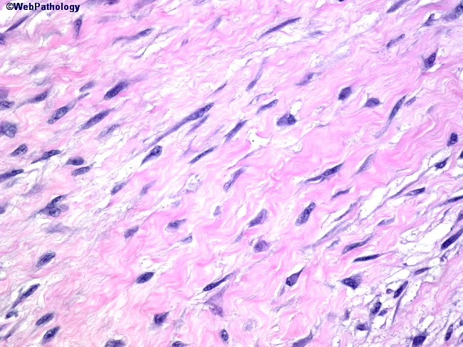
High-power light microscopy of a benign gastrointestinal stromal tumor (GIST) reveals spindle-shaped cells arranged in interlacing fascicles. The neoplastic cells are slender with occasional tapered ends and minimal cytoplasm, producing a homogeneous, eosinophilic background. Nuclei are elongated, with small, inconspicuous nucleoli and lack of pronounced nuclear waviness, pleomorphism, or hyperchromasia. The stroma is dominated by bland, eosinophilic collagen, and mitotic figures are not evident in the field, supporting a low proliferative activity. This spindle cell GIST subtype typically shows uniform cytology and cohesive fascicles, distinguishing it from schwannian or myogenic tumors, which may exhibit nuclear palisading or wavy nuclei. The absence of jagged nuclear contours or notable cytoplasmic vacuolization further supports a benign course in small lesions. Clinically, small GISTs are frequently asymptomatic and discovered incidentally during imaging or surgery. Immunohistochemical confirmation (CD117/c-KIT and DOG1 positivity) and mutational analysis (KIT or PDGFRA) are routinely pursued to establish diagnosis and guide risk stratification, though histology alone can strongly suggest GIST when classic spindle morphology is present. The image highlights diagnostic features relevant to surgical pathology, including tumor size, cellular blandness, and microenvironment, which influence management decisions and prognosis in gastrointestinal stromal tumors. This context supports accurate diagnosis and appropriate surgical planning.
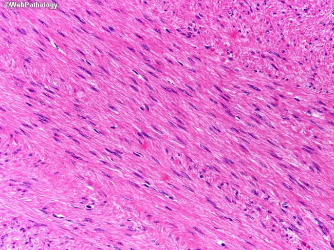
Histopathology: A formalin-fixed, paraffin-embedded tissue section stained with Hematoxylin and Eosin (H&E) shows interlacing fascicles of spindle-shaped smooth muscle cells with elongated, blunt-ended or cigar-shaped nuclei and abundant eosinophilic cytoplasm. The cells are uniform in size, with minimal nuclear atypia and rare mitotic figures; there is no evident necrosis. The architectural pattern is consistent with a benign leiomyoma arising from the smooth muscle layer, such as the muscularis propria of the gastrointestinal tract or uterine myometrium, depending on anatomic origin. Immunohistochemistry demonstrates tumor cells that are strongly positive for desmin and smooth muscle actin (SMA), confirming smooth muscle differentiation. The neoplastic cells are negative for gastrointestinal stromal tumor markers CD34, CD117 (c-KIT), and DOG1, effectively excluding GIST. Diagnostic significance: the combination of characteristic spindle cell morphology and desmin/SMA positivity with CD34/CD117/DOG1 negativity supports a diagnosis of leiomyoma over GIST or leiomyosarcoma. Clinical correlation: these lesions are typically benign with favorable prognosis following complete excision; differential diagnoses include leiomyosarcoma (increased mitotic activity, atypia, necrosis) and GIST (c-KIT or DOG1 positivity). This image exemplifies classical smooth muscle tumor histology and IHC profile, and is useful for educational purposes in surgical pathology, GI/hepatic pathology, and mesenchymal tumor differential diagnosis. Educational diagnostic utility.
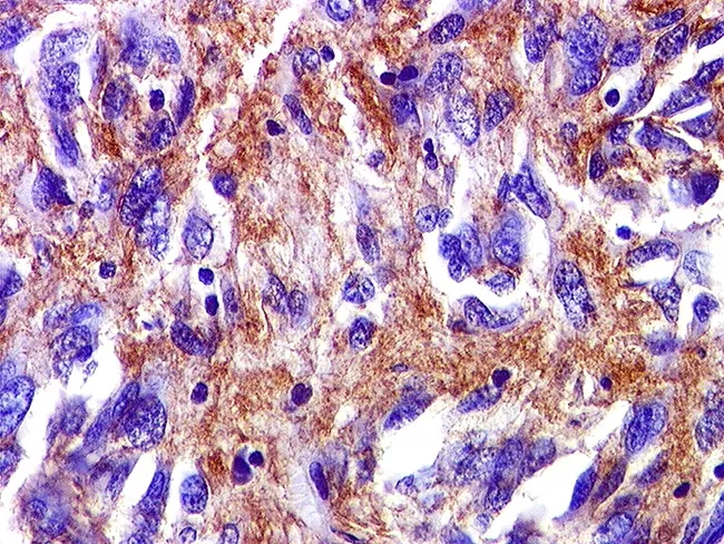
Immunohistochemical slide of a gastrointestinal stromal tumor (GIST) showing robust cytoplasmic/membranous brown staining on a hematoxylin counterstain background. The image likely represents a tissue section stained for CD117 (c-KIT) and/or DOG1, markers that confer high sensitivity and specificity for GIST. In this field, CD34 may be variably positive and SMA positivity can be focal, complicating differential diagnosis with leiomyosarcoma. The tissue is FFPE, sectioned at 4 μm, and stained with DAB chromogen to yield brown immunoreactivity. The tumor cells display spindle-cell morphology in fascicles embedded in a collagenous stroma; nuclei are ovoid to elongated with variable mitotic activity. The immunophenotype—predominant CD117 (c-KIT) and DOG1 positivity—supports the diagnosis of GIST, with DOG1 often providing superior specificity. The combined histology and IHC pattern differentiates GIST from smooth muscle and neural neoplasms. Clinically, this immunoprofile directs targeted therapy options (tyrosine kinase inhibitors such as imatinib or sunitinib) and informs mutational testing for KIT or PDGFRA to predict treatment response. This image is valuable for education in oncologic pathology, GI pathology, and diagnostic immunohistochemistry, illustrating how a mesenchymal GI tumor is identified by characteristic immunostains. It is suitable as a reference for immunophenotype, differential diagnosis, and molecular correlation in GIST research and teaching.
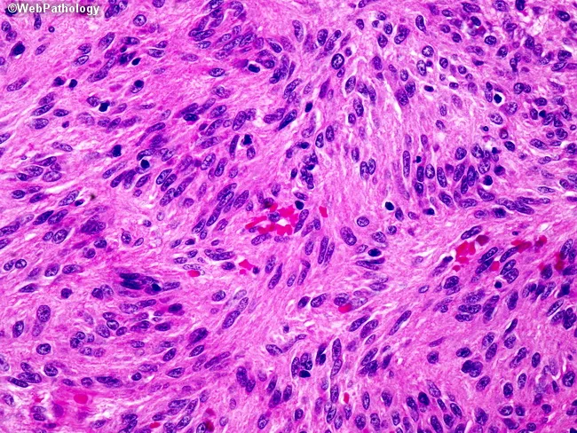
Imaging modality: Histopathology with bright-field light microscopy of a hematoxylin and eosin (H&E) stained section from a gastrointestinal tract neoplasm. Anatomic context: gastrointestinal tract wall (GI tract), smooth muscle tissue, muscularis propria region. Visual features: densely cellular fascicles of spindle-shaped cells with elongated, blunt-ended cigar-shaped nuclei and eosinophilic cytoplasm; minimal pleomorphism; absence of conspicuous mitotic figures within this field; variable stromal collagen and small, serpiginous blood vessels. The cells exhibit classic smooth muscle morphology consistent with leiomyosarcoma; adjacent areas elsewhere in the tumor may reveal higher mitotic activity and pleomorphism. Pathology: malignant neoplasm with smooth muscle differentiation; immunohistochemistry typically shows desmin and smooth muscle actin (SMA) positivity with h-caldesmon expression; in this field, morphological features strongly support leiomyosarcoma over benign leiomyoma or other spindle cell tumors such as GIST. Diagnostic significance: histology indicates a primary GI smooth muscle sarcoma; prognosis is influenced by mitotic rate, tumor size, necrosis, and location; clinical correlation includes surgical resection planning and potential adjuvant therapy; differential diagnoses include GIST, leiomyoma, fibrosarcoma, and sarcoma NOS. Potential clinical use: reference for tumor morphology in GI spindle cell neoplasms, educational cases in surgical oncology, pathology sign-out, and radiopathologic correlation in GI oncology.
PMID: 38840030
GIST (Gastrointestinal Stromal Tumor) - Complete Management Guide
1. Overview & Cell of Origin
- Sabiston Textbook of Surgery, p. 1325
- Quick Compendium of Clinical Pathology 5th ed., p. 404
2. Molecular Subtypes (Critical for Treatment)
| Mutation | Frequency | Imatinib Response |
|---|---|---|
| KIT exon 11 | ~70% | High (>80% response) |
| KIT exon 9 | ~10% | Moderate (40-50%); higher dose may help |
| KIT exons 13, 17 | Rare | 40-50% |
| PDGFRA (non-D842V) | ~5% | Variable |
| PDGFRA D842V | ~5% | Resistant to imatinib; avapritinib effective |
| SDH-deficient (wild-type) | ~15% | Imatinib-insensitive |
| NF1-associated | Rare | KIT overexpressed but unmutated |
- Familial GIST syndrome: germline KIT mutations
- Carney triad: GIST + pulmonary chondromas + extra-adrenal paragangliomas
3. Diagnosis & Workup
Imaging
- CT abdomen/pelvis with IV contrast is the primary staging modality
- GISTs appear as well-circumscribed hypervascular masses, often exophytic
- PET-CT is useful for monitoring TKI response (early metabolic response before size changes)
Endoscopy & EUS
- Endoscopically: smooth submucosal masses, normal overlying mucosa (ulceration = high-risk feature)
- EUS: hypoechoic, homogeneous, well-defined borders in muscularis propria
- High-risk EUS features: irregular borders, cystic spaces, ulceration, echogenic foci, heterogeneity
Biopsy
- EUS-guided CNB (core-needle biopsy) preferred over FNA for superior diagnostic yield
- Percutaneous biopsy: valid alternative; risk of peritoneal seeding <1%
- Mucosal biopsy alone is insufficient for submucosal tumors
Pathology Panel
- Histology: spindle cell (70%), epithelioid (20%), mixed (10%)
- IHC: CD117 (c-KIT)+, DOG1+, CD34 variable
- Mandatory: tumor site, size, focality, mitotic rate, margin status, SDHB expression
- Molecular: KIT/PDGFRA sequencing to guide systemic therapy
4. Risk Stratification
| Risk | Size | Mitotic Rate | Site |
|---|---|---|---|
| Very Low | <2 cm | <5/50 HPF | Any |
| Low | 2-5 cm | <5/50 HPF | Any |
| Intermediate | <5 cm | 6-10/50 HPF | Gastric |
| High | >10 cm, or any size | >10/50 HPF | Any |
| High | >5 cm | >5/50 HPF | Non-gastric |
5. NCCN Management Algorithm
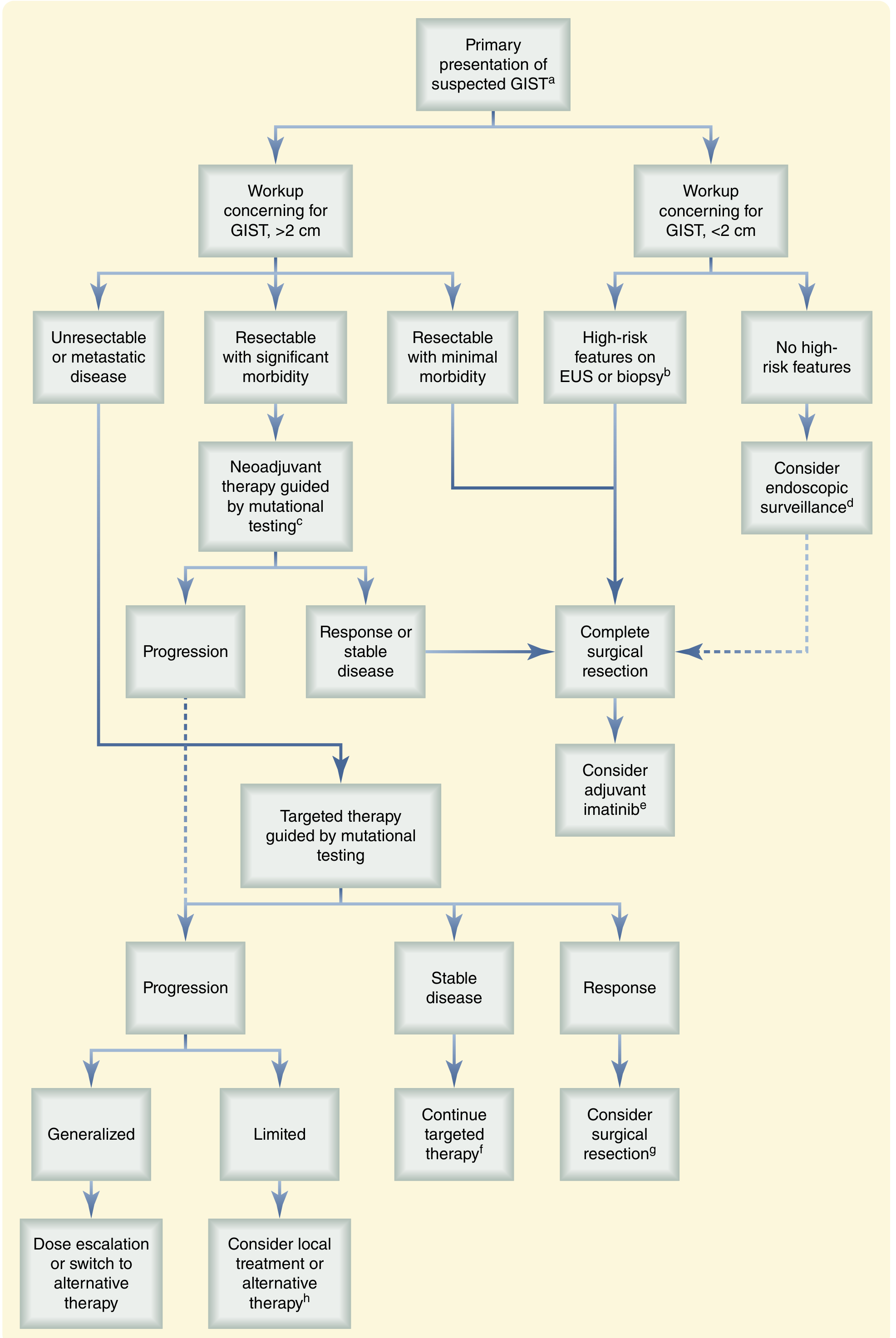
6. Surgical Management
Small GISTs (<2 cm)
- Esophageal, small bowel, colorectal tumors: resect regardless of size (more aggressive biology)
- Gastric tumors <2 cm with no high-risk EUS/biopsy features: endoscopic surveillance is acceptable
- Gastric tumors <2 cm with high-risk features: resect
Localized Resectable GISTs (>2 cm)
- Complete surgical resection (R0) is the only potentially curative treatment
- Oncologic principles:
- Full macroscopic and microscopic resection
- Avoid capsule disruption (prevents peritoneal seeding)
- Explore abdomen for peritoneal/hepatic metastases
- Lymphadenectomy is not required (lymph node metastases are rare in GIST, unlike carcinoma)
- Laparoscopic resection is appropriate for gastric GISTs; data now support it even for tumors >5 cm at experienced centers (2023 meta-analysis)
Site-Specific Considerations
-
Stomach: Wedge resection preserving function; avoid formal gastrectomy unless required for margins
-
Small bowel: Segmental resection
-
Rectum: Can require multi-organ resection; neoadjuvant imatinib strongly considered to downstage and preserve sphincter function
-
Duodenum: Technically challenging; neoadjuvant imatinib often used to reduce operative morbidity
-
Sabiston Textbook of Surgery, p. 1327; Maingot's Abdominal Operations, p. 520
7. Neoadjuvant Therapy
- Resectable tumor with significant expected morbidity (e.g., rectal GIST requiring APR, duodenal GIST)
- Borderline resectable or locally advanced disease
- Imatinib 600 mg/day for 8-12 weeks preoperatively
- Objective response in 90% of primary GISTs
- 92% R0/R1 resection rate; 2-year RFS = 83%
- Optimal duration: 6+ months if continued radiographic response observed (maximal response to imatinib typically takes 6-9 months)
- KIT or PDGFRA-mutant (non-D842V): imatinib
- PDGFRA D842V: avapritinib
- SDH-deficient: may not benefit from standard TKI
8. Adjuvant Therapy
| Risk Group | Adjuvant Imatinib |
|---|---|
| Low/Very Low risk | Not indicated |
| Intermediate risk | Consider 1 year |
| High risk | 3 years (standard of care - SSGXVIII trial: 3 yr > 1 yr) |
- Dose: 400 mg/day (imatinib)
- Must confirm KIT/PDGFRA mutation before prescribing (wild-type GIST may not benefit)
- Imatinib is not effective for PDGFRA D842V-mutant tumors
9. Advanced/Metastatic Disease
First-line: Imatinib
- 400 mg/day standard starting dose
- 800 mg/day for KIT exon 9 mutations (higher dose improves PFS)
- Up to 80% achieve complete/partial response or stable disease
- Continue imatinib indefinitely - stopping leads to rapid progression even in complete responders
Second-line: Sunitinib
- After imatinib progression or intolerance
- Multi-targeted TKI (KIT, PDGFRA, VEGFR)
Third-line: Regorafenib
- After imatinib + sunitinib failure
Fourth-line/Special cases:
- Ripretinib: Broad-spectrum KIT/PDGFRA inhibitor; approved 4th-line
- Avapritinib: Specifically for PDGFRA D842V mutations (any line); also 4th-line+ for other GISTs
Surgery in Metastatic Disease
- Resection of limited metastases with stable or responsive disease on imatinib can be considered
- Liver metastases resection + TKI: 2023 meta-analysis confirms improved survival benefit over TKI alone
- Palliative debulking may relieve symptoms (obstruction, bleeding) but is not curative
10. Monitoring Response
- CT abdomen/pelvis is the standard modality
- Response by Choi criteria (density change on CT), not just size (RECIST underestimates TKI response - tumors may become cystic/hypodense while responding)
- PET-CT: very sensitive for early metabolic response (within weeks of starting imatinib)
11. Surveillance After Resection
- High/intermediate risk: CT abdomen/pelvis with IV contrast every 3-6 months for 3-5 years, then annually
- Low/very low risk: CT every 6-12 months for 5 years, then consider annual or as clinically indicated
12. Summary of TKI Lines
1st line → Imatinib 400 mg (800 mg for exon 9)
2nd line → Sunitinib
3rd line → Regorafenib
4th line → Ripretinib (or Avapritinib for D842V at any line)
Key References
- Sabiston Textbook of Surgery, 21st ed., Chapter 65
- Maingot's Abdominal Operations, Chapter 24
- Quick Compendium of Clinical Pathology 5th ed., Section 7.4.1.2
- British Sarcoma Group GIST Guidelines 2025 (PMID 38840030)
- Chinese Expert Consensus on Surgical Management of GIST 2025 (PMID 40866315)
- Liver resection for metastatic GIST - Meta-Analysis 2023 (PMID 37740754)