
Classification of congenital heart disease
classification of congenital heart disease diagram
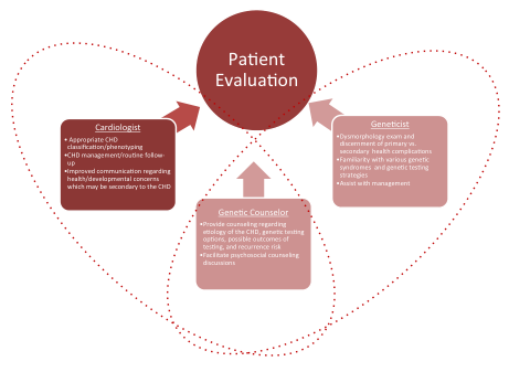
Summary : This flowchart illustrates the multidisciplinary process of patient evaluation involving three key roles: Cardiologist, Geneticist, and Genetic Counselor, each contributing specific expertise to the assessment and management of patients, particularly those with congenital heart disease (CHD).
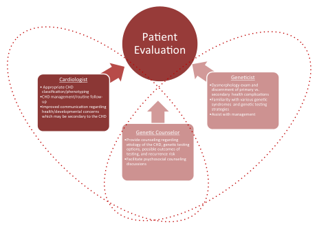
Summary : This flowchart illustrates the multidisciplinary process of patient evaluation involving a cardiologist, geneticist, and genetic counselor, each contributing specific expertise to the assessment and management of patients, particularly those with congenital heart disease (CHD).
acyanotic cyanotic congenital heart disease left to right right to left shunt
ventricular septal defect patent ductus arteriosus atrial septal defect congenital
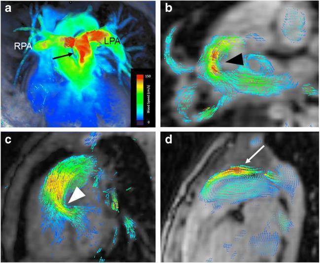
This diagnostic imaging panel demonstrates 4-D flow MRI visualizations in pediatric cardiovascular cases, utilizing color-coded velocity maps and vector fields to illustrate hemodynamic patterns. Image (a) is a coronal velocity map showing accelerated, high-velocity flow (red) in the main pulmonary artery, right pulmonary artery (RPA), and left pulmonary artery (LPA) consistent with pulmonary valve stenosis. Images (b) and (c) utilize axial and sagittal vector fields respectively to visualize shunt flow through septal defects; (b) highlights an atrial septal defect (ASD) with swirling vectors, and (c) shows a ventricular septal defect (VSD). Image (d) provides a sagittal view of a patent ductus arteriosus (PDA), demonstrating abnormal reverse and accelerated flow in the pulmonary artery represented by high-velocity color-coded vectors. The visualizations use a color scale ranging from blue (slow flow) to red (high velocity, up to 150 cm/s), facilitating the identification of stenosis, shunts, and turbulent flow patterns in congenital heart disease.
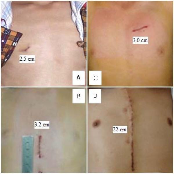
This clinical photograph set provides a side-by-side comparison of surgical incisions on the chest for treating congenital heart defects (CHD). The image illustrates the difference between Off-Pump Occlusion via Transthoracic Minimally Invasive Surgery (OPOTTMIS) and Traditional Open Surgery (TOS). Panels A, B, and C demonstrate the minimally invasive approach used for Atrial Septal Defect (ASD), Ventricular Septal Defect (VSD), and Patent Ductus Arteriosus (PDA) respectively, featuring small, localized incisions ranging from 2.5 cm to 3.2 cm in length. These are located in various thoracic regions (lateral and parasternal). In contrast, Panel D shows a Traditional Open Surgery incision, which is a significant 22 cm midline median sternotomy scar extending vertically down the center of the chest. The visual highlights the pedagogical concept of 'minimally invasive' by contrasting the cosmetic and anatomical impact of contemporary occlusion techniques versus traditional longitudinal sternotomy for pediatric cardiac procedures.
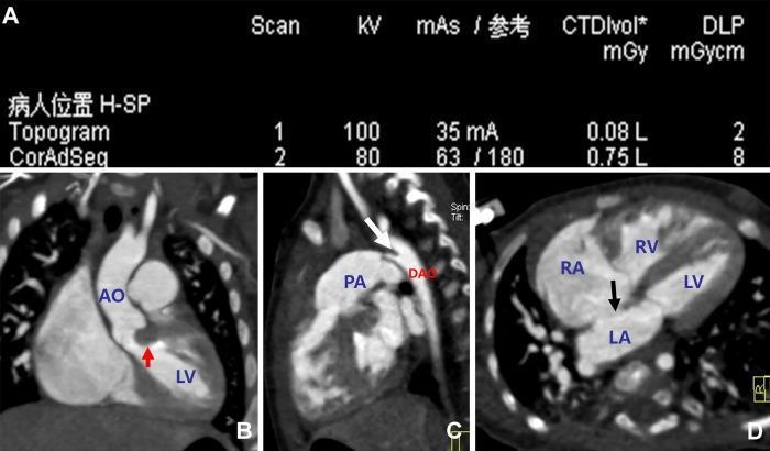
This diagnostic visual includes a panel of multi-slice spiral computed tomography (MSCT) images (B-D) and scan parameters (A) from a 4-month-old female patient with congenital heart disease. Panel B presents a multiplanar reconstructed (MPR) oblique view of the left ventricle (LV) and aorta (AO), where a red arrow highlights a focal protrusion of the ventricular septum into the left ventricular outflow tract (LVOT), consistent with subvalvular aortic stenosis. Panel C is a sagittal MPR view showing the pulmonary artery (PA) and descending aorta (DAO); a white arrow indicates a tubular connection at the aortic isthmus, diagnostic of a patent ductus arteriosus (PDA). Panel D displays an axial slice through the four-chamber view of the heart (RA, RV, LA, LV), where a black arrow identifies a defect in the atrial septum, representing an atrial septal defect (ASD). This image is an educational resource for pediatric cardiology and radiology, demonstrating the use of low-dose MSCT in evaluating complex congenital cardiac anomalies.
Tetralogy of Fallot anatomy diagram cyanotic congenital heart
Classification of Congenital Heart Disease (CHD)
I. Classic Hemodynamic Classification (Most Widely Used)
A. Lesions Causing Outflow Obstruction (Obstructive Lesions)
| Ventricle Affected | Lesion |
|---|---|
| Left ventricle | Coarctation of the aorta, Aortic stenosis (valvular/subvalvular/supravalvular) |
| Right ventricle | Pulmonary valve stenosis |
B. Lesions Causing Left-to-Right Shunting (Acyanotic Shunts)
| Lesion | Key Feature |
|---|---|
| Ventricular Septal Defect (VSD) | Most common CHD overall |
| Atrial Septal Defect (ASD) | Ostium secundum most common type |
| Patent Ductus Arteriosus (PDA) | Failure of fetal ductus to close |
| Endocardial Cushion Defect (AVSD) | Associated with Down syndrome |
| Partial Anomalous Pulmonary Venous Return | One or more pulmonary veins drain into right heart |
C. Lesions Causing Right-to-Left Shunting (Cyanotic Lesions)
With Decreased Pulmonary Blood Flow
| Lesion | Mechanism |
|---|---|
| Tetralogy of Fallot (ToF) | VSD + RV outflow obstruction + RV hypertrophy + overriding aorta |
| Pulmonary atresia | Complete obstruction of RV outflow |
| Tricuspid atresia | Absent tricuspid valve; requires ASD/VSD for survival |
With Increased Pulmonary Blood Flow
| Lesion | Mechanism |
|---|---|
| Transposition of the Great Vessels (TGA) | Aorta from RV, PA from LV - parallel circulations; survival needs ASD/PDA |
| Truncus arteriosus | Single great vessel; mixing of blood |
| Double-outlet right ventricle (DORV) | Both great arteries arise from RV |
| Total Anomalous Pulmonary Venous Return (TAPVR) | All pulmonary veins drain into systemic veins |
| Hypoplastic left heart syndrome (HLHS) | Severely underdeveloped left heart |
| Single ventricle | Only one functional pumping chamber |
- Morgan and Mikhail's Clinical Anesthesiology, 7e, pp. 780-781
II. Simpler Clinical Classification (Acyanotic vs. Cyanotic)
| Category | Subgroup | Examples |
|---|---|---|
| Acyanotic | Left-to-right shunt | VSD, ASD, PDA, AVSD |
| Acyanotic | Obstructive | Pulmonary stenosis, Aortic stenosis, Coarctation of aorta |
| Cyanotic | Decreased pulmonary flow | Tetralogy of Fallot, Tricuspid atresia, Pulmonary atresia |
| Cyanotic | Increased pulmonary flow | TGA, Truncus arteriosus, TAPVR, HLHS |
Key point: Right-to-left shunts produce cyanosis by allowing deoxygenated blood into the systemic circulation. Left-to-right shunts cause heart failure by volume-overloading the pulmonary circulation. With prolonged L→R shunting, Eisenmenger physiology can develop (pulmonary hypertension + shunt reversal = acquired cyanosis).
III. By Lesion Complexity (AHA/ACC Framework)
| Complexity | Examples |
|---|---|
| Simple | Isolated small ASD, small VSD, mild pulmonary valve stenosis, bicuspid aortic valve |
| Moderate | ASD (large), VSD (moderate/large), AVSD, coarctation, Ebstein anomaly, Tetralogy of Fallot (repaired) |
| Complex/Severe | Eisenmenger syndrome, Fontan circulation, unrepaired cyanotic CHD, TGA, HLHS, DORV, single ventricle |
IV. AHA/ACC Physiological Stage Classification (ACHD)
| Stage | Key Features |
|---|---|
| A | NYHA Class I; no hemodynamic/anatomic sequelae; normal exercise capacity; normal organ function |
| B | NYHA Class II; mild valvular disease; arrhythmias not requiring treatment; trivial/small shunts |
| C | NYHA Class III; significant valvular disease; moderate ventricular dysfunction; hemodynamically significant shunt; mild/moderate hypoxemia; pulmonary hypertension |
| D | NYHA Class IV; severe hypoxemia/cyanosis; Eisenmenger syndrome; severe pulmonary hypertension; refractory end-organ dysfunction |
- Braunwald's Heart Disease, 11e - Modified from AHA/ACC 2018 ACHD Guidelines (Stout et al., J Am Coll Cardiol. 2019;73:1494)
V. Pathophysiologic Consequences Summary
| Mechanism | Consequence | Examples |
|---|---|---|
| L→R shunt | Volume overload of pulmonary circulation → CHF, pulmonary HTN | VSD, ASD, PDA |
| R→L shunt | Systemic arterial desaturation → cyanosis, erythrocytosis, hyperviscosity | ToF, TGA, Tricuspid atresia |
| Outflow obstruction | Pressure overload → ventricular hypertrophy, CHF | Aortic stenosis, Coarctation, Pulmonary stenosis |
| Mixed/complex | Both cyanosis and pulmonary overcirculation | TGA, Truncus arteriosus, TAPVR |
VI. Special Complications of Cyanotic CHD
-
Compensated erythrocytosis: Hematocrit stable (usually <65%), no hyperviscosity symptoms - no treatment needed
-
Uncompensated erythrocytosis: Hyperviscosity symptoms, thrombotic risk (especially stroke in children <4 years), risk worsened by dehydration
-
Coagulopathy: Low-normal platelets, coagulation cascade defects
-
Hyperuricemia: From increased renal urate reabsorption due to hypoperfusion
-
Paradoxical embolism: Venous thrombi can bypass lungs via R→L shunt
-
Morgan and Mikhail's Clinical Anesthesiology, 7e, p. 781
Survival Dependence of Some Lesions
- Transposition of great vessels - survives via PDA, ASD, or VSD
- Pulmonary atresia - depends on PDA for pulmonary flow
- Tricuspid atresia - depends on ASD + VSD