
Explain the complete histology of the digestive system with each minor detail and it should cover the following topics thoroughly: Lips Cheeks Palate Tongue complete Teeth complete Gums Oropharynx General structure plan of alimentary canal Enteric nervous system Esophagus Stomach Small intestine Large intestine Along with their clinical correlates You can also divide it in parts as you're feasible but I want the complete detail so I wouldn't need to read the book
Reading File
Finding Sources
Finding Sources
Finding Sources
Good - I have both books. Now let me read the key sections in parallel. Given budget constraints, I'll do large reads of the most comprehensive sections.
Reading File
Reading File
Excellent - I'm getting a lot of data. Let me continue reading in parallel - the Wheater's text (block5) from offset 4459 onward for oral cavity, and the Junqueira block2 from a different offset for the detailed content.
Reading File
Reading File
Excellent content coming in. Let me now read the deeper sections - continuing Junqueira block2 and the Wheater's block6 for GI tract details.
Reading File
Reading File
I need the actual GI tract content from Wheater's block6. Let me find the right offset.
Finding Sources
Reading File
Now I have the line numbers for the key GI sections. Let me read them in parallel.
Reading File
Reading File
I now have a wealth of detailed content. Let me get more from both blocks to fill the remaining topics - especially more on tongue, teeth details from Wheater's block5, and more on esophagus/stomach/intestines from block6.
Reading File
Reading File
I now have extensive detailed content from both major textbooks. I have enough to write a comprehensive answer covering all topics. Let me now compile the complete histology of the digestive system.Here is the complete, detailed histology of the digestive system drawn directly from Wheater's Functional Histology (Wheater's) and Junqueira's Basic Histology, 17e (Junqueira).
Complete Histology of the Digestive System
PART 1: ORAL CAVITY - OVERVIEW
The oral cavity is lined by oral mucosa, which is subdivided into three functional types based on location:
| Type | Location | Epithelium |
|---|---|---|
| Masticatory mucosa | Gingiva (gums) + hard palate | Keratinized OR parakeratinized stratified squamous |
| Lining mucosa | Cheeks, lips (inner), soft palate, floor of mouth, oropharynx | Non-keratinized stratified squamous |
| Specialized mucosa | Dorsum of tongue | Stratified squamous + taste buds |
The oral cavity proper is bounded superiorly by hard and soft palates, inferiorly by the tongue and floor of mouth, and posteriorly by the entrance to the oropharynx. - Junqueira, p.1412
PART 2: THE LIPS (LABIA)
The lip has a well-developed core of orbicularis oris striated muscle, making it highly mobile for ingestion, speech, and communication. Each lip has three distinct surfaces:
1. Internal Mucous Surface (Oral Mucosa)
- Lined by thick, non-keratinized stratified squamous epithelium (lining mucosa)
- Lamina propria underlies the epithelium - loose connective tissue with blood vessels and nerves
- Submucosa is present and contains numerous minor labial salivary glands (mucous and mixed in character), which secrete continuously keeping the surface moist
2. Vermilion Zone (Red Zone)
- The prominent red/pink zone visible on the exterior of the lip
- Covered by very thin, keratinized stratified squamous epithelium - this is the transition from oral mucosa to skin
- No salivary glands, sweat glands, or hair follicles - this is clinically important
- The connective tissue papillae are extremely tall and vascular, with a rich capillary network that imparts the characteristic pink/red color visible through the thin epithelium
- Richly innervated with sensory endings (explains the exquisite sensitivity of the lips)
- Moistened by the tongue licking saliva onto it - since there are no glands, it dries rapidly in arid conditions
3. External Skin Surface
- Covered by thin, keratinized stratified squamous epithelium typical of skin
- Contains hair follicles, sebaceous glands, and sweat glands
Clinical Correlate - Cheilitis & Herpes Labialis
The absence of glands in the vermilion makes it prone to drying and cracking (cheilitis). Herpes simplex virus type 1 (HSV-1) resides latently in trigeminal ganglia and reactivates to infect the stratified squamous epithelium at the vermilion, producing "cold sores" or "fever blisters" - painful vesicles that rupture and crust. Recurrence is triggered by UV light, fever, stress, or immunosuppression. - Junqueira, p.736
PART 3: THE CHEEKS (BUCCAL MUCOSA)
Histological Structure
- Inner surface lined by non-keratinized stratified squamous epithelium (lining mucosa) - thick, resilient, and flexible
- Lamina propria: loose connective tissue with blood vessels, lymphatics, sensory nerve endings, scattered lymphocytes and plasma cells
- Submucosa: dense connective tissue containing minor buccal salivary glands (mixed serous and mucous), diffuse lymphoid tissue, and adipose tissue
- Deep to the submucosa: buccinator muscle (striated), responsible for keeping food between the teeth during chewing
- Parotid (Stensen) duct opens into the vestibule of the mouth at a small elevation on the mucosal surface opposite the upper second molar tooth
- Throughout the epithelium: scattered antigen-presenting Langerhans cells and rich sensory innervation
Clinical Correlate - Aphthous Ulcers / Leukoplakia
The non-keratinized buccal epithelium is vulnerable to minor trauma and inflammatory conditions. Aphthous ulcers (canker sores) are painful shallow ulcerations of the lining mucosa with unclear etiology (immune-mediated). Leukoplakia appears as a white patch on the buccal mucosa resulting from hyperkeratosis; it is a potentially premalignant lesion. Biopsy is required to rule out dysplasia or squamous cell carcinoma. - Junqueira, p.736
PART 4: THE PALATE
The palate forms the roof of the oral cavity and has two distinct parts with different histology.
Hard Palate
The hard palate is supported by bone (palatine processes of the maxilla and horizontal plates of the palatine bones).
Epithelium: Keratinized stratified squamous (masticatory mucosa) - the most heavily keratinized part of the oral cavity (resists abrasion from food)
Lamina propria: Thick, dense connective tissue with tall papillary projections into the epithelium; directly fused to the periosteum of underlying bone (no submucosa in some areas = mucoperiosteum)
Submucosa: Present only in the anterior (fatty) zone and posterior (glandular) zone of the hard palate:
- Fatty zone (anterior): contains adipose tissue
- Glandular zone (posterior): contains numerous mucous palatine salivary glands (minor glands)
- Along the midline raphe: no submucosa; epithelium attaches directly to periosteum
Incisive papilla: At the anterior midline, a small elevation marks the nasopalatine canal
Soft Palate
The soft palate lacks bone; its core is striated muscle (palatoglossus, palatopharyngeus, levator and tensor veli palatini, and musculus uvulae).
Oral surface (inferior): Non-keratinized stratified squamous epithelium - lining mucosa; the lamina propria contains mucous minor salivary glands in the submucosa
Nasal surface (superior): Pseudostratified ciliated columnar epithelium (respiratory type) with goblet cells
Transition: Abrupt transition from oral-type to respiratory-type epithelium occurs on the soft palate; this is a unique zone
Lamina propria of both surfaces: loose connective tissue with scattered lymphoid nodules, mast cells, and lymphocytes
Clinical Correlate - Cleft Palate
Failure of fusion of the palatine shelves during embryonic development results in cleft palate. The anatomy of the mucosa and submucosa at the cleft edges determines surgical repair strategies. Submucosal cleft palate (intact surface mucosa but absent muscle) can cause speech and swallowing difficulties. - Wheater's, p.1414
PART 5: THE TONGUE
The tongue is a complex muscular organ critical for mastication, swallowing, speech, and taste.
Core - Striated Muscle
- The body consists of interlacing bundles of striated muscle running in three perpendicular planes: horizontal, vertical, and longitudinal
- This orthogonal arrangement provides the tongue's remarkable mobility and flexibility
- Between the muscle bundles: fibroadipose connective tissue, blood vessels, nerve bundles (including chorda tympani and lingual nerve)
Surfaces of the Tongue
Inferior Surface
- Thin non-keratinized stratified squamous epithelium (lining mucosa)
- Smooth, no papillae
- Thin lamina propria
Dorsal Surface
- Covered by specialized mucosa - the most complex oral mucosa
- Divided by the sulcus terminalis (V-shaped groove) into:
- Anterior two-thirds (oral/body): studded with papillae
- Posterior one-third (pharyngeal/root): no papillae, has lingual tonsil (aggregated lymphoid nodules)
Lingual Papillae (Anterior 2/3)
1. Filiform Papillae (most numerous)
- Small, conical/thread-like projections
- Covered by keratinized stratified squamous epithelium at the tip
- No taste buds
- Function: give the tongue its rough texture, aid in food manipulation (rasping)
- Arranged in rows parallel to the sulcus terminalis
2. Fungiform Papillae
- Mushroom-shaped (broad top, narrow stalk)
- Scattered among filiform papillae, more numerous at the tip and margins
- Covered by non-keratinized or lightly keratinized stratified squamous epithelium
- Contain taste buds on their superior surface (dorsal surface)
- Appear as red dots visible to the naked eye due to rich vascular connective tissue core (reflecting through thin epithelium)
3. Circumvallate (Vallate) Papillae
- 8-12 large papillae arranged in a V-shaped row just anterior to the sulcus terminalis
- Each papilla is wide and flat-topped, surrounded by a deep circular moat (furrow/trench)
- The walls of the furrow are densely packed with taste buds (hundreds per papilla) - the highest density of taste buds in the mouth
- Von Ebner's serous glands open into the bottom of the furrow via ducts - their watery secretion flushes taste substances into contact with taste buds and washes them away to allow detection of new substances
- Covered by non-keratinized stratified squamous epithelium
4. Foliate Papillae
- Leaf-like folds (5-8 parallel ridges) on the lateral margins of the tongue posteriorly
- Taste buds present in the epithelium lining the folds
- Well-developed in children; often rudimentary in adults
- Minor salivary glands drain into the furrows between the folds
Taste Buds
- Oval-shaped neuroepithelial sensory organs - about 50-100 micrometers tall
- Found in: circumvallate papillae (walls of furrow), fungiform papillae (dorsal surface), foliate papillae, soft palate, epiglottis
- Total number in humans: ~10,000 taste buds
- Open to the surface via a taste pore - a small apical opening filled with microvilli
- Cell types within a taste bud:
- Type I (dark/supporting cells): Most numerous; elongated cells with dark nuclei; wrap around other cells; secrete a glycoprotein matrix
- Type II (light/receptor cells): Express taste receptor proteins (GPCRs) on microvilli for sweet, bitter, and umami detection; do NOT form synapses directly
- Type III (intermediate cells): Spindle-shaped; form conventional synapses with afferent nerve fibers; detect sour (acidic) stimuli via proton channels
- Type IV (basal cells): Small, round; located at base; are stem cells that give rise to the other types; taste bud cells turn over every 10-14 days
- Salty and sour detected via ion channels; sweet, bitter, umami detected via GPCRs (T1R and T2R receptor families)
- Innervation: Anterior 2/3 by chorda tympani (VII); posterior 1/3 by glossopharyngeal (IX); epiglottis by vagus (X)
Clinical Correlate - Taste Disorders & Genetic Basis
The genetic basis of taste involves the TAS1R gene family (for sweet/umami) and TAS2R gene family (for bitter). Polymorphisms in TAS2R38 determine the ability to taste PROP (6-n-propylthiouracil) - "supertasters" have more circumvallate papillae and taste buds. Chemotherapy, zinc deficiency, and viral infections (including COVID-19 anosmia/ageusia) can damage taste bud neuroepithelium. Zinc is required for gustin (carbonic anhydrase VI), a protein secreted by von Ebner's glands essential for taste bud maintenance. - Wheater's, p.1409
Posterior 1/3 of Tongue (Lingual Tonsil)
- No papillae
- Covered by non-keratinized stratified squamous epithelium
- Mucosa has irregular crypts (invaginations into the connective tissue) surrounded by lymphoid nodules (forming the lingual tonsil)
- Mucous glands (posterior lingual glands) in the submucosa open into the crypts
PART 6: TEETH
A complete tooth consists of: enamel, dentin, cementum, pulp cavity, and supporting tissues (periodontal ligament, alveolar bone, gingiva).
1. ENAMEL
- The hardest substance in the body - 96-98% inorganic (hydroxyapatite crystals), 1-2% organic (mostly enamelins and amelogenins)
- Produced by ameloblasts (from oral ectoderm) during tooth development; once the tooth erupts, ameloblasts are lost - enamel cannot be regenerated
- Structure: Enamel is organized into enamel rods (prisms) - tightly packed, keyhole-shaped cylindrical structures running from the dentinoenamel junction to the surface
- Each enamel rod is a column of tightly packed hydroxyapatite crystals (30 nm in diameter) with their long axes parallel to the rod
- Incremental lines of Retzius: Brown striae visible in ground sections - represent periodic rhythmic appositional growth of enamel during development
- Neonatal line: A pronounced line of Retzius marking the stress of birth (present in primary and some permanent teeth)
- Hunter-Schreger bands: Alternating light and dark bands in longitudinal sections; represent alternating directions of enamel rod orientation - provides resistance to shear
- Enamel spindles: Short, club-shaped processes of odontoblasts that cross the dentinoenamel junction into enamel
- Enamel tufts: Defective, hypocalcified groups of enamel rods at the dentinoenamel junction - appear as grass-like tufts on cross section
- Enamel lamellae: Thin sheet-like faults running from the surface inward, partially filled with organic material - potential pathways for bacterial invasion
2. DENTIN
- Forms the bulk of the tooth - 70% inorganic (hydroxyapatite), 20% organic (mostly type I collagen), 10% water
- Produced by odontoblasts (from neural crest-derived mesenchyme) - these cells persist throughout life at the inner surface of dentin adjacent to the pulp
- Dentin is traversed by dentinal tubules running from the pulp cavity to the periphery (dentinoenamel junction at crown, dentinocemental junction at root)
- Each dentinal tubule contains an odontoblastic process (cytoplasmic extension of an odontoblast) - these processes are the basis of dentinal sensitivity
- Peritubular dentin: Highly mineralized dentin immediately surrounding each tubule - forms a cuff
- Intertubular dentin: Less mineralized dentin between the tubules; contains type I collagen fibers
- Incremental lines (von Ebner lines): Represent periodic growth cycles - visible in ground sections as fine lines perpendicular to tubules
- Owen contour lines: Accentuated lines of von Ebner, correspond to disturbance lines in dentin matrix formation
- Interglobular dentin: Areas of incompletely mineralized dentin between calcification globules - seen near dentinoenamel junction; prominent in vitamin D deficiency (rickets)
- Tomes granular layer: A thin zone of granular appearance at the periphery of root dentin
3. CEMENTUM
- Calcified connective tissue covering the root of the tooth (analogous to bone)
- ~65% inorganic (hydroxyapatite), ~35% organic (mostly type I collagen)
- Acellular (primary) cementum: Located in the coronal portion of the root; formed first; no cells embedded in matrix; Sharpey's fibers (periodontal ligament fiber ends) are anchored in it
- Cellular (secondary) cementum: Located at the apical portion of the root; contains cementocytes in lacunae within the matrix; formed throughout life; can repair root surface damage
- Produced by cementoblasts (from periodontal ligament)
- Unlike bone, cementum has no blood vessels (avascular) and undergoes minimal remodeling
4. DENTAL PULP
- Occupies the central pulp cavity (pulp chamber in crown + root canals)
- Loose connective tissue - gelatinous matrix rich in ground substance
- Contains: odontoblasts (at periphery, lining the walls), fibroblasts (most abundant cell), undifferentiated mesenchymal stem cells, macrophages, lymphocytes
- Nerve fibers: Myelinated and unmyelinated fibers enter via the apical foramen; unmyelinated fibers accompany odontoblastic processes into dentinal tubules (basis of dentin hypersensitivity)
- Blood vessels: Arterioles, venules, and capillaries enter via the apical foramen
- Ground substance: Rich in glycosaminoglycans (particularly hyaluronic acid and chondroitin sulfate)
- Pulp stones (denticles): Calcified deposits within the pulp - may be free-floating or attached to the wall
5. SUPPORTING TISSUES OF THE TEETH
Periodontal Ligament (PDL)
- Dense fibrous connective tissue occupying the periodontal space between the root cementum and alveolar bone
- Width: ~0.2 mm (narrows with age and disuse)
- Composed of principal fiber bundles (type I collagen, with some type III):
- Alveolar crest fibers: Most coronal; prevent tooth from being pulled out
- Horizontal fibers: Resist lateral forces
- Oblique fibers: Most numerous; run obliquely from cementum (apical) to alveolar bone (coronal); resist apical forces (main weight-bearing fibers)
- Apical fibers: Around apex; prevent tipping
- Interradicular fibers: Between roots of multirooted teeth
- Sharpey's fibers: Collagen bundle ends embedded in both cementum and alveolar bone
- Cells: Fibroblasts (predominant), cementoblasts, osteoblasts, osteoclasts, undifferentiated mesenchymal cells, rests of Malassez (epithelial remnants of Hertwig root sheath - may give rise to cysts), macrophages, mast cells
- Oxytalan fibers: Thin, elastic-like fibers running parallel to long axis of root; associated with blood vessel adventitia
- Blood vessels and nerve endings: Rich neurovascular supply - Meissner corpuscles and Ruffini endings for proprioception (important for control of bite force)
Alveolar Bone (Alveolar Process)
- Specialized bone forming the sockets (alveoli) that house the teeth
- Bundle bone (alveolar bone proper): The inner socket wall into which Sharpey's fibers insert; highly compact; lamellated; lines the socket
- Supporting alveolar bone: Cancellous and cortical bone surrounding the bundle bone
Clinical Correlate - Dental Caries
Caries results from bacterial acid demineralization of enamel and dentin. Streptococcus mutans ferments sucrose to lactic acid, which demineralizes hydroxyapatite crystals. The process begins in enamel (initially reversible if remineralized with fluoride) and progresses through dentin toward the pulp. Once bacteria reach the pulp via exposed dentinal tubules, irreversible pulpitis (pulp inflammation) occurs, requiring root canal therapy. Fluoride substitutes for hydroxyl groups in hydroxyapatite forming fluorapatite, which is more acid-resistant. - Wheater's, p.1462
PART 7: GUMS (GINGIVA)
The gingiva is the specialized part of the masticatory oral mucosa surrounding the neck of each tooth.
Structure
Gingival mucosa (= masticatory mucosa):
- Keratinized or parakeratinized stratified squamous epithelium
- Lamina propria: thick papillary layer of dense connective tissue with collagen fiber bundles (gingival fibers) that radiate from the alveolar crest and cementum to maintain gingival attachment
- No submucosa - the lamina propria attaches directly to the periosteum of alveolar bone = mucoperiosteum
- Dentogingivial fibers (transseptal and alveologingival fibers): anchor the free gingiva to the tooth and alveolar bone
Junctional epithelium (attachment epithelium):
- Thin, non-keratinized epithelium that adheres directly to the enamel or cementum surface
- Secretes a basal lamina-like material on its inner surface (facing the tooth)
- Cells attach to this material via hemidesmosomes - this is called the epithelial attachment
- In young individuals: attachment is to enamel; in older individuals with gingival recession: attachment migrates onto cementum
- Extremely permeable - allows passage of gingival fluid (crevicular fluid) into the sulcus
Gingival sulcus:
- A shallow groove (normally ≤2 mm deep) between the free gingiva and the tooth
- Lined by crevicular epithelium (non-keratinized stratified squamous)
- The crevicular fluid contains immunoglobulins, complement, and neutrophils - first line of defense
Periodontium: The entire attachment apparatus - junctional epithelium + cementum + PDL + alveolar bone
Clinical Correlate - Periodontitis & Stem Cell Therapy
Gingivitis is inflammation of the superficial gingival tissues (reversible); if untreated, it progresses to periodontitis with destruction of the periodontium. Pathogenic bacteria such as Porphyromonas gingivalis colonize the deepened periodontal pocket (>3 mm), producing lipopolysaccharide and collagenases. Inflammatory cytokines activate osteoclasts, resulting in irreversible alveolar bone loss and tooth mobility. The discovery of periodontal ligament stem cells (PDLSCs) - multipotent cells within the PDL - has opened the possibility of regenerative periodontal therapy. Transplanted human PDLSCs in animal models differentiate into cementum, periodontal ligament, alveolar bone, peripheral nerves, and blood vessels. - Wheater's, p.1463
PART 8: OROPHARYNX
The oropharynx is the posterior region of the oral cavity connecting the mouth with the esophagus and larynx.
Epithelium
- Non-keratinized stratified squamous epithelium (lining mucosa) - continuous with buccal and soft palate mucosa
- Reflects the functional need for flexibility and resistance to abrasion as food passes through
Lamina Propria
- Loose connective tissue with numerous lymphoid nodules and diffuse lymphoid tissue
- IgA-secreting plasma cells are abundant - the epithelial cells above them express the polymeric immunoglobulin receptor (pIgR), which binds IgA dimers and performs transcytosis into the lumen, releasing secretory IgA (sIgA) - the major immunological defense of mucosal surfaces
Submucosa
- Dense connective tissue with minor mucous salivary glands that continuously secrete to keep the surface moist
- Rich lymphoid tissue
Waldeyer's Tonsillar Ring
The oropharynx is guarded by the Waldeyer's ring - a circular arrangement of lymphoid tissue:
- Palatine tonsils: Located at the entrance to the oropharynx between the palatopharyngeal and palatoglossal arches
- Pharyngeal tonsil (adenoid): On the roof of the nasopharynx
- Tubal tonsils: Lateral nasopharynx near the auditory tube opening
- Lingual tonsil: Base of the tongue
Palatine Tonsils - Detailed Histology
- Surface epithelium: Non-keratinized stratified squamous
- Deep invaginations called crypts penetrate into the lymphoid tissue - provide huge surface area for antigen sampling
- Lymphoid follicles: Secondary follicles with germinal centers (B-cell zones) in the lamina propria and filling the core of the tonsil
- T lymphocytes in the interfollicular zones
- Reticular epithelium: Specialized epithelium lining the crypts with M-cell-like properties for antigen uptake
- Capsule of dense fibrous connective tissue on the external aspect
- No afferent lymphatics - antigen sampling occurs directly across the crypt epithelium
Clinical Correlate - Tonsillitis, Peritonsillar Abscess, Adenoid Hypertrophy
Repeated bacterial tonsillitis (typically Streptococcus pyogenes) causes reactive hyperplasia of germinal centers. Food debris and bacteria may accumulate in the crypts forming tonsilloliths (tonsil stones). Peritonsillar abscess (quinsy) forms between the tonsil capsule and the superior pharyngeal constrictor - requires surgical drainage. Adenoid hypertrophy in children narrows the nasopharyngeal airway, causing nasal obstruction, recurrent otitis media (obstructing the Eustachian tube), and sleep-disordered breathing. - Junqueira, p.1413
PART 9: GENERAL STRUCTURE PLAN OF THE ALIMENTARY CANAL
From the esophagus to the anal canal, the digestive tract wall has a uniform basic organization of four concentric layers:
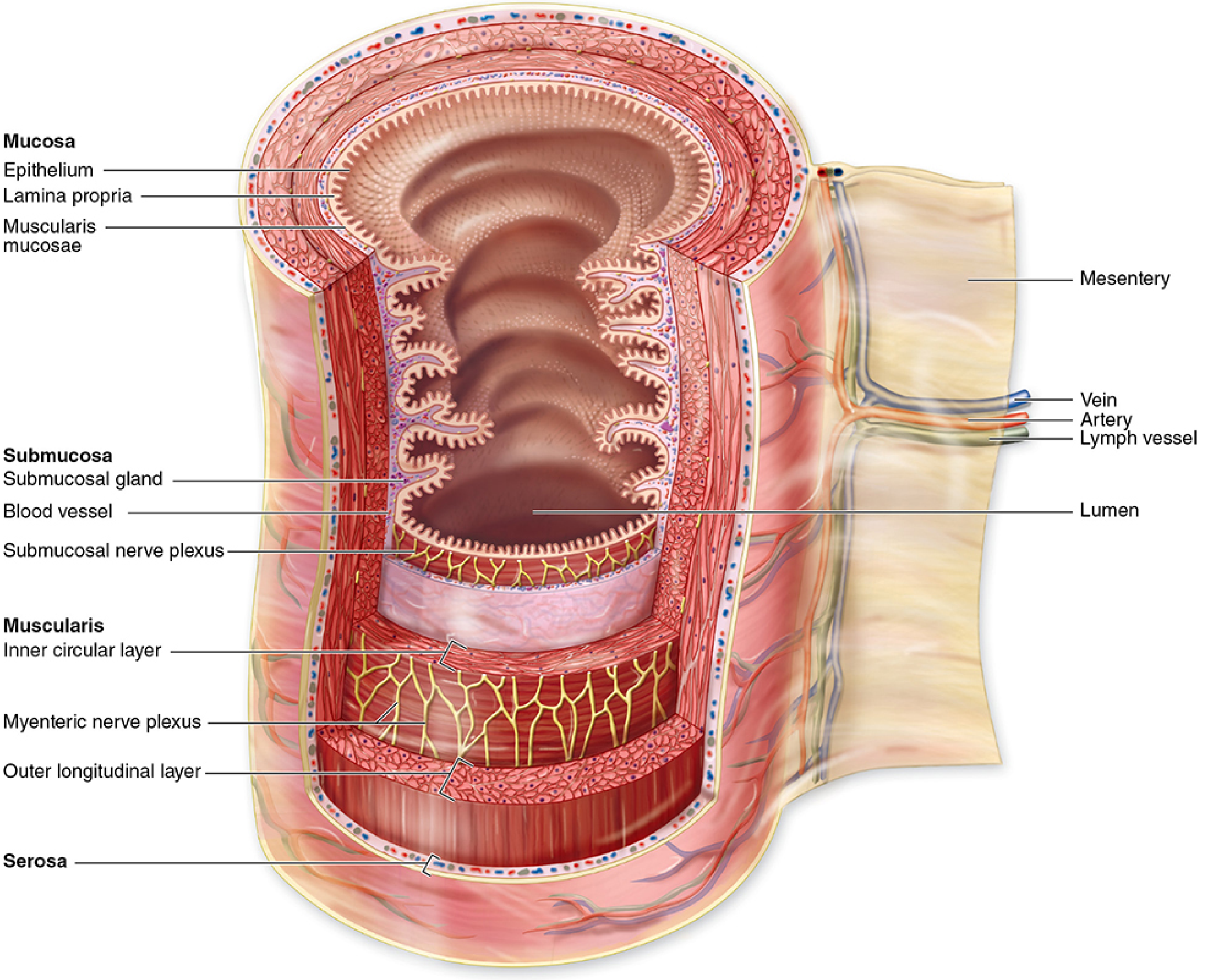
Layer 1: MUCOSA (Innermost)
The mucosa has three sub-layers:
a) Epithelium
- Type varies by region (discussed per organ below)
- Rests on a basement membrane
- Functions: protection, secretion, absorption
b) Lamina Propria
- Loose connective tissue immediately beneath the epithelium
- Contains: capillaries, lymphatics (lacteals in small intestine), nerve fibers, plasma cells (especially IgA-secreting), lymphocytes, eosinophils, mast cells
- May contain mucosal glands (crypts of Lieberkühn in intestines, gastric glands in stomach)
- Contains MALT (mucosa-associated lymphoid tissue) - lymphoid nodules and diffuse lymphoid cells
- Secretory IgA is the dominant immunoglobulin of the gut lumen
c) Muscularis Mucosae
- A thin layer of smooth muscle (usually 2 sub-layers: inner circular + outer longitudinal)
- Separates mucosa from submucosa
- Permits local independent movements of the mucosa (independent of outer muscularis externa) - agitates the mucosal surface to enhance mixing and absorption
Layer 2: SUBMUCOSA
- Dense irregular connective tissue
- Contains: larger blood vessels (submucosal arterial plexus), lymphatics, adipose cells
- The submucosal (Meissner's) plexus of autonomic nerves resides here (see ENS below)
- Submucosal glands: Present only in the esophagus (esophageal glands proper) and duodenum (Brunner's glands) - nowhere else
- Lymphoid nodules (especially in ileum - Peyer's patches extend from mucosa into submucosa)
Layer 3: MUSCULARIS EXTERNA (Muscularis Propria)
- Thick layer of smooth muscle in two distinct orientations:
- Inner circular layer: Muscle fibers run circumferentially - when it contracts, it narrows the lumen
- Outer longitudinal layer: Muscle fibers run along the length - when it contracts, it shortens the tube
- These two layers work together to generate peristalsis (coordinated propulsive waves) and segmentation (mixing contractions)
- Exceptions to smooth muscle rule:
- Esophagus (upper 1/3): striated (voluntary) muscle
- Esophagus (middle 1/3): mixed striated + smooth
- Esophagus (lower 1/3): smooth muscle only
- External anal sphincter: striated (voluntary)
- Stomach has an additional oblique (inner) layer - giving it 3 muscle layers
- The myenteric (Auerbach's) plexus lies between the inner circular and outer longitudinal layers
Layer 4: SEROSA / ADVENTITIA (Outermost)
Serosa:
- In regions suspended within the peritoneal cavity (stomach, small intestine, most of large intestine)
- Thin layer of loose connective tissue + simple squamous mesothelium (visceral peritoneum)
- Rich in blood vessels, lymphatics, adipose cells
- The serosa is continuous with the mesentery, which suspends the intestines in the abdominal cavity and transmits nerves and vessels
Adventitia:
- In regions attached to surrounding structures (esophagus, retroperitoneal portions of GI tract)
- Dense connective tissue without a mesothelial covering
- Blends with surrounding tissues
Regional Specializations Summary Table
| Feature | Esophagus | Stomach | Small Intestine | Large Intestine |
|---|---|---|---|---|
| Epithelium | Non-keratinized SSE | Simple columnar (mucous) | Simple columnar | Simple columnar |
| Villi | Absent | Absent | Present | Absent |
| Crypts/Glands | Mucosal glands (sparse) | Gastric glands | Crypts of Lieberkühn | Crypts of Lieberkühn |
| Submucosal glands | Esophageal glands proper | Absent | Brunner's (duodenum only) | Absent |
| Muscularis (outer) | 2 layers (mixed/smooth) | 3 layers (oblique added) | 2 layers smooth | 2 layers (outer = taeniae coli) |
PART 10: ENTERIC NERVOUS SYSTEM (ENS)
The ENS is often called the "second brain" - it contains approximately 100 million neurons (more than the spinal cord) and can function independently of the CNS.
Submucosal (Meissner's) Plexus
- Located in the submucosa, between the muscularis mucosae and muscularis externa
- Functions: Regulates secretion by mucosal glands, controls local blood flow, coordinates absorptive activities
- Neurons are smaller and less densely packed than in the myenteric plexus
- Contains sensory, interneurons, and secretomotor neurons
- Postganglionic parasympathetic and local enteric neurons
Myenteric (Auerbach's) Plexus
- Located between the inner circular and outer longitudinal smooth muscle layers of the muscularis externa
- Functions: Generates and coordinates contractile activity of the muscularis (peristalsis + segmentation); determines the direction and speed of peristaltic waves
- Contains ganglia (aggregations of neuron cell bodies) interconnected by nerve fiber bundles running in all directions
- Contains all three neuron types: sensory (IPAN - intrinsic primary afferent neurons), interneurons, and motor neurons
Neurotransmitters of the ENS
The ENS uses a wide variety of neurotransmitters:
- Acetylcholine (ACh): Stimulates muscle contraction (excitatory motor neurons), stimulates secretion
- Vasoactive intestinal peptide (VIP): Inhibits smooth muscle - key inhibitory neurotransmitter of the gut
- Substance P: Excitatory; stimulates contraction
- Nitric oxide (NO): Key inhibitory neurotransmitter; mediates relaxation of circular muscle ahead of a peristaltic wave
- Serotonin (5-HT): Initiates peristaltic reflex; stimulates secretion - produced by enterochromaffin cells in the mucosa
- CGRP, enkephalins, somatostatin, neuropeptide Y: Various regulatory functions
Peristaltic Reflex (Law of the Intestine)
- Distension of a segment activates IPANs
- Above the bolus: ACh/Substance P released → circular muscle contracts, longitudinal muscle relaxes → propulsive wave moves aborally
- Below the bolus: VIP/NO released → circular muscle relaxes, longitudinal muscle contracts → accommodation
Clinical Correlates
Hirschsprung Disease (Congenital Aganglionic Megacolon):
- Failure of neural crest cell migration to distal colon results in an aganglionic segment (absent myenteric and submucosal plexuses)
- The affected segment cannot relax → functionally obstructed, causing massive proximal dilation (megacolon)
- Most commonly affects the rectosigmoid region
- Diagnosis: rectal suction biopsy showing absence of ganglion cells + increased acetylcholinesterase staining in nerve fibers
- Treatment: surgical resection of the aganglionic segment
Chagas Disease:
- Trypanosoma cruzi infection destroys the ganglion cells of the ENS
- Results in chagasic megaesophagus (achalasia-like) and chagasic megacolon
- Pathology: chronic inflammation with lymphocytic infiltration → ganglion cell destruction → denervation dilation
Irritable Bowel Syndrome (IBS):
-
Alterations in ENS serotonin signaling (altered 5-HT3 and 5-HT4 receptor activity)
-
Abnormal visceral sensation and gut motility without structural damage
-
Junqueira, p.734 | Wheater's, p.1520
PART 11: ESOPHAGUS
Overview
The esophagus is a 25 cm muscular tube extending from the pharynx (at C6) to the stomach (at T11). It courses through the neck, posterior mediastinum, and diaphragm. In its collapsed state, the lumen has a branched (stellate) appearance due to longitudinal mucosal folds.
Layer-by-Layer Histology
Mucosa
Epithelium: Non-keratinized stratified squamous epithelium - the thickest epithelium of the GI tract (except oral cavity). Human esophageal epithelium does not normally keratinize (though surface cells may show some keratohyalin granules). The non-keratinized squamous epithelium is well adapted for the abrasive forces of swallowing solid food.
Lamina Propria: Loose connective tissue; contains diffuse lymphoid tissue, occasional lymphoid nodules (often near ducts of mucous glands), blood vessels, and nerves.
Esophageal Cardiac Glands (Mucous glands of the lamina propria): Small groups of mucous glands present in the lamina propria at two locations:
- Upper esophagus (near pharynx)
- Lower esophagus (near stomach/cardia) These are simple tubular or branched glands producing neutral mucus to protect the epithelium.
Muscularis Mucosae: Composed of longitudinally-arranged smooth muscle fibers - begins near the level of the cricoid cartilage. Unusually thick in the proximal esophagus (aids in swallowing).
Submucosa
- Dense connective tissue
- Contains esophageal glands proper (the TRUE submucosal glands of the esophagus) - compound tubuloacinar mucous glands that open via long ducts through the mucosa into the lumen. These glands secrete acidic mucus for lubrication.
- Submucosal plexus (Meissner's)
- Lymphoid nodules clustered near gland ducts
Muscularis Externa
Critical regional variation - the type of muscle changes along the length:
- Upper 1/3 (proximal): Skeletal (striated) muscle - under voluntary control; involved in the initiation of swallowing
- Middle 1/3: Mix of skeletal and smooth muscle
- Lower 1/3 (distal): Smooth muscle only - controlled by the ENS and ANS
The muscularis externa has:
- Inner circular layer (prominent)
- Outer longitudinal layer
- Myenteric (Auerbach's) plexus between the two layers
Lower Esophageal Sphincter (LES): A functional sphincter (no anatomically distinct muscle) in the distal esophagus. Maintained by neurohormonal mechanisms; normally contracted preventing gastric reflux. Histologically, the circular smooth muscle layer is slightly thickened here.
Adventitia (NOT Serosa)
- The esophagus does NOT have a serosa because it is not suspended in the peritoneal cavity
- Has only an adventitia - loose connective tissue blending with surrounding structures
Esophagogastric Junction (Z-line/Ora Serrata)
- Abrupt transition from stratified squamous epithelium of esophagus to simple columnar mucous epithelium of stomach
- Visible endoscopically as a distinct Z-line (zigzag line) located at the level of the LES
- This transition is normally at or below the diaphragmatic hiatus
Clinical Correlates
Gastroesophageal Reflux Disease (GERD) and Barrett's Esophagus:
- Chronic reflux of acidic gastric contents damages the squamous epithelium of the lower esophagus
- The squamous epithelium undergoes metaplasia - replaced by intestinal-type columnar epithelium (with goblet cells) = Barrett's esophagus
- Barrett's is a premalignant condition - risk of adenocarcinoma is 30-125x higher than in the general population
- Histologically: intestinal metaplasia with goblet cells (PAS/Alcian blue positive); biopsy graded for dysplasia
- Treatment: proton pump inhibitors, surveillance endoscopy, ablative therapy
Achalasia:
- Degeneration of inhibitory neurons (VIP/NO-containing) of the myenteric plexus in the lower esophagus
- Results in failure of LES relaxation and loss of peristalsis
- Manometry shows high LES pressure and aperistalsis
- Endoscopy: bird-beak appearance; biopsy shows reduced/absent myenteric ganglion cells
Esophageal Varices:
-
Portal hypertension causes dilation of the submucosal venous plexus of the lower esophagus
-
Thin-walled varices are prone to catastrophic hemorrhage
-
Wheater's, p.1527
PART 12: STOMACH
The stomach is a J-shaped reservoir that stores food, initiates protein digestion, and controls the rate of gastric emptying. It is divided anatomically and histologically into: cardia, fundus, body (corpus), pyloric antrum, and pylorus.
General Features of the Gastric Mucosa
- Lined by simple columnar mucous epithelium (surface mucous cells)
- No villi in the stomach (unlike the small intestine)
- Surface studded with small depressions called gastric pits (foveolae) - openings of the gastric glands
- The ratio of pit depth to gland depth varies by region (short pits in fundus/body, deep pits in pylorus)
- The entire mucosal surface is covered by an alkaline mucus layer (produced by surface mucous cells) that protects against HCl
Gastric Mucosal Zones
A. Cardia
- Narrow ring (0.5-3 cm wide) at the esophagogastric junction
- Gastric pits: Short
- Glands: Branched tubular mucous glands (cardiac glands) - primarily mucus-secreting; simple branched tubular
- Contains scattered enteroendocrine cells (G cells, ECL cells)
- The lamina propria shows mucous glands similar to the cardiac glands of the lower esophagus
- Transition zone - may contain glands of mixed esophageal and gastric type
B. Fundus and Body (Corpus) - Oxyntic Mucosa
This is the main acid-secreting region - histologically the most distinctive:
Gastric pits: Short (occupy top 25% of mucosal thickness)
Glands (fundic/oxyntic glands): Long, straight, branched tubular glands occupying the remaining 75% of the mucosa. Densely packed; 3-7 glands per pit. They are divided into:
- Neck (upper, isthmus): mitotically active zone containing mucous neck cells and stem cells
- Body/middle: Contains the main secretory cells
- Base: Richest in chief cells
Cell Types in Fundic Glands (the most important and tested):
1. Surface mucous cells (mucous columnar cells)
- Line the pit (foveolae) and surface
- Tall columnar cells with pale, foamy supranuclear cytoplasm (due to mucus-filled vesicles)
- Secrete neutral mucopolysaccharide (mucin) - forms the protective mucus layer
- Nuclei: basal, flattened
- High rate of turnover: replaced every 3-5 days from stem cells in the neck/isthmus
2. Mucous Neck Cells
- Located in the neck of the glands
- Short, irregular cells with irregular nuclei
- Secrete acidic (sulfomucin) mucus - different from surface mucous cells
- Contains few small granules
- Represent progenitor/transit amplifying cells - they divide and migrate up (to become surface mucous cells) or down (to become chief or parietal cells)
3. Parietal Cells (Oxyntic Cells)
- Large, pyramidal cells - most characteristic cell of the gastric body
- Nuclei: central, round, prominent (binucleate in some)
- Cytoplasm: intensely eosinophilic (PAS-negative) due to enormous numbers of mitochondria (which provide energy for active proton pumping)
- Ultrastructure: Deep intracellular canaliculi (branched invaginations of apical plasma membrane) lined with microvilli greatly increase secretory surface area; tubulovesicular system (cytoplasmic membrane vesicles carrying H+/K+-ATPase in the resting state)
- H+/K+-ATPase (the proton pump): Pumps H+ into the canaliculus in exchange for K+; coupled with Cl- channels to produce HCl
- Also secrete intrinsic factor (IF) - a glycoprotein essential for vitamin B12 absorption in the ileum
- Located in the neck and body of the gland; bulge toward the lumen and toward the basement membrane
4. Chief Cells (Zymogenic/Peptic Cells)
- Predominate in the base of fundic glands
- Basophilic cytoplasm (abundant rER in the basal cytoplasm for protein synthesis)
- Apical cytoplasm full of zymogen granules - eosinophilic in well-fixed tissue, but often appear empty in routine sections due to extraction of loosely bound pepsinogen
- Secrete pepsinogen I and II (precursors of pepsin) - activated by HCl in the lumen
- Also secrete gastric lipase and small amounts of rennin (chymosin - curdles milk)
5. Enteroendocrine (APUD) Cells
- Scattered throughout the glands; most numerous at the base
- Small, clear cells with basal granules - "clear cells" or "D cells," "G cells" etc.
- Part of the diffuse neuroendocrine system (DNES) - also called APUD (Amine Precursor Uptake and Decarboxylation) cells
- Types in the stomach:
- ECL cells (Enterochromaffin-Like): In body/fundus; secrete histamine in response to gastrin and vagal stimulation; histamine binds H2 receptors on parietal cells to stimulate HCl secretion
- G cells: In the antrum (and duodenum); secrete gastrin in response to protein digestion products, distension, and vagal stimulation → stimulates ECL cells and parietal cells
- D cells: In fundus and antrum; secrete somatostatin → inhibits gastrin release and acid secretion (paracrine feedback)
- A cells (X/A cells): Secrete ghrelin ("hunger hormone") - stimulates appetite and GH release
- Enterochromaffin (EC) cells: Secrete serotonin (5-HT) - stimulates peristalsis
6. Stem Cells (Undifferentiated)
- Located in the isthmus/neck region
- Source of all other gastric mucosal cells
C. Pyloric Antrum / Pylorus
- Gastric pits: Deep (occupy top 50-70% of mucosal thickness)
- Glands: Pyloric glands - branched, coiled tubular glands; shorter than fundic glands; mucous-secreting (acidic mucin); some contain G cells
- No parietal cells, no chief cells in the pyloric glands
- The pyloric sphincter: Thickened inner circular smooth muscle layer separating stomach from duodenum
- Pyloric mucosa transitions into duodenal mucosa at the pyloric valve
Submucosa, Muscularis, and Serosa
Submucosa: Dense connective tissue; no submucosal glands; contains the submucosal (Meissner's) plexus; large blood and lymph vessels
Muscularis Externa: Three layers (unique to the stomach):
- Inner oblique layer (innermost) - aids in churning
- Middle circular layer (forms the pyloric sphincter)
- Outer longitudinal layer
Serosa: Thin connective tissue + mesothelium - stomach is completely covered by peritoneum
Clinical Correlates
Pernicious Anemia and Autoimmune Gastritis:
- Autoimmune destruction of parietal cells → loss of HCl + loss of intrinsic factor
- Without IF, vitamin B12 cannot be absorbed in the ileum → megaloblastic anemia
- Histology: atrophic gastritis with intestinal metaplasia of the gastric body; absence of parietal cells; lymphocytic infiltrate; compensatory hypergastrinemia
- Risk factor for gastric carcinoid (from ECL cell hyperplasia due to high gastrin)
Peptic Ulcer Disease (PUD):
- Mucosal defense fails (mucus layer, bicarbonate secretion, mucosal blood flow)
- Major cause: H. pylori infection - gram-negative spiral bacterium that colonizes the gastric antrum, induces chronic gastritis, increases gastrin production and HCl secretion
- Antral (H. pylori) ulcers: Associated with hypergastrinemia and hypersecretion; duodenal ulcers more common
- Gastric ulcers: Mucosal defense failure; often associated with NSAID use (inhibit COX → ↓ prostaglandins → ↓ mucus + bicarbonate secretion)
- Histology: ulcer base shows necrosis, granulation tissue, fibrosis; edges may show intestinal metaplasia
Zollinger-Ellison Syndrome:
- Gastrinoma - neuroendocrine tumor (usually in pancreas/duodenum) secreting massive amounts of gastrin
- → Extreme parietal cell hyperplasia and gastric acid hypersecretion
- → Multiple refractory peptic ulcers, often in unusual locations
- Histology: fundic mucosa shows parietal cell hyperplasia; no H. pylori
Gastric Carcinoma:
-
Intestinal type (associated with H. pylori): chronic gastritis → atrophy → intestinal metaplasia → dysplasia → carcinoma; gland-forming
-
Diffuse type: Poorly cohesive cells (loss of E-cadherin); "signet ring cells" (mucin-filled cells pushing nucleus to periphery); linitis plastica pattern with desmoplasia
-
Wheater's, p.1576 | Junqueira, p.755
PART 13: SMALL INTESTINE
The small intestine (duodenum + jejunum + ileum) is the primary site of digestion and absorption. It is ~6-7 m long with an enormous absorptive surface area created by three levels of mucosal folding.
Surface Area Amplification - Three Levels
| Level | Structure | Increase | Cumulative |
|---|---|---|---|
| 1 | Plicae circulares (valves of Kerckring) | 3x | 3x |
| 2 | Villi | 10x | 30x |
| 3 | Microvilli (brush border) | 20x | 600x |
Total surface area of small intestine: approximately 200 m²
Mucosa - Unique Features
Plicae Circulares (Circular Folds / Valves of Kerckring)
- Permanent, circular or helical folds of both mucosa AND submucosa
- They do NOT disappear when the intestine is distended (unlike gastric rugae)
- Most prominent in the jejunum and proximal ileum; sparse in the duodenum proximally and ileum distally
Villi
- Finger-like or leaf-like projections of mucosa (epithelium + lamina propria core) extending 0.5-1.5 mm into the lumen
- Each villus has a central lacteal (blind-ending lymphatic capillary) surrounded by a network of capillaries and a supporting core of smooth muscle fibers from the muscularis mucosae (causes villi to contract and pump lymph)
- Shape varies by region:
- Duodenum: Broad, leaf-shaped
- Jejunum: Finger-shaped (tallest)
- Ileum: Shorter, more finger-like
Crypts of Lieberkühn (Intestinal Glands)
- Straight tubular glands that extend from the base of villi down to the muscularis mucosae
- Open at the base of each villus
- Site of cell proliferation and renewal
- Ratio villus:crypt = approximately 5:1 in the jejunum
Cell Types of the Small Intestinal Epithelium
1. Enterocytes (Absorptive cells) - most numerous (~80%)
- Tall columnar cells with oval basal nuclei
- Apical brush border: Dense layer of microvilli (1-2 µm tall) coated with glycocalyx (heavily glycosylated integral membrane proteins)
- The glycocalyx contains key brush border enzymes anchored into the microvillus membrane:
- Peptidases (enterokinase/enteropeptidase, aminopeptidases, dipeptidases)
- Disaccharidases (lactase, sucrase-isomaltase, maltase)
- Alkaline phosphatase
- Lateral membranes connected by tight junctions (zonula occludens), adherens junctions, and desmosomes - the junctional complex
- Intracellular digestion: Fatty acids and glycerol absorbed into SER → reassembled into triglycerides → packaged with apoprotein → chylomicrons → exocytosed into the lacteal
- Rapid turnover: replaced every 3-5 days (fastest cell renewal in the body)
2. Goblet Cells - second most common
- Scattered among enterocytes; increase in number distally (most in ileum > jejunum > duodenum)
- Classic goblet shape: narrow base, distended apical portion filled with mucus granules
- Secrete mucin (MUC2 predominantly) forming a protective gel layer over the epithelium
- In H&E sections: apical mucus appears pale/clear
3. Paneth Cells
- Located exclusively at the base of the crypts
- Conspicuous large, brightly eosinophilic, pyramidal apical granules - the most dramatic secretory granules in the GI tract
- Secrete:
- Defensins (cryptdins) - antimicrobial peptides active against bacteria, fungi, some viruses
- Lysozyme - cleaves bacterial cell wall peptidoglycan
- Phospholipase A2 - antimicrobial
- Tumor necrosis factor-α (TNF-α)
- Function: regulate the composition of the intestinal microbiome; maintain the intestinal stem cell niche by producing Wnt3 and Notch ligands
- Long-lived cells (~20 days)
4. Enteroendocrine Cells (EEC)
- Scattered throughout the crypts; part of the DNES
- Key types in the small intestine:
- S cells (duodenum): Secrete secretin in response to acid → stimulates pancreatic bicarbonate secretion
- I cells (duodenum/jejunum): Secrete cholecystokinin (CCK) in response to fats and proteins → stimulates pancreatic enzyme secretion + gallbladder contraction
- K cells: Secrete GIP (glucose-dependent insulinotropic peptide/gastric inhibitory peptide) → stimulates insulin release
- L cells (ileum): Secrete GLP-1 (glucagon-like peptide-1) - major incretin; PYY - inhibits appetite
- EC cells: Secrete serotonin - promotes peristalsis
- Mo cells: Secrete motilin - stimulates MMC (migrating motor complex)
5. M cells (Microfold cells)
- Located in the follicle-associated epithelium (FAE) overlying Peyer's patches
- Lack well-developed brush border; instead have irregular microfolds
- Specialized for transcytosis of antigens from the intestinal lumen to underlying lymphoid tissue
- Central to intestinal immune surveillance - present antigens to underlying dendritic cells, B and T cells in Peyer's patches
- After antigen exposure, B cells leave the Peyer's patches via lymphatics → travel to mesenteric lymph nodes → enter circulation → home back to the intestinal lamina propria as IgA-secreting plasma cells
6. Stem Cells (Intestinal Epithelial Stem Cells)
- Located at the base of the crypts between Paneth cells (position 1-4 from crypt base = Lgr5+ crypt base columnar cells)
- Express Lgr5 (leucine-rich repeat-containing G protein-coupled receptor 5)
- Divide rapidly; generate transit amplifying cells that migrate upward
- Regulated by Wnt signaling (Paneth cells provide Wnt3), Notch signaling, and BMP signaling (from subepithelial myofibroblasts)
Regional Differences Along the Small Intestine
| Feature | Duodenum | Jejunum | Ileum |
|---|---|---|---|
| Villi | Broad, leaf-shaped | Tallest finger-like | Shorter finger-like |
| Goblet cells | Few | Moderate | Many |
| Plicae circulares | Few (proximal) | Most prominent | Fewer distally |
| Submucosal glands | Brunner's glands | None | None |
| Lymphoid tissue | Scattered nodules | Scattered nodules | Peyer's patches |
| Paneth cells | Present | Present | Present (most) |
| Enzyme activity | Highest | High | Lower |
Brunner's Glands (duodenum only):
- Located in the submucosa of the duodenum (proximal more than distal)
- Branched tubuloacinar compound mucous glands
- Secrete: alkaline (bicarbonate-rich) viscous mucus that neutralizes gastric acid entering the duodenum; also secrete urogastrone (EGF) which promotes mucosal proliferation
- Ducts pass through the muscularis mucosae to open into the base of the crypts
Peyer's Patches (ileum):
- Aggregated lymphoid nodules in the lamina propria and submucosa of the ileum
- Each patch contains 10-200 follicles; follicles have germinal centers (B-cell zones) surrounded by T-cell zones
- The overlying epithelium is the follicle-associated epithelium with abundant M cells
- Major site of gut-associated immune responses; initiates mucosal IgA responses
Submucosa of the Small Intestine
- Dense connective tissue
- Brunner's glands (duodenum only)
- Meissner's plexus
- Blood vessels and lymphatics
Muscularis Externa
- Inner circular + outer longitudinal smooth muscle layers
- Auerbach's myenteric plexus between the layers
- Generates both peristalsis and segmentation (non-propulsive mixing contractions)
Clinical Correlates
Celiac Disease (Gluten-Sensitive Enteropathy):
- Autoimmune reaction to gliadin (component of gluten) → T-cell mediated injury to small intestinal mucosa
- Histological changes (Marsh classification):
- Type 0: Normal
- Type 1: Increased intraepithelial lymphocytes (>25 per 100 enterocytes)
- Type 2: + crypt hyperplasia
- Type 3: + villous atrophy (subtotal to total)
- Type 3c: Total villous atrophy + crypt hyperplasia - mucosa appears flat
- Loss of brush border enzymes → malabsorption of fats, carbohydrates, proteins, vitamins A, D, E, K, B12, folate, iron, calcium
- Anti-tissue transglutaminase (anti-tTG) antibodies are the serological marker
- Risk of small intestinal lymphoma (EATL) if untreated
Crohn's Disease (Terminal Ileum):
- Transmural granulomatous inflammation affecting any part of the GI tract but most commonly the terminal ileum and proximal colon
- Histology: non-caseating granulomas (Langerhans giant cells), transmural lymphoid aggregates, skip lesions, fissuring ulcers, submucosal fibrosis, neural hyperplasia
- Complications: strictures, fistulas, abscess, malabsorption
Carcinoid Tumors (NETs):
- Arise from enteroendocrine cells (EC cells); most common in ileum
- Histology: nests of uniform round cells with granular cytoplasm; chromogranin A positive; synaptophysin positive
- Carcinoid syndrome: serotonin hypersecretion causing flushing, diarrhea, bronchoconstriction, right heart valve disease (hepatic metastases required for systemic syndrome)
Lactase Deficiency:
-
Reduced expression of lactase (brush border disaccharidase) - most common enzyme deficiency worldwide (especially in East Asian, African, Indigenous populations)
-
Undigested lactose passes to colon → bacterial fermentation → osmotic diarrhea, bloating, flatulence
-
Wheater's, p.976 | Junqueira, p.776
PART 14: LARGE INTESTINE
The large intestine (cecum + ascending, transverse, descending, sigmoid colon + rectum + anal canal) is approximately 1.5 m long. Its primary functions are: absorption of water and electrolytes (converting liquid chyme to solid feces), and storage and expulsion of feces.
Key Distinguishing Features vs. Small Intestine
| Feature | Small Intestine | Large Intestine |
|---|---|---|
| Villi | Present | Absent |
| Goblet cells | Moderate | Very abundant |
| Crypts | Present | Present (deeper) |
| Plicae circulares | Present | Absent |
| Paneth cells | Present | Absent (or rare) |
| Outer longitudinal muscle | Complete layer | Taeniae coli (3 bands) |
| Appendices epiploicae | Absent | Present |
| Haustra (sacculations) | Absent | Present |
Mucosa of the Colon
Epithelium: Simple columnar epithelium lining the flat (non-villous) surface
Surface cells: Columnar absorptive cells with a brush border (shorter microvilli than in the small intestine); absorb Na+, Cl-, and water; the brush border here is NOT as enzyme-rich as in the small intestine
Goblet cells: Extremely numerous - the colonic mucosa is dominated by goblet cells. They secrete mucin forming a thick mucus layer that lubricates the passage of feces and protects the epithelium from bacterial toxins and mechanical damage.
Crypts of Lieberkühn (Colonic crypts):
- Long, straight, test tube-shaped crypts (longer than in the small intestine)
- Open directly onto the flat surface (no villi)
- Cells: Absorptive cells (surface), goblet cells (throughout), enteroendocrine cells, stem cells (base)
- No Paneth cells (or very few at base in the proximal colon only)
- Stem cells at crypt base express Lgr5+
Lamina Propria of the Colon
- Loose connective tissue between the crypts
- Rich in plasma cells (predominantly IgA-secreting), lymphocytes, eosinophils, macrophages, mast cells
- Lymphoid nodules - scattered throughout but most prominent in the cecum and appendix
- The entire lamina propria forms the innate immune barrier between the microbiome and systemic circulation
Muscularis Mucosae
- Two thin layers of smooth muscle (inner circular + outer longitudinal) as elsewhere
Submucosa
- Dense connective tissue
- Meissner's plexus
- No submucosal glands
- Abundant lymphoid nodules (extending from lamina propria)
- Fat cells may be present
Muscularis Externa
Inner circular layer: Complete ring around the colon; responsible for segmentation contractions (haustral churning)
Outer longitudinal layer: Modified into three thick longitudinal bands called taeniae coli (anterior tenia, omental tenia, mesocolic tenia) - the three bands are shorter than the colon itself, causing the characteristic sacculations (haustra). Auerbach's myenteric plexus lies between the two layers.
Serosa / Appendices Epiploicae
- The colon has a serosa (mesothelium-covered) where it is suspended by mesentery
- Appendices epiploicae: Small fat-filled pendulous projections of the serosa; prominent in the transverse and sigmoid colon
Regional Differences Within the Large Intestine
Cecum
- Relatively wide caliber, blind-ending pouch
- Histology similar to the rest of the colon
- Contains many lymphoid follicles
Vermiform Appendix
- A narrow tube with a very small, irregular lumen (often partially or completely obliterated by fibrosis in adults)
- Mucosa: Similar to colon; crypts of Lieberkühn present but few; few goblet cells relative to colon
- Lamina propria and submucosa: Massively infiltrated with lymphoid nodules (often forms a near-complete ring of lymphoid tissue around the lumen) - analogous to a tonsil; this is the immunological role of the appendix
- Muscularis externa: Complete inner circular + outer longitudinal (unlike the taeniae of the colon)
- Serosa: Present (mesoappendix)
Rectum
- Lacks taeniae coli - the outer longitudinal muscle is again complete (like the appendix)
- Mucosa similar to sigmoid colon: no villi, crypts, abundant goblet cells
- Rectal columns of Morgagni: Longitudinal folds of mucosa in the upper rectum; at their bases are transverse folds = anal valves; below the valves are anal sinuses containing anal glands
Anal Canal - Zone Transitions
The anal canal has the most complex epithelial transitions in the GI tract:
- Upper zone (above pectinate/dentate line): Mucosa continuous with rectum; simple columnar epithelium; anal columns and sinuses here
- Pectinate line (Dentate line): Key anatomical landmark at the level of the anal valves
- Transitional zone (0.5-1 cm above dentate line): Stratified cuboidal or columnar epithelium - transitional epithelium
- Below pectinate line: Non-keratinized stratified squamous epithelium (anoderm) - very sensitive to pain (important clinically)
- Perianal skin: Keratinized stratified squamous epithelium with hair follicles and sweat glands
Internal anal sphincter: Thickened inner circular smooth muscle of the muscularis externa - involuntary control
External anal sphincter: Striated (skeletal) muscle - voluntary control (pudendal nerve, S2-S4)
Anal glands: Simple tubular mucous glands opening into the anal sinuses; their ducts pass through the internal sphincter into the sphincteric space (site of origin of anorectal fistulas)
Clinical Correlates
Colorectal Cancer (CRC):
- Most arise from adenomatous polyps (tubular, villous, or tubulovillous) - the adenoma-carcinoma sequence
- Adenoma histology: Dysplastic crypts with crowded, pseudo-stratified, hyperchromatic nuclei; mitoses; loss of goblet cell differentiation
- Molecular pathways:
- Chromosomal instability pathway (most common, ~80%): APC → KRAS → SMAD4 → TP53 mutations (Vogelstein sequence)
- Microsatellite instability (MSI) pathway (~15%): Lynch syndrome (germline MLH1/MSH2 mutations) or sporadic MLH1 methylation; right-sided tumors, mucinous histology, lymphocytic infiltrate; better prognosis; MSI-H tumors respond to immunotherapy
- Histology of CRC: Gland-forming adenocarcinoma; mucin production; desmoplastic stroma; lymphovascular invasion
Ulcerative Colitis (UC):
- Diffuse, continuous inflammation limited to the mucosa and submucosa of the colon
- Begins in the rectum and extends proximally
- Histology: crypt architecture distortion (branching, dropout), crypt abscesses (neutrophils filling crypts), basal plasmacytosis, goblet cell depletion, surface ulceration
- No transmural inflammation, no granulomas (distinguishes from Crohn's)
- Pseudopolyps: Islands of regenerating mucosa surrounded by ulceration
- Risk: colorectal cancer after 10+ years of pancolitis
Hirschsprung Disease (also relevant here - see ENS section above)
Appendicitis:
- Obstruction of the appendiceal lumen (usually by fecalith or lymphoid hyperplasia) → bacterial overgrowth → mucosal ulceration → transmural inflammation
- Histology: neutrophilic infiltration of the muscularis, mucosal ulceration, periappendiceal inflammation
- Peritonitis risk if perforated
Hemorrhoids:
- Internal hemorrhoids: Arise above the pectinate line from the superior hemorrhoidal venous plexus; covered by columnar mucosa; not painful (no somatic sensation above dentate line)
- External hemorrhoids: Arise below the pectinate line; covered by squamous anoderm; exquisitely painful (somatic innervation below dentate line)
Diverticular Disease:
- Herniations of mucosa and submucosa through gaps in the muscularis (where blood vessels penetrate)
- Most common in the sigmoid colon
- Risk: low-fiber diet reducing stool bulk, increased intraluminal pressure
- Diverticulitis: Inflammation of diverticula; microperforation possible; abscess, fistula, peritonitis
Lymphatic Distribution in Large Intestinal Disease - Clinical importance:
-
Lymphatics in the colon do not extend into the mucosa above the muscularis mucosae; therefore, in situ carcinoma and intramucosal adenocarcinoma cannot metastasize
-
Invasion through the muscularis mucosae into the submucosa is the threshold for metastatic potential (pT1 staging)
-
Wheater's, p.346 | Junqueira, p.800
SUMMARY TABLE - Key Staining and Identification Features
| Structure | Key Histological ID Features |
|---|---|
| Esophagus | Non-keratinized SSE; submucosal mucous glands; mixed skeletal+smooth muscle |
| Stomach body | Short pits, long glands; parietal cells (pink, central nucleus); chief cells (basal basophilia, apical granules) |
| Pylorus | Deep pits (50-70%), coiled mucous glands, G cells, no parietal/chief cells |
| Duodenum | Villi + Brunner's glands in submucosa |
| Jejunum | Tallest villi; most plicae circulares; few goblet cells |
| Ileum | Peyer's patches; more goblet cells; shorter villi |
| Colon | Flat surface; no villi; abundant goblet cells; crypts; taeniae coli |
| Appendix | Abundant lymphoid tissue nearly filling submucosa; small irregular lumen |
| Anal canal | Transition SSE → transitional → columnar at pectinate line |
Sources: Wheater's Functional Histology (Histology: A Text and Atlas with Correlated Cell and Molecular Biology) | Junqueira's Basic Histology, 17th EditionThis is the complete, book-level histology of the entire digestive system. Here is a quick navigation guide to what was covered:
Quick Navigation Guide
| Part | Topic |
|---|---|
| Part 1 | Oral Cavity Overview - 3 mucosa types |
| Part 2 | Lips - 3 surfaces, vermilion zone, clinical correlates |
| Part 3 | Cheeks - buccal mucosa, parotid duct |
| Part 4 | Palate - hard vs. soft, mucoperiosteum, respiratory epithelium transition |
| Part 5 | Tongue - 4 papilla types, taste bud cell types, lingual tonsil |
| Part 6 | Teeth - enamel rods, dentinal tubules, cementum, pulp, all incremental lines |
| Part 7 | Gums - junctional epithelium, epithelial attachment, periodontitis |
| Part 8 | Oropharynx - Waldeyer's ring, palatine tonsils, secretory IgA |
| Part 9 | General Plan - 4 layers in detail, serosa vs adventitia, regional table |
| Part 10 | ENS - Meissner's + Auerbach's plexuses, neurotransmitters, Hirschsprung's |
| Part 11 | Esophagus - muscle zone transitions, LES, Z-line, Barrett's, achalasia |
| Part 12 | Stomach - 6 cell types of fundic glands, 3 gastric regions, Zollinger-Ellison |
| Part 13 | Small intestine - 6 cell types, Brunner's glands, Peyer's patches, celiac disease |
| Part 14 | Large intestine - taeniae coli, anal canal zones, UC vs Crohn's, CRC pathways |
Every major clinical correlate has been integrated directly into the relevant section, including Barrett's esophagus, pernicious anemia, celiac disease, Crohn's disease, ulcerative colitis, colorectal cancer (both molecular pathways), Hirschsprung disease, dental caries, periodontitis, and more.
This is a shared conversation. Sign in to Orris to start your own chat.