
Acute pancreatitis
Reading File
Now let me search the library for acute pancreatitis content in parallel:
Finding Sources
Searching PubMed
"acute pancreatitis"[MeSH Terms] AND management
Excellent - Sleisenger & Fordtran's GI and Liver Disease has the most comprehensive coverage. Let me read from the main chapters in parallel:
Reading File
Reading File
Good content. Let me now read the main Sleisenger block for comprehensive management and pathophysiology, plus get more from Rosen's Emergency Medicine:
Reading File
Finding Sources
Excellent. Let me read Schwartz's Surgery for pathophysiology and management, plus continue reading Rosen's:
Reading File
Reading File
Great - I have excellent content. Let me read the management/treatment section and scoring systems from Rosen's:
Reading File
Reading File
I now have very rich content from multiple authoritative sources. Let me get the management section and Ranson's criteria:
Reading File
Finding Sources
I now have comprehensive content from multiple authoritative textbooks. Let me fetch the key anatomical illustration and compile the full answer:
Loading Image
I now have all the key content needed for a comprehensive response. Let me compile the full answer.
Acute Pancreatitis
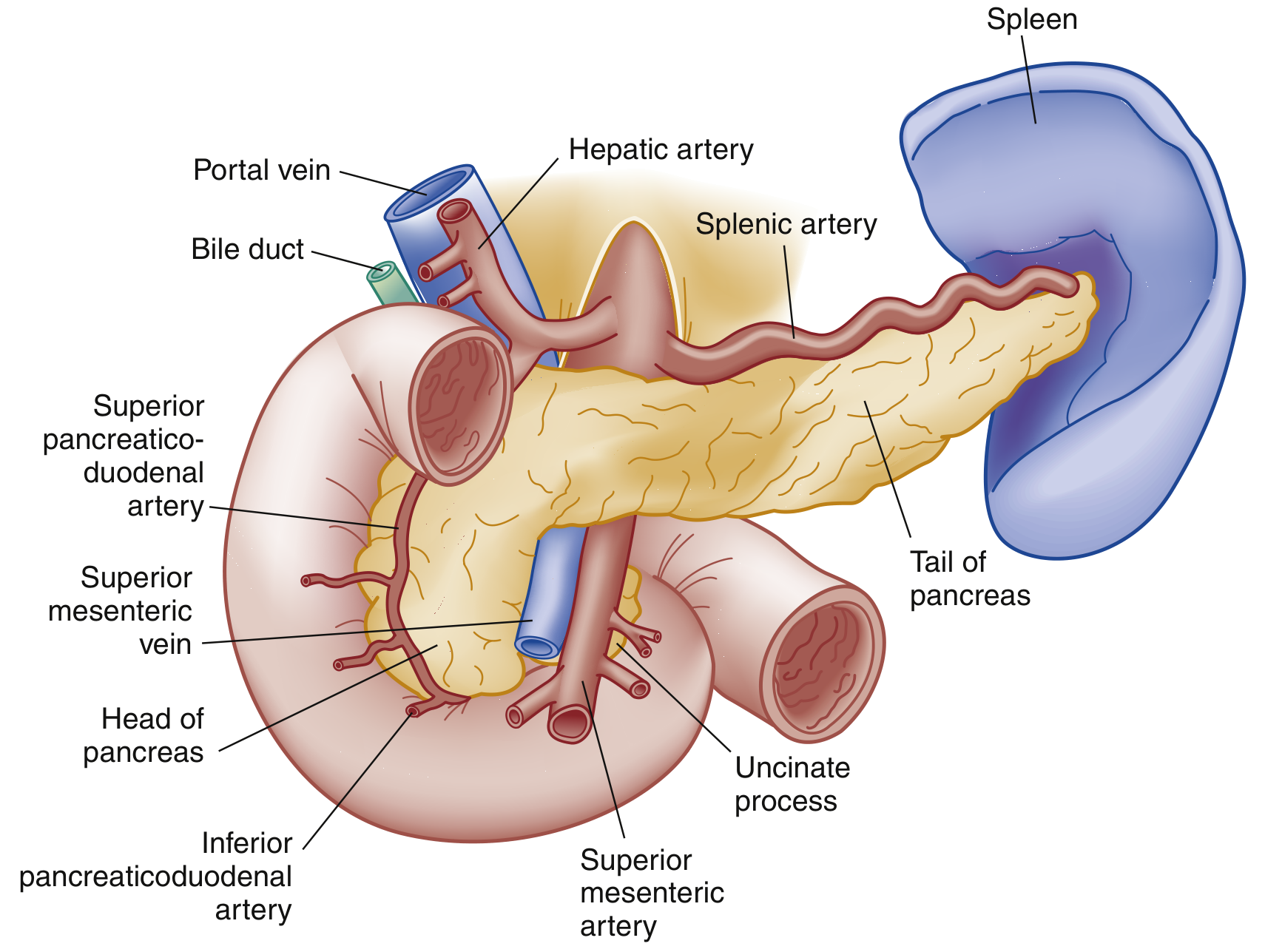
Definition & Epidemiology
Acute pancreatitis (AP) is an inflammatory condition leading to enzymatic autodigestion and destruction of pancreatic tissue. Its presentation ranges from mild, self-limited disease to sepsis and multiorgan failure. It is the most common pancreatic disease worldwide and one of the leading reasons for gastrointestinal hospitalization in the United States. Mortality reaches up to 30% in severe cases, though overall mortality has declined despite rising admissions. - Rosen's Emergency Medicine, p. 1265
Etiology
The causes of AP are numerous and can be grouped into categories:
Most common overall causes:
- Gallstones - 40% to 70% of all cases
- Chronic alcohol use - 25% to 35%
Other significant causes:
| Category | Specific Causes |
|---|---|
| Toxic-Metabolic | Alcohol, drugs, hypertriglyceridemia (TG >1000 mg/dL), hypercalcemia, uremia, scorpion venom |
| Mechanical-Obstructive | Biliary stones, pancreas divisum, annular pancreas, tumors (ampullary, pancreatic, neuroendocrine), post-ERCP, trauma |
| Infectious | Viral (mumps, coxsackie, HIV, CMV, EBV, varicella), bacterial (TB, Salmonella, Legionella, Mycoplasma), parasitic (Ascaris) |
| Vascular | Vasculitis, embolism, hypoperfusion/ischemia, hypercoagulability |
| Other | Idiopathic, hereditary (PRSS1, SPINK1, CFTR mutations), autoimmune, diabetes/DKA |
Smoking and diabetes are independent risk factors. Many idiopathic cases may be caused by occult microlithiasis. - Rosen's Emergency Medicine, p. 1266
Pathophysiology
The core mechanism, first proposed by Chiari in 1896, is premature intrapancreatic activation of digestive enzymes leading to autodigestion.
Key cascade:
- Precipitating event (gallstone obstruction, alcohol, etc.) triggers sustained cytosolic calcium increase in acinar cells
- This causes colocalization of zymogens (trypsinogen) with lysosomal enzymes (cathepsin B) in cytoplasmic vacuoles
- Cathepsin B activates trypsinogen to trypsin within these vacuoles
- Trypsin permeabilizes the vacuoles, releasing cathepsin B into the cytosol
- Cathepsin B permeabilizes mitochondrial membranes, releasing cytochrome C and triggering apoptotic cell death
- Mice lacking trypsinogen-7 (the key isoform activated during AP) show significantly less injury - confirming trypsin's central role
Protective mechanisms that become overwhelmed:
- Synthesis of enzymes as inactive precursors (zymogens)
- Separation of production and activation sites
- Trypsin inhibitors (SPINK1) within the pancreas
Intrapancreatic events lead to local inflammation, peripancreatic fat necrosis, and in severe cases, vascular disruption and hemorrhagic necrosis.
Systemic events are driven by the spread of activated enzymes and inflammatory mediators (cytokines, IL-1, TNF-α) into the systemic circulation, triggering SIRS. - Schwartz's Principles of Surgery, p. 1469
Classification (Revised Atlanta Criteria 2012)
| Grade | Features |
|---|---|
| Mild | No organ failure; no local or systemic complications |
| Moderately Severe | Transient organ failure (<48 h); local or systemic complications without persistent organ failure |
| Severe | Persistent organ failure (>48 h); may have local complications |
Types by morphology:
- Interstitial edematous pancreatitis - the majority (~90-95%); usually resolves within the first week
- Necrotizing pancreatitis - ~5-10% of cases; involves pancreatic parenchyma +/- peripancreatic tissue; necrotic tissue may remain sterile, liquefy, or become infected
Local complications (by timing):
- Within 4 weeks: Acute peripancreatic fluid collection (APFC), Acute necrotic collection (ANC)
- After 4 weeks: Pancreatic pseudocyst (APFC with wall), Walled-off necrosis (WON - ANC with wall)
- Rosen's Emergency Medicine, Box 77.2; Schwartz's Surgery, Table 33-8
Clinical Features
Symptoms:
- Persistent epigastric or left upper quadrant pain, often radiating to the back, chest, or flanks
- Pain may be relieved by sitting up or leaning forward
- Nausea, vomiting, anorexia
- Severity of pain does NOT correlate with disease severity
Signs:
- Vital signs: may be normal early; tachycardia, fever, tachypnea; hypotension in severe/complicated cases
- Jaundice suggests obstructive cause (gallstone or tumor)
- Abdominal distension, epigastric tenderness, guarding; bowel sounds may be absent (ileus)
- Right upper quadrant tenderness and Murphy's sign in gallstone pancreatitis
- Cullen sign - periumbilical bluish discoloration (hemoperitoneum) - rare, poor prognosis
- Grey Turner sign - reddish-brown flank discoloration (retroperitoneal bleeding) - rare, poor prognosis
- Pleural effusion (left-sided predominance) in up to 50% of patients
Systemic complications:
- ARDS, atelectasis, pleural effusion (pulmonary)
- Cardiovascular collapse, shock (fluid shifts)
- Acute kidney injury (hypoperfusion + inflammatory mediators)
- DIC (cytokine-mediated coagulation cascade activation)
- Hyperglycemia (decreased insulin), hypocalcemia (low albumin and Mg)
- Rosen's Emergency Medicine, p. 1265-1266
Diagnosis
Atlanta diagnostic criteria - at least 2 of 3 must be present:
- Abdominal pain characteristic of AP
- Serum lipase or amylase > 3x upper limit of normal
- Characteristic findings on abdominal imaging
Laboratory Tests:
- Lipase is preferred - more sensitive AND specific than amylase; remains elevated ~1-2 weeks
- Amylase - rises within hours, normalizes in 3-5 days; falsely negative in alcohol and hypertriglyceridemia-induced AP; elevated in many non-pancreatic conditions
- ALT - positive predictive value 95% for biliary pancreatitis when elevated
- Triglycerides - if >1000 mg/dL without gallstones or alcohol history, points to hypertriglyceridemic etiology
- CBC, BMP (assess for SIRS, organ failure)
- Calcium (hypocalcemia = poor prognosis)
- Testing both enzymes simultaneously does NOT improve sensitivity or specificity
Imaging:
- Ultrasound - first-line; evaluates for gallstones, biliary dilation; limited by bowel gas obscuring pancreas
- CT scan with contrast - not routinely needed for diagnosis; used when diagnosis is uncertain or to assess complications; best performed 48-72 hours after onset (earlier imaging may underestimate necrosis)
- CT Severity Index (CTSI) / Modified CTSI - grades pancreatic inflammation (0-4) + necrosis (0-4); total score predicts morbidity
- MRI/MRCP - better for biliary pathology; useful to assess pancreatic duct integrity
- Rosen's Emergency Medicine, p. 1267
Severity Scoring Systems
| System | Key Variables | Notes |
|---|---|---|
| Ranson Criteria | At admission: age >55, WBC >16,000, glucose >200, AST >250, LDH >350. At 48h: Hct drop >10%, BUN rise >5, Ca <8, PaO2 <60, base deficit >4, fluid sequestration >6L | Score ≥3 = severe; widely used since 1974 |
| APACHE II | Age, temperature, MAP, HR, RR, PaO2, pH, Na, K, Cr, Hct, WBC, GCS, chronic health | Can be calculated at admission; updated daily |
| BISAP | BUN >25, Impaired mental status, SIRS criteria, Age >60, Pleural effusion | Simple 5-point score; evaluated at 24h |
| Modified CTSI | Pancreatic inflammation grade + necrosis grade on CT | Radiologic severity assessment |
Scoring systems should augment clinical judgment, not replace it. Accuracy of any single or combined predictor is approximately 70%. Key early risk markers: elevated BUN/creatinine and persistent SIRS after adequate fluid resuscitation. - Schwartz's Surgery, p. 1473
Management
Treatment is primarily supportive.
1. Fluid Resuscitation (most critical early intervention)
- Patients are volume-depleted from decreased intake, vomiting, and third-spacing due to inflammatory mediators
- IAP/APA recommendation: Goal-directed resuscitation at 5-10 mL/kg/h targeting:
- HR < 120/min
- MAP 65-85 mmHg
- Urine output > 0.5-1 mL/kg/h
- ACG recommendation: 250-500 mL/h
- Lactated Ringer's (LR) preferred over normal saline - large volumes of NS cause hyperchloremic metabolic acidosis, which worsens the inflammatory response and activates trypsinogen
- Colloids not routinely recommended (may help if Hct <24 or albumin <2 g/dL)
- Inadequate resuscitation in first 24h increases SIRS, organ failure, pancreatic necrosis, and ICU admissions
- Monitor with serial Hct, BUN, creatinine
2. Pain Control
- Adequate analgesia is essential; opioids are generally required for moderate-to-severe pain
- IV morphine or hydromorphone; the historical concern about morphine causing sphincter of Oddi spasm has not been clinically validated
3. Nutritional Support
- Early enteral nutrition (EN) is preferred - within 24-48 hours when tolerated
- Nasojejunal or nasogastric feeding are equivalent in most studies
- EN maintains gut mucosal barrier, reduces bacterial translocation, reduces infectious complications
- Total parenteral nutrition (TPN) only if enteral route is not tolerated or not feasible
- "NPO until symptoms improve" is outdated in mild-moderate disease; early feeding reduces hospital stay and complications
4. Antibiotics
- Not indicated prophylactically in sterile necrotizing pancreatitis (multiple RCTs have shown no benefit)
- Use only when infection is documented or strongly suspected (infected necrosis, cholangitis)
- Fine-needle aspiration (FNA) of pancreatic necrosis under CT guidance can confirm infection (gram stain + culture)
5. Management of Biliary Pancreatitis
- ERCP indicated within 24-72 hours if concurrent cholangitis or persistent biliary obstruction
- Early ERCP for uncomplicated gallstone pancreatitis does NOT improve outcomes
- Cholecystectomy should be performed during the same hospitalization (mild disease) or after recovery (severe disease) to prevent recurrence
6. Management of Infected Necrosis
- Step-up approach: antibiotics first, then minimally invasive drainage/debridement
- Percutaneous or endoscopic drainage before surgical necrosectomy (reduces mortality)
- Surgical necrosectomy reserved for failure of less invasive approaches
- Optimal timing: delay intervention >4 weeks to allow walled-off necrosis to mature
7. Hypertriglyceridemia-Induced AP
- Insulin infusion, plasmapheresis, or gemfibrozil to reduce TG levels rapidly
- Heparin activates lipoprotein lipase and may help acutely
- Rosen's Emergency Medicine, p. 1269; Schwartz's Surgery
Differential Diagnosis
Conditions that can mimic AP include:
Abdominal: Peptic ulcer disease/perforation, acute cholecystitis, choledocholithiasis, cholangitis, mesenteric ischemia, bowel obstruction, AAA, ectopic pregnancy
Cardiopulmonary: Myocardial infarction, pneumonia, pericarditis, pleural effusion
Systemic: Sickle cell crisis, diabetic ketoacidosis
- Rosen's Emergency Medicine, Box 77.3
Prognosis & Complications
Predictors of poor prognosis:
- Persistent SIRS (>48h)
- Organ failure (especially multi-organ)
- Infected pancreatic necrosis
- Obesity (BMI >30)
- Age >55
- Ranson score ≥3
Key complications:
- Pseudocyst (>4 weeks - most common late local complication)
- Walled-off necrosis
- Pancreatic fistula
- Splenic or portal vein thrombosis
- Bowel necrosis
- Gastrointestinal bleeding
- Chronic pancreatitis (after recurrent episodes)
- Pancreatic exocrine/endocrine insufficiency
Overall mortality has decreased over recent decades despite rising incidence. Most patients (~80%) have mild disease that resolves within 1 week. The 5-10% with necrotizing pancreatitis carry significantly higher morbidity and mortality.
Sources: Rosen's Emergency Medicine 9e | Schwartz's Principles of Surgery 11e | Sleisenger & Fordtran's GI and Liver Disease | Textbook of Family Medicine 9e
This is a shared conversation. Sign in to Orris to start your own chat.