
Antibody its classification , structure and function of each antibody ..... 15 marks
Reading File
Now let me search the medical library for comprehensive antibody content in parallel.
Finding Sources
Finding Sources
Finding Sources
Reading File
Excellent content found. Let me now read more from this section and get the Henry's Clinical Diagnosis content for the individual Ig classes.
Reading File
Reading File
Excellent content. Let me get the full Roitt's section on structure and the early part of the chapter for antibody classification, plus fetch key images.
Reading File
Reading File
Now let me fetch the key antibody structure images from Roitt's, and also get the Roitt's basic structure section.
Loading Image
Loading Image
Reading File
I have very rich content. Let me also fetch the IgG basic structure diagram and get the IgE/IgD section from Henry's.
Loading Image
Reading File
I now have comprehensive content from multiple authoritative textbooks. Let me compose the complete 15-mark answer.
Antibody (Immunoglobulin): Classification, Structure, and Function
Definition
An antibody (immunoglobulin, Ig) is a glycoprotein produced by plasma cells (differentiated B lymphocytes) in response to antigenic stimulation. Antibodies belong to the gamma-globulin fraction of serum proteins and are the mediators of humoral immunity.
Basic Structure of an Antibody
All antibodies share a common four-chain monomer unit as the basic structural element.
Four-Chain Unit
The basic immunoglobulin monomer consists of:
- Two identical heavy (H) chains (~55,000 Da for IgG/IgA/IgD; ~70,000 Da for IgM/IgE)
- Two identical light (L) chains (~24,000 Da each)
- Chains held together by inter-chain disulfide bonds and non-covalent forces
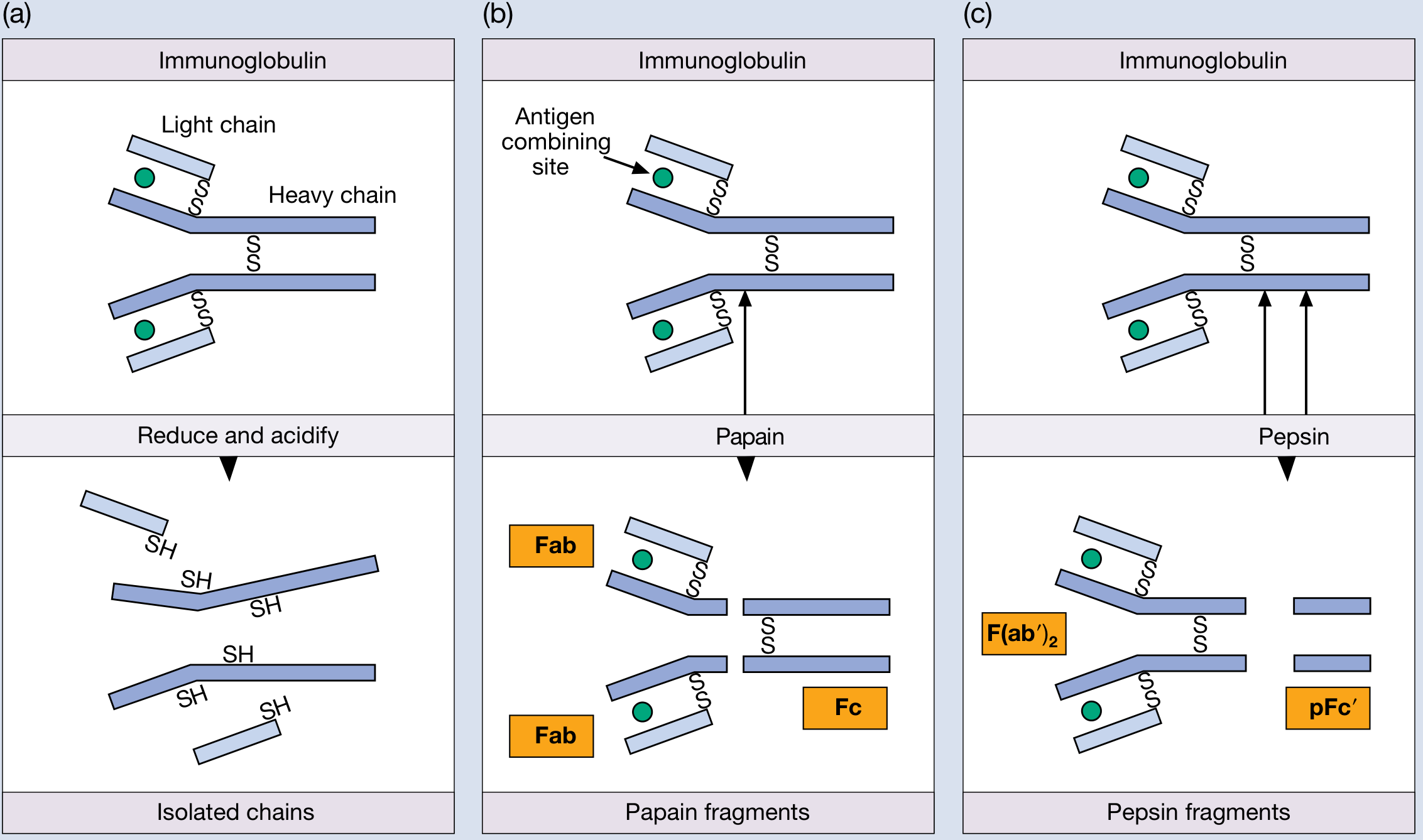
Figure: (a) Chain assembly showing heavy and light chains. (b) Papain digestion yields two Fab fragments + one Fc fragment. (c) Pepsin digestion yields F(ab')2 + pFc' fragment.
Regions of the Antibody
| Region | Full Name | Contents | Function |
|---|---|---|---|
| Fab | Fragment Antigen Binding | VH + VL + CH1 + CL | Antigen binding |
| Fc | Fragment Crystallizable | CH2 + CH3 (or CH4) | Effector functions (complement, Fc receptor binding) |
| Hinge | - | Between Fab and Fc | Flexibility, protease sensitivity |
| CDRs | Complementarity Determining Regions | 3 loops in VH + 3 in VL | Direct antigen contact |
Variable vs. Constant Regions
- The N-terminal ~110 amino acids of both H and L chains show high sequence variability - these are the Variable (V) domains, containing three hypervariable regions (CDRs) that form the antigen-binding site.
- The C-terminal portions are the Constant (C) domains - these are conserved within each class and determine isotype and effector function.
Light Chains
Two types exist: kappa (κ) and lambda (λ). In humans, κ chains are slightly more common. The same two light chain types are shared across all five antibody classes.
Heavy Chains
The heavy chain type defines the antibody class (isotype):
| Heavy Chain | Antibody Class |
|---|---|
| γ (gamma) | IgG |
| μ (mu) | IgM |
| α (alpha) | IgA |
| δ (delta) | IgD |
| ε (epsilon) | IgE |
Classification of Antibodies
1. By Class (Isotype) - 5 Classes
Based on the constant region of the heavy chain:
IgG, IgM, IgA, IgD, IgE
2. By Subclass
- IgG: 4 subclasses - IgG1, IgG2, IgG3, IgG4 (differ mainly in hinge region)
- IgA: 2 subclasses - IgA1 (long hinge, susceptible to bacterial proteases), IgA2 (short hinge, protease-resistant)
- IgM: 2 subclasses described but less clinically significant
3. By Light Chain Type (Type)
- Kappa (κ) type or Lambda (λ) type - applies to all classes
4. By Form
- Monomeric: IgG, IgE, IgD, serum IgA
- Dimeric: Secretory IgA (with J chain + secretory component)
- Pentameric: IgM (with J chain)
Structure and Function of Each Antibody Class
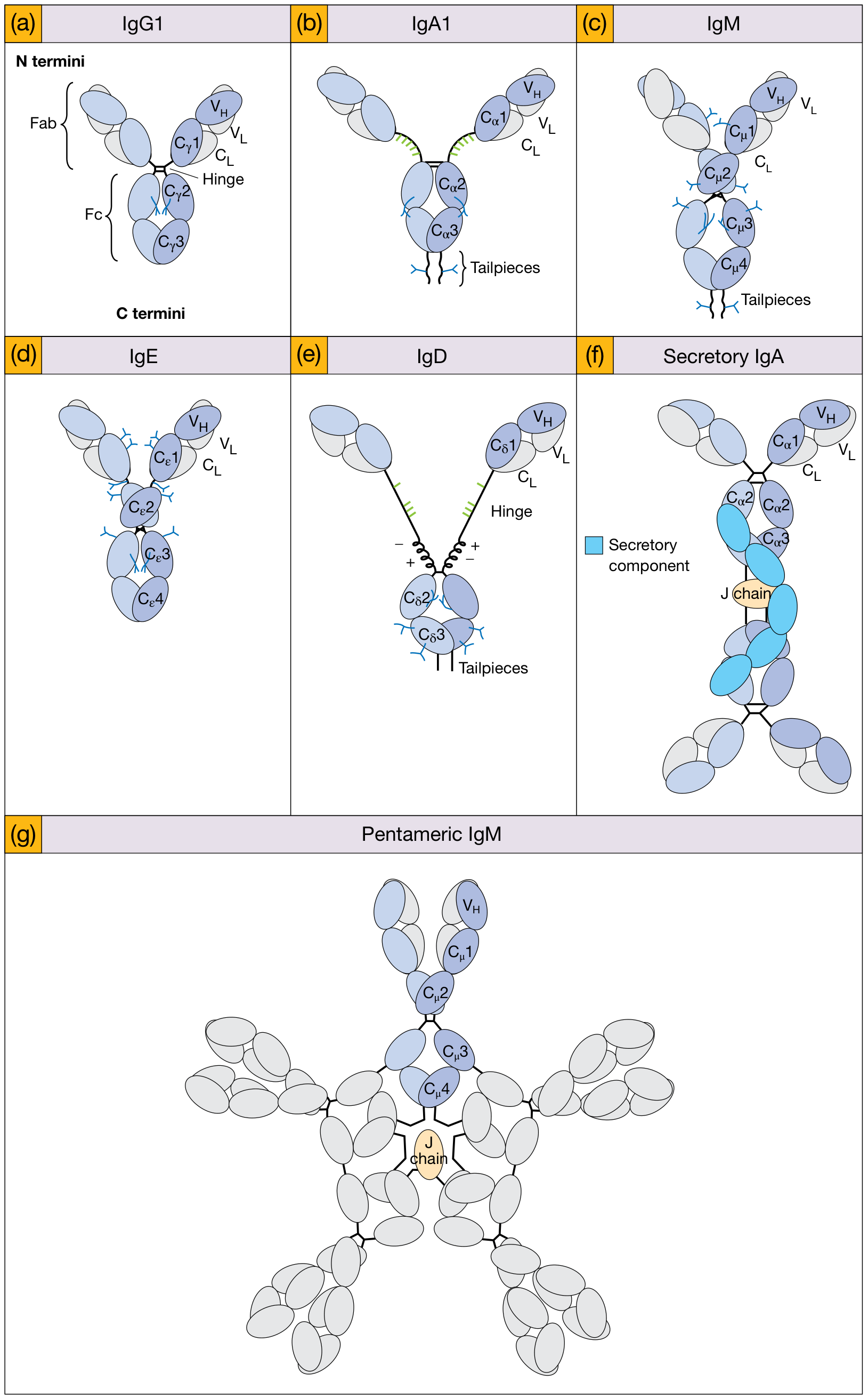
Figure 3.12 from Roitt's Essential Immunology: Schematic structures of IgG1, IgA1, IgM (monomer), IgE, IgD, Secretory IgA, and Pentameric IgM. Heavy chains shown in dark/pale blue; light chains in gray; N-linked carbohydrate (branched, blue); O-linked carbohydrate (linear, green); J chain (orange); Secretory component (blue block).
IgG
Structure:
- Monomeric four-chain unit (2 γ heavy chains + 2 light chains)
- Molecular weight: ~150 kDa
- Hinge region connects Fab arms to Fc (flexible, protease-sensitive)
- 3 constant domains in heavy chain (Cγ1, Cγ2, Cγ3)
- Cγ2 domains do NOT closely interact but carry N-linked carbohydrate between them
- Cγ2 domain carries binding sites for complement C1q and Fc receptors
- 4 subclasses (IgG1-4): IgG1 and IgG3 most effective at complement activation and ADCC; IgG4 can undergo Fab arm exchange to produce bispecific molecules
- Serum concentration: 8-16 mg/mL (constitutes ~80% of total serum immunoglobulin)
- Half-life: 18-23 days (longest of all isotypes, maintained by neonatal Fc receptor, FcRn)
Functions:
- Major antibody of secondary (anamnestic) immune response - produced copiously on re-exposure to antigen
- Opsonization - Fc region binds to FcγR on macrophages and neutrophils, enhancing phagocytosis of IgG-coated microorganisms
- Complement activation (classical pathway) via C1q binding to Cγ2 - IgG1 and IgG3 > IgG2 >> IgG4
- Antibody-Dependent Cell-mediated Cytotoxicity (ADCC) - guides NK cells to destroy IgG-coated target cells
- Placental transfer - the ONLY immunoglobulin that crosses the placenta (via FcRn), providing passive immunity to the neonate
- Neutralization of toxins, viruses, and bacteria
- Widely distributed in blood and extravascular tissue fluids (48% intravascular)
IgM
Structure:
- Secreted form is a pentamer of five four-chain units joined by a J (joining) chain (15 kDa acidic glycoprotein)
- Molecular weight: ~900 kDa; sedimentation rate: 19S
- Heavy chain: μ, with 5 constant domains (Cμ1-Cμ4 + extra domain replaces hinge region)
- 10 antigen-binding sites (though due to steric constraints, functional valency for large antigens may be lower)
- J chain links two adjacent Fc regions at carboxy-terminal tailpieces via disulfide bonds
- Monomeric IgM is expressed as B-cell receptor (BCR) on naive B lymphocytes (membrane-anchored form with transmembrane sequence)
- Serum concentration: 1.2-4.0 mg/mL (~13% of total Ig); half-life: 5-6 days
Functions:
- First antibody produced in primary immune response - first isotype expressed by developing B cells and by the body upon initial infection
- Most efficient complement activator (classical pathway) - a single IgM molecule bound to antigen can activate C1q (requires two adjacent Fc regions; IgG requires two nearby molecules)
- Powerful agglutinating antibody (+4 potency) due to 10 binding sites - effective at cross-linking particulate antigens, bacteria, and red blood cells
- B-cell antigen receptor - monomeric surface IgM is the primary antigen recognition molecule on naïve B cells
- Confined largely to blood (41% intravascular) due to large size; does NOT cross the placenta
- Phylogenetically the most primitive immunoglobulin; μ chain gene is believed to be the evolutionary precursor of other heavy chain genes
IgA
Structure:
- Exists in three forms:
- Serum IgA: mainly monomeric (small amounts of dimer); molecular weight ~160 kDa
- Secretory IgA (sIgA): dimer - two monomers linked by J chain + secretory component (SC)
- SC is derived from the poly-Ig receptor on epithelial cells; it protects IgA from proteolytic degradation in secretions
- Heavy chain: α; 3 constant domains + hinge region
- IgA1: long O-linked glycosylated hinge; susceptible to bacterial IgA1 proteases
- IgA2: very short hinge; more resistant to bacterial proteases; predominates in lower GI tract
- Serum concentration: 0.4-2.2 mg/mL (~6% of total Ig); half-life: 5-6.5 days
- Fastest catabolized of the major immunoglobulins (24%/day)
Functions:
- Primary defense of mucosal surfaces - sIgA is the dominant antibody in saliva, tears, breast milk, colostrum, intestinal secretions, respiratory and urogenital secretions
- Immune exclusion - sIgA prevents attachment of pathogens to epithelial surfaces (first line of mucosal defense)
- Provides passive immunity to neonates via breast milk
- Serum IgA can link pathogens to effector cells via FcαR (Fc receptor for IgA)
- Does NOT activate complement via classical pathway; may activate the alternative pathway
- Present in external secretions at high levels (+4 potency)
- IgA deficiency is the most common primary immunodeficiency (1 in 600 persons)
IgE
Structure:
- Monomeric four-chain unit
- Molecular weight: ~190 kDa (heavier than IgG due to extra domain)
- Heavy chain: ε; 4 constant domains (Cε1-Cε4) - NO hinge region (replaced by extra domain pair, similar to IgM)
- Lowest serum concentration of all Igs: 17-450 ng/mL (~0.002% of total Ig)
- Half-life in serum: very short (~2-3 days), but when bound to mast cell FcεRI receptors, half-life extends to weeks
- Most IgE in the body exists bound to high-affinity FcεRI receptors on mast cells and basophils rather than free in serum
Functions:
- Mediator of immediate (Type I) hypersensitivity reactions (allergy and anaphylaxis): Antigen (allergen) cross-links IgE bound to mast cell FcεRI → mast cell degranulation → release of histamine, leukotrienes, prostaglandins → acute inflammatory reaction (urticaria, bronchoconstriction, anaphylaxis)
- Anti-parasitic defense - particularly against helminth (worm) infections; IgE-mediated eosinophil activation and mast cell responses are protective against large extracellular parasites
- Fixation to homologous mast cells and basophils (+4 potency)
- Elevated in atopic conditions: asthma, allergic rhinitis, atopic dermatitis, and parasitic infections
- Does NOT activate complement and does not cross the placenta
IgD
Structure:
- Monomeric four-chain unit
- Molecular weight: ~185 kDa
- Heavy chain: δ; 3 constant domains (Cδ1-Cδ3) with a hinge region bearing O-linked carbohydrates and a unique polyanionic region (alternating positive/negative charges)
- Has tailpieces at the C-termini of heavy chains
- Extremely low serum concentration: ~0.03 mg/mL (~1% of total Ig)
- Shortest half-life: ~2.8 days (fastest catabolized along with IgE; ~37%/day)
- Very sensitive to proteolytic degradation
Functions:
- B-cell antigen receptor (BCR) - the primary function of IgD is as a surface receptor on mature naive B lymphocytes (co-expressed with surface IgM); it participates in B-cell activation and signaling
- As a co-receptor with IgM on B cells, IgD may lower the threshold for B-cell activation or participate in B-cell tolerance
- Serum function is poorly understood - it does not fix complement, does not cross the placenta, and does not bind to mast cells
- Recent research suggests IgD may play a role in upper respiratory immunity and in amplifying immune surveillance at mucosal sites
- Principally bound to B cells (75% intravascular)
Summary Comparison Table
| Property | IgG | IgM | IgA | IgE | IgD |
|---|---|---|---|---|---|
| Heavy chain | γ | μ | α | ε | δ |
| Serum conc. (mg/mL) | 8-16 | 1.2-4.0 | 0.4-2.2 | 0.00002-0.00045 | 0.03 |
| % of total Ig | 80% | 13% | 6% | 0.002% | 1% |
| Molecular form | Monomer | Pentamer | Monomer/Dimer | Monomer | Monomer |
| MW (kDa) | 150 | 900 | 160/385 | 190 | 185 |
| Half-life (days) | 18-23 | 5-6 | 5-6.5 | ~2-3 | 2.8 |
| Crosses placenta | Yes | No | No | No | No |
| Complement (classical) | Yes (IgG1,3) | Yes (potent) | No | No | No |
| Agglutination | ± | ++++ | ++ | - | - |
| Present in secretions | No | ± | ++++ | - | - |
| Binds mast cells/basophils | ± | - | ± | ++++ | - |
| ADCC | Yes | - | ± | - | - |
Key Points for 15-Mark Answer
- All antibodies share the basic four-chain Y-shaped monomer of 2H + 2L chains linked by disulfide bonds
- Fab binds antigen; Fc mediates effector functions
- CDRs (hypervariable regions) within V domains are the direct antigen contact points
- Five classes (isotypes) defined by heavy chain constant regions: γ, μ, α, ε, δ
- IgG = major serum Ig, secondary response, opsonization, ADCC, placental transfer
- IgM = primary response, best complement activator, confined to blood, B-cell receptor
- IgA = mucosal immunity, secretory form (sIgA) with J chain + secretory component
- IgE = allergy/anaphylaxis (type I hypersensitivity), anti-parasitic defense
- IgD = mainly B-cell surface receptor; function in serum unclear
Sources:
- Roitt's Essential Immunology, Chapter 3 - The structure and function of immunoglobulin classes, pp. 92-98
- Henry's Clinical Diagnosis and Management by Laboratory Methods, Chapter 47 - General Properties of Immunoglobulins, pp. 1091-1094 (Tables 47.3 & 47.4)
This is a shared conversation. Sign in to Orris to start your own chat.