I have comprehensive data from multiple authoritative sources. Let me now compile the full, detailed answer.
Classification of Oral Ulcerative Lesions
Oral ulcers are among the most common mucosal lesions encountered in clinical practice. They represent a final common endpoint of a wide spectrum of etiologies - from simple trauma to life-threatening malignancy. Classification can be approached from several frameworks:
I. Classification by Etiology (Most Widely Used)
1. Traumatic / Reactive
The commonest cause of oral ulceration. Usually has an identifiable precipitating factor and resolves once the cause is removed.
| Subtype | Examples |
|---|
| Mechanical | Sharp tooth cusps, ill-fitting dentures, toothbrush trauma, cheek bite |
| Chemical | Aspirin burns, acid/alkali injury, phenol application |
| Thermal | Hot food/beverage burns |
| Factitious (self-inflicted) | Habitual picking, psychogenic |
| Radiation-induced | Post-radiotherapy mucositis |
| Eosinophilic ulcer (TUGSE) | Traumatic Ulcerative Granuloma with Stromal Eosinophilia - slow-healing, deep pseudoinvasive lesion, often on the tongue; often resolves after incisional biopsy |
Traumatic ulcers are typically single, painful, located at sites of trauma, with a yellow-white sloughing base and erythematous halo. They heal within 1-2 weeks.
2. Infective
a. Viral
- Herpes simplex virus (HSV-1): Primary herpetic gingivostomatitis - multiple small ulcers on attached gingiva and hard palate; recurrent herpes labialis (lip vermilion)
- Varicella zoster: Intraoral vesicles rupturing to form painful ulcers
- Herpes zoster: Unilateral distribution following trigeminal dermatome
- Herpangina / Hand-foot-mouth disease (Coxsackievirus): Posterior oropharyngeal ulcers in children
- Infectious mononucleosis (EBV): Tonsillar/pharyngeal ulcers with petechiae
- HIV: Predisposes to recurrent aphthae, CMV ulcers, and HSV
b. Bacterial
- Syphilis (Treponema pallidum): Primary chancre - single indurated, painless ulcer; secondary - "snail track" ulcers; tertiary - gumma
- Tuberculosis: Chronic, indurated, painful ulcer; usually secondary to pulmonary TB
- Acute necrotizing ulcerative gingivitis (ANUG / Vincent's angina): Pseudomembranous, punched-out ulcers of interdental papillae; fetid odor; Fusobacterium + spirochetes
- Actinomycosis: Soft tissue swelling or ulcerative lesion, often cervicofacial
- Noma (Cancrum oris): Rapidly spreading gangrenous ulceration in malnourished children
c. Fungal
- Histoplasmosis, Blastomycosis, Cryptococcosis: Chronic granulomatous ulcers in immunocompromised patients
- Candidiasis (chronic): Erythematous/ulcerative forms in immunosuppression
3. Idiopathic (Recurrent Aphthous Stomatitis - RAS)
RAS is the most common non-traumatic oral ulceration, affecting 20-40% of the population. It arises from T-cell mediated immunological damage to the mucosal basal layer. It is classified into three types per clinical features:
| Type | Size | Number | Duration | Site | Scarring | Key Features |
|---|
| Minor (MiRAS) | <10 mm | <10 at a time | 2-3 weeks | Non-keratinized mucosa only (labial, buccal, floor of mouth) | None | Most common; front of mouth; often trauma-related sites |
| Major (MaRAS / Sutton's disease) | >10 mm | 1-3 at a time | Up to 3 months | Keratinized AND non-keratinized; posterior sites (soft palate, tonsillar pillars) | May scar | Deep, severe; can mimic carcinoma |
| Herpetiform (HU) | <5 mm (1-3 mm) | Up to 100 | Up to 2 weeks | Keratinized and non-keratinized; widespread | None | Least common; coalesce into large irregular ulcers; mimic herpetic stomatitis (but no virus) |
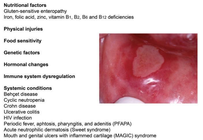
Common triggers: Haematinic deficiencies (ferritin, folate, B12 - can occur with a normal blood count), stress, trauma, sodium lauryl sulphate (SLS) in toothpaste, foods (chocolate, tomatoes, nuts), menstrual cycle, systemic disease (Crohn's, coeliac, Behcet's, HIV, PFAPA syndrome).
Scott-Brown's Otorhinolaryngology Head & Neck Surgery, p. 710
4. Associated with Systemic Disease
A key diagnostic category - oral ulcers may be the presenting or sole feature of an underlying systemic condition.
| System | Disease | Notes |
|---|
| Haematological | Leukaemia, neutropenia, anaemia, other WBC dyscrasias | Impaired immunity leads to ulceration |
| Haematinic deficiency | Iron deficiency (low ferritin), folate deficiency, B12 deficiency | Even without anaemia |
| Gastrointestinal | Coeliac disease, Crohn's disease, ulcerative colitis | Aphthous-type ulcers; may precede GI diagnosis |
| Connective tissue / vasculitis | Behcet's disease (oral + genital ulcers + uveitis), SLE | Characteristic recurrent painful ulcers |
| Skin diseases (see below) | Lichen planus, pemphigus, pemphigoid, erythema multiforme | Mucocutaneous overlap |
| Endocrine | Rapid growth periods, menstrual cycle | Hormonal triggers |
| Iatrogenic | NSAIDs, beta-blockers, antirheumatic drugs, chemotherapy, radiotherapy | Drug-induced ulcers; resolve on withdrawal |
5. Immune-Mediated / Mucocutaneous Diseases
These involve vesicle/blister formation that ruptures to leave erosions or ulcers:
| Condition | Mechanism | Key Oral Features |
|---|
| Erosive Lichen Planus | T-cell mediated basement membrane attack | Reticular (Wickham's striae), atrophic, and erosive forms on buccal mucosa/tongue; recurrent; low malignant potential |
| Pemphigus vulgaris | IgG autoantibody against desmoglein (intercellular) | Oral lesions often first; fragile, irregular ulcers; Nikolsky's sign positive |
| Mucous membrane pemphigoid (MMP / cicatricial pemphigoid) | IgG against basement membrane (subepithelial) | Desquamative gingivitis; scarring; requires ophthalmology referral |
| Erythema multiforme | Immune complex hypersensitivity (often post-HSV or drug) | "Target" lip lesions; hemorrhagic crusting; may have widespread oral ulcers |
| Linear IgA disease / Dermatitis herpetiformis | Subepidermal blistering | Oral ulcers may accompany skin vesicles |
| Epidermolysis bullosa | Genetic collagen defect | Fragile mucosa with recurrent ulceration; progressive scarring |
| Paraneoplastic pemphigus | Associated with lymphoproliferative disease or thymoma | Resistant to treatment; biopsy essential |
Scott-Brown's Otorhinolaryngology Head & Neck Surgery, p. 711-712
6. Neoplastic
Any persistent (>3 weeks), non-healing oral ulcer must be considered malignant until proven otherwise.
| Type | Features |
|---|
| Squamous cell carcinoma (SCC) | Most common oral malignancy; ulcero-proliferative or indurated edge; painless initially; floor of mouth, lateral tongue, retromolar area; risk factors: tobacco, alcohol, HPV |
| Lymphoma | Submucosal mass or ulceration; may resemble aphthae |
| Salivary gland malignancies | Ulceration of minor gland sites (palate) |
| Metastatic tumours | Rare; jaw or mucosal deposits |
Early oral SCC classically presents as a nodule or ulcerative lesion of the lip or mucosa, lasting >2 weeks - Cummings Otolaryngology, p. 2199
II. Classification by Duration (Clinical Utility)
| Category | Duration | Examples |
|---|
| Acute | Days to weeks | Traumatic ulcers, herpetic stomatitis, RAS (minor/herpetiform), ANUG |
| Chronic | Weeks to months | Oral lichen planus, pemphigus, pemphigoid, oral SCC, major RAS, TB, drug-induced |
| Recurrent | Episodic pattern | RAS (all types), herpes labialis, Behcet's disease |
III. Classification by Presence of Preceding Vesicles/Bullae
This is a useful clinical framework since it narrows the differential:
| Category | Examples |
|---|
| Non-vesicular ulcers (no preceding blister) | Traumatic ulcer, RAS, ANUG, TB, SCC, neutropenic ulcer |
| Vesiculobullous ulcers (blister ruptures to form ulcer) | HSV, HZ, hand-foot-mouth, pemphigus, pemphigoid, erythema multiforme, lichen planus (erosive) |
IV. University of Baghdad Etiological Classification (Oral Pathology Teaching Format)
A commonly used teaching classification:
- Infective - Bacterial, Viral, Fungal
- Traumatic - Mechanical, Chemical, Thermal, Factitious, Radiation, Eosinophilic ulcer
- Idiopathic (RAS) - Minor, Major, Herpetiform
- Associated with systemic disease - Haematological, GI tract diseases, Behcet's disease, HIV
Key Diagnostic Clues
| Feature | Suggests |
|---|
| Single, self-limited, site of trauma | Traumatic ulcer |
| Multiple small ulcers, clustered, vesicular prodrome | Viral (HSV) |
| Recurrent, non-keratinized mucosa, <10mm | Minor RAS |
| Large, posterior, >3 weeks | Major RAS vs. malignancy |
| Indurated, painless, >3 weeks, tobacco/alcohol user | SCC - urgent biopsy |
| Bilateral white striae + ulceration | Erosive lichen planus |
| Nikolsky's sign positive | Pemphigus |
| Oral + genital ulcers + uveitis | Behcet's disease |
| Punched-out interdental papillae, fetid breath | ANUG |
| Unilateral, follows trigeminal dermatome | Herpes zoster |
Visual Reference
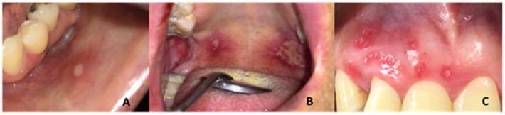
Here is a composite showing the three RAS subtypes for comparison:
Minor RAS (A), Major RAS (B), and Herpetiform RAS (C) - note size, number, and site differences
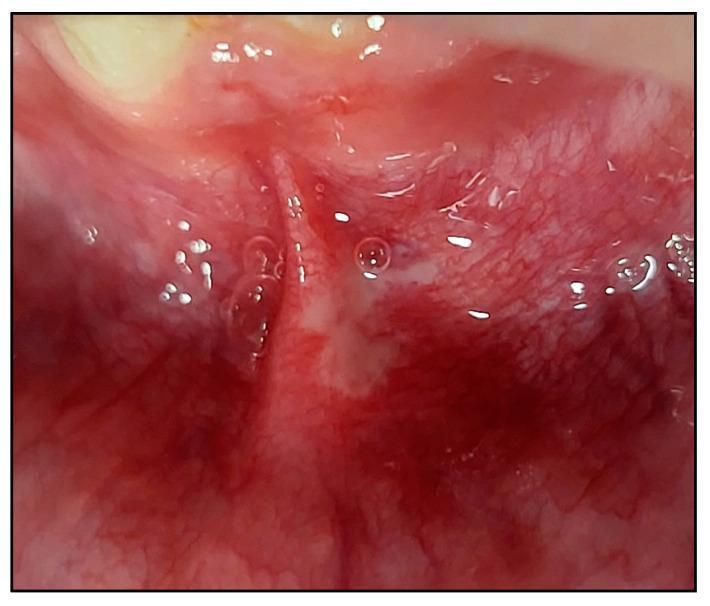
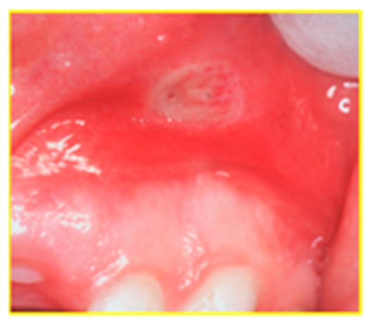
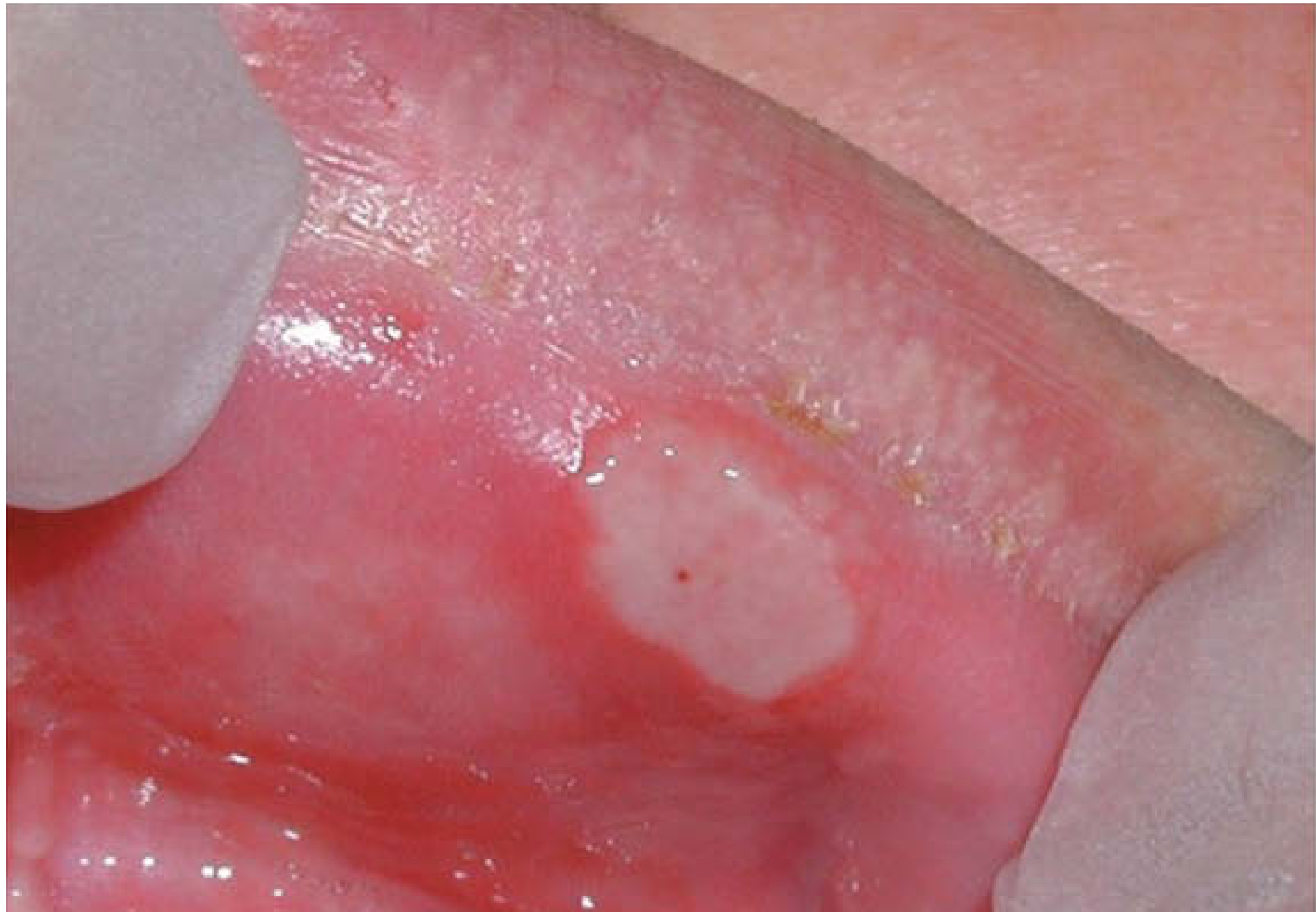
Typical aphthous ulcer with central white base and erythematous halo - Scott-Brown's Otorhinolaryngology
Sources:
- Scott-Brown's Otorhinolaryngology Head & Neck Surgery (Table 42.8 - Oral ulceration with systemic disease; Table 42.9 - Aphthous types)
- Cummings Otolaryngology Head and Neck Surgery (RAS, oral SCC)
- SADJ 2016 - Oral mucosal ulceration: a clinician's guide
- J Oral Med Oral Surg Oral Pathol Oral Radiol - Brief review of oral ulcerative lesions