
Muscles of forearm
Reading File
Now let me search the medical library for forearm muscles content.
Finding Sources
Finding Sources
Excellent! I have multiple anatomy textbooks. Let me read the key sections from the THIEME Atlas and also Gray's for comprehensive coverage.
Reading File
Finding Sources
Now let me read the detailed muscle tables from the THIEME Atlas (the 3192+ area has detailed info on radialis muscles, and 3824+ has more tables):
Reading File
Reading File
I can see there are detailed muscle descriptions. Let me now read the detailed sections on each muscle group (posterior extensors and anterior flexors):
Reading File
Finding Sources
Reading File
Reading File
I now have all the key data. Let me also fetch a couple of the diagrams to include in the answer:
Loading Image
Loading Image
Reading File
I also need the posterior deep extensors section. Let me fetch the data from lines 3300-3400:
Reading File
Loading Image
Excellent! I now have comprehensive information from the THIEME Atlas textbook. Let me also fetch the anterior view diagram:
Loading Image
I now have comprehensive data from the THIEME Atlas. Let me compile the full answer.
Muscles of the Forearm
The forearm (antebrachium) muscles are organized into three groups: anterior (flexor) compartment, posterior (extensor) compartment, and the radial (lateral) group. All are innervated by branches of the brachial plexus (C5-T1).
Anterior (Flexor) Compartment
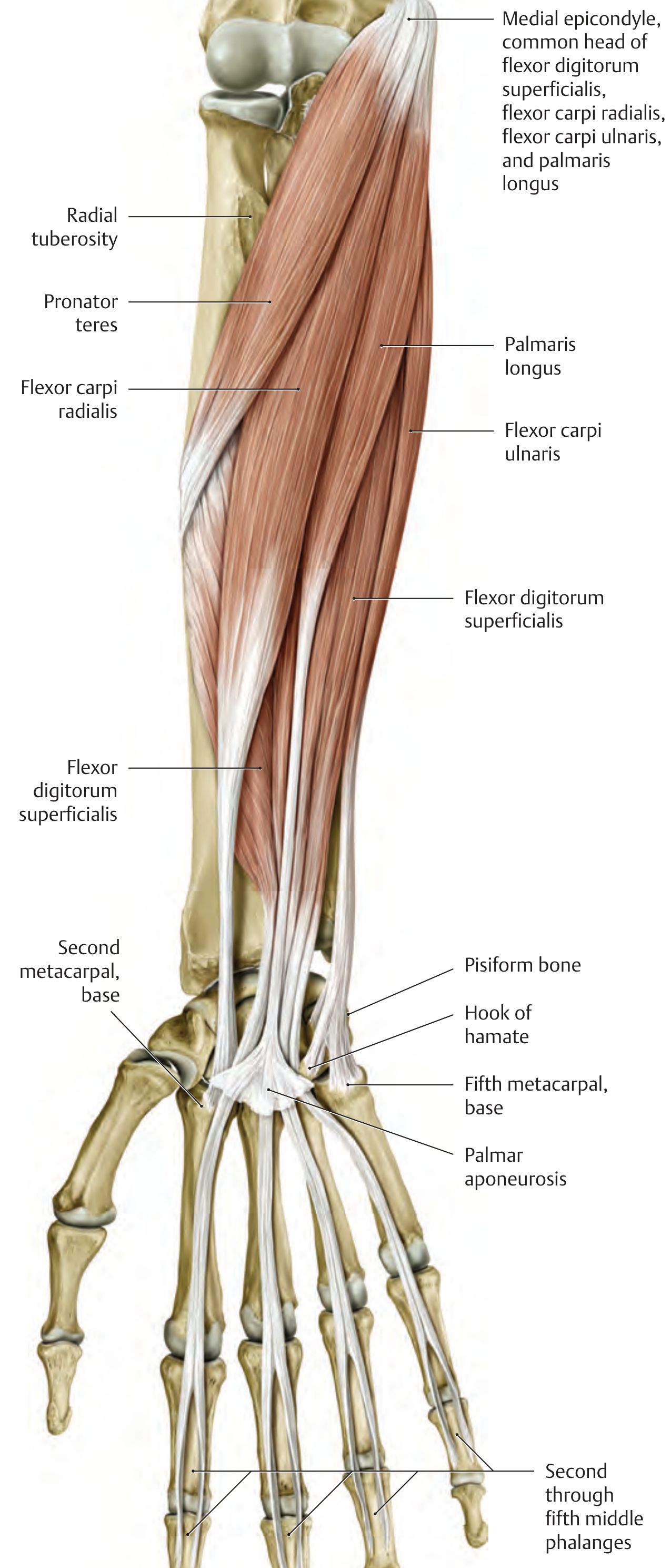
Anterior view - superficial flexors:
Superficial Layer (5 muscles)
All 5 share a common origin from the medial epicondyle of the humerus ("common flexor head").
| Muscle | Origin | Insertion | Action | Nerve |
|---|---|---|---|---|
| Pronator teres | Medial epicondyle (humeral head) + coronoid process of ulna (ulnar head) | Lateral surface of radius (mid-shaft) | Pronation; weak elbow flexion | Median (C6) |
| Flexor carpi radialis | Medial epicondyle | Base of 2nd (±3rd) metacarpal | Wrist flexion, radial deviation; weak pronation | Median (C6-C8) |
| Palmaris longus | Medial epicondyle | Palmar aponeurosis | Wrist flexion; tightens palmar aponeurosis | Median (C8-T1) |
| Flexor carpi ulnaris | Medial epicondyle (humeral head) + olecranon (ulnar head) | Pisiform, hook of hamate, base of 5th metacarpal | Wrist flexion, ulnar deviation | Ulnar (C7-T1) |
| Flexor digitorum superficialis | Medial epicondyle + coronoid process + proximal radius | Sides of middle phalanges (digits 2-5) | Flexion at PIP, MCP, wrist; weak elbow flexion | Median (C7-T1) |
Note: Flexor digitorum superficialis is the only muscle that forms an intermediate layer (between superficial and deep groups), as it covers the deep flexors.
Deep Layer (3 muscles)
| Muscle | Origin | Insertion | Action | Nerve |
|---|---|---|---|---|
| Flexor digitorum profundus | Proximal 2/3 of anterior ulna + interosseous membrane | Palmar surface of distal phalanges (digits 2-5) | Flexion at DIP, PIP, MCP, wrist | Median (radial half: digits 2-3, C8-T1); Ulnar (ulnar half: digits 4-5, C8-T1) |
| Flexor pollicis longus | Anterior radius (mid-shaft) + interosseous membrane | Palmar distal phalanx of thumb | Flexion at MCP + IP of thumb; wrist flexion, radial deviation | Median (C8-T1) |
| Pronator quadratus | Distal 1/4 of anterior ulna | Distal 1/4 of anterior radius | Pronation; stabilizes distal radioulnar joint | Median (C8-T1) |
Posterior (Extensor) Compartment
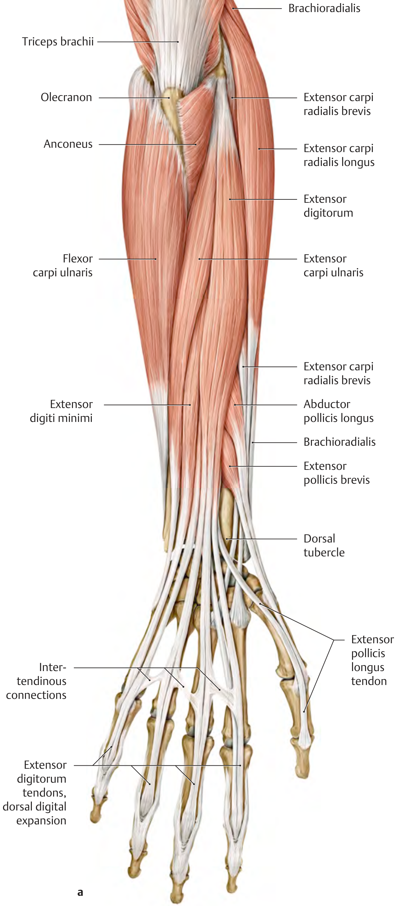
Posterior view - superficial + deep extensors:
Superficial Layer (3 muscles)
All arise from the lateral epicondyle via the common extensor head. All are innervated by the radial nerve (C7, C8).
| Muscle | Insertion | Action |
|---|---|---|
| Extensor digitorum | Dorsal digital expansion of digits 2-5 | Extension of MCP, PIP, DIP (digits 2-5); wrist extension |
| Extensor digiti minimi | Dorsal digital expansion of digit 5 | Extension and abduction of digit 5; wrist extension + ulnar deviation |
| Extensor carpi ulnaris | Base of 5th metacarpal | Wrist extension, ulnar deviation |
Deep Layer (5 muscles)
All are innervated by the radial nerve (C7, C8) (deep branch/posterior interosseous nerve).
| Muscle | Origin | Insertion | Action |
|---|---|---|---|
| Supinator | Lateral epicondyle + olecranon/supinator crest of ulna + radial collateral ligament + anular ligament | Radius (between radial tuberosity and pronator teres insertion) | Supination |
| Abductor pollicis longus | Dorsal radius, ulna + interosseous membrane | Base of 1st metacarpal | Thumb abduction; radial deviation of wrist |
| Extensor pollicis brevis | Posterior radius + interosseous membrane | Base of proximal phalanx of thumb | Extension of thumb (CMC, MCP); radial deviation |
| Extensor pollicis longus | Posterior ulna + interosseous membrane | Base of distal phalanx of thumb | Full thumb extension (CMC, MCP, IP); wrist radial deviation |
| Extensor indicis | Posterior ulna + interosseous membrane | Dorsal digital expansion of digit 2 | Extension of index finger (MCP, PIP, DIP); wrist extension |
The tendons of abductor pollicis longus and extensor pollicis brevis form the anatomical snuffbox (anterior boundary), while extensor pollicis longus forms its posterior boundary.
Radial (Lateral) Group
These muscles sit between the anterior and posterior compartments and are innervated by the radial nerve. They are sometimes grouped with the posterior compartment.
| Muscle | Origin | Insertion | Action | Nerve |
|---|---|---|---|---|
| Brachioradialis | Lateral surface of distal humerus (lateral supracondylar ridge) + lateral intermuscular septum | Styloid process of radius | Elbow flexion; semipronation (brings forearm to neutral) | Radial (C5-C6) |
| Extensor carpi radialis longus | Lateral supracondylar ridge + lateral intermuscular septum | Dorsal base of 2nd metacarpal | Wrist extension + radial deviation; assists in fist closure | Radial (C6-C7) |
| Extensor carpi radialis brevis | Lateral epicondyle | Dorsal base of 3rd metacarpal | Wrist extension + radial deviation; assists in fist closure | Radial (C7-C8) |
Quick Reference: Innervation Summary
| Nerve | Muscles Supplied |
|---|---|
| Median nerve | All anterior compartment muscles EXCEPT flexor carpi ulnaris and medial half of flexor digitorum profundus |
| Ulnar nerve | Flexor carpi ulnaris + medial half of flexor digitorum profundus (digits 4-5) |
| Radial nerve | All posterior compartment muscles + radial group |
Key Clinical Points
- Medial epicondyle is the common origin for all superficial flexors - inflammation at this point is golfer's elbow (medial epicondylitis)
- Lateral epicondyle is the common origin for superficial extensors - inflammation is tennis elbow (lateral epicondylitis)
- Pronator teres syndrome: median nerve compressed between the two heads of pronator teres, causing weakness of all median-innervated forearm/hand muscles
- Palmaris longus is absent in ~14% of people and is the most common tendon used for grafts
- Anterior interosseous nerve (branch of median) supplies the deep flexors - its injury causes inability to make an "OK" sign (no flexion at DIP of index + IP of thumb)
Source: General Anatomy and Musculoskeletal System, THIEME Atlas of Anatomy, pp. 333-340
This is a shared conversation. Sign in to Orris to start your own chat.