
Chicken Pox - epidemiology, prevention, control Also provide image for this
chickenpox varicella rash skin lesions
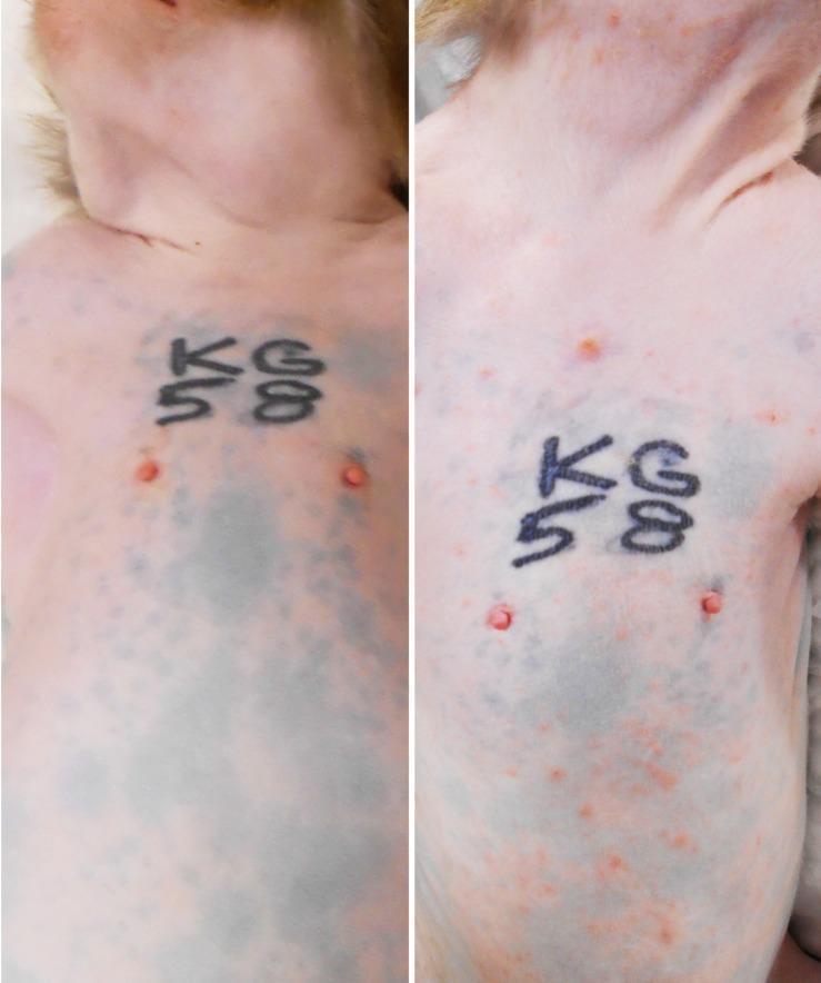
A comparative side-by-side clinical photograph illustrating the progression of primary varicella (chickenpox) rash in a rhesus macaque model. The left panel shows the baseline pre-inoculation state, displaying clear skin on the neck, torso, and abdomen with normal anatomical markings and identifying tattoos. The right panel, captured at 9 days post-inoculation (dpi), demonstrates a severe, generalized acute skin rash. The morphology of the lesions includes a dense distribution of small, erythematous (reddish/pinkish) macules and papules. The rash is widespread, involving the neck, shoulders, chest, and ventral abdomen. This comparison serves to highlight the clinical manifestation of Simian Varicella Virus (SVV), which mirrors the presentation of Varicella-Zoster Virus (VZV) in humans, focusing on the density, distribution, and characteristic appearance of primary varicella lesions.
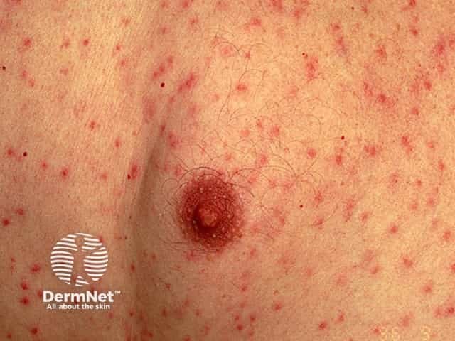
Clinical photography of skin lesions depicting a varicella (chickenpox)–like vesicular exanthem concentrated on the trunk with scattered papules and crusted lesions. The image shows numerous small erythematous papules with central vesiculation and crust formation on a backdrop of unremarkable skin. In classic varicella, lesions appear in crops, evolving from macules to vesicles (dew drops on a rose petal) and eventually crusting, often involving the trunk more than the face, and accompanied by pruritus, fever, and malaise. The depicted lesion is in a later crusted stage, while adjacent lesions are in various stages, reflecting the dynamic evolution of the eruption. Relevant imaging: not radiologic; this is dermatologic clinical photography. Diagnostic relevance: a hallmark varicella rash that aids rapid clinical diagnosis in outpatient or inpatient settings, allowing differential with disseminated herpes simplex, pityriasis rosea, or pityriasis versicolor; contagious until crusting. Clinically, management includes supportive care, antipyretics, antihistamines for itch, and antiviral therapy in high-risk patients; infection control measures to prevent transmission. Potential educational uses include dermatology training, telemedicine triage, clinical case discussions, and AI-based image retrieval for vesicular exanthems.
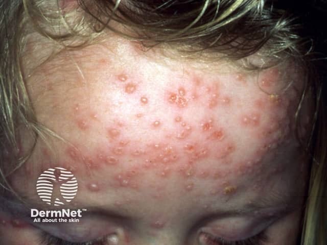
Clinical photography of active varicella (chickenpox) rash on the frontal scalp and forehead of a pediatric patient. Modality: digital color photograph taken under ambient lighting; standard clinical technique without dermoscopy or fluorescence. The image shows numerous small vesicles on an erythematous, slightly raised base distributed across the forehead and extending toward the temple. Lesions are in multiple evolutionary stages, with some vesicles capturing clear serous content, others showing crusted surfaces, and crusts around the margins. The pattern is characteristic of varicella: centripetal distribution with trunk and face involvement, crops of lesions appearing sequentially, and a tendency for vesicles to be more numerous on exposed skin areas. The epidermis and superficial dermis contain superficial fluid-filled vesicles, each about 2-4 mm in diameter, some conforming to the classic dew drop on a rose petal description. Facial involvement may accompany fever and malaise in pediatric patients. Although lesions may be pruritic, Nikolsky sign is typically absent; scarring is uncommon in children. Diagnosis is clinical, supported by history of varicella exposure or vaccination status; differential includes disseminated herpes zoster, pityriasis rosea, and vesicular dermatitis. Recognizing multistage lesions in a contagious exanthem is clinically significant for isolation, supportive therapy, and public health reporting as Varicella-zoster virus infection.
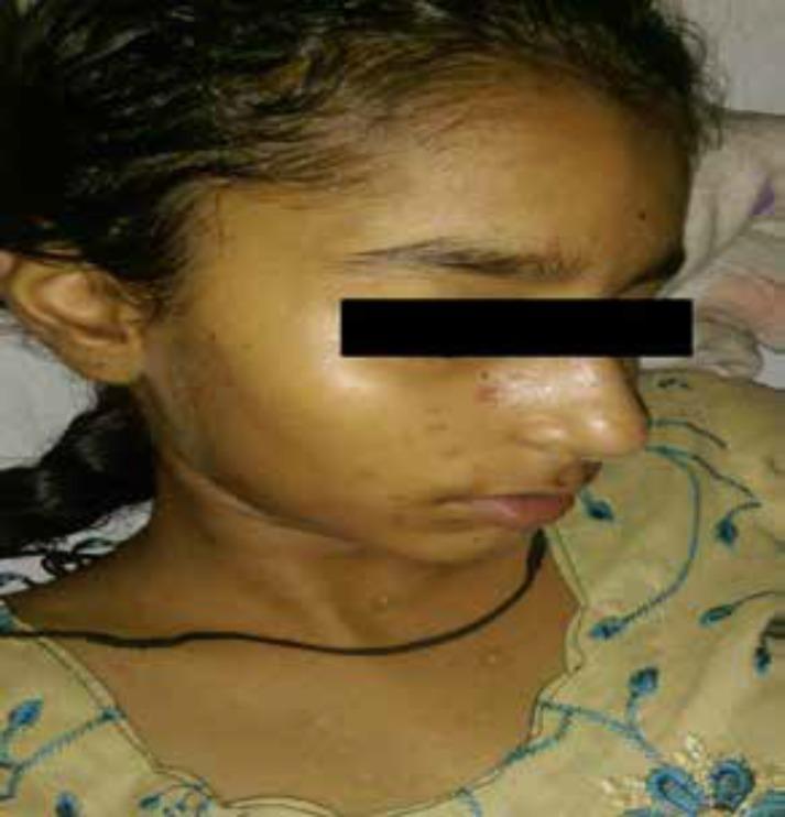
Clinical photograph of a pediatric patient's face and upper neck demonstrating a pleomorphic skin eruption characteristic of varicella-zoster virus (chickenpox) infection. The image shows multiple small lesions in varying stages of evolution, including erythematous and reddish-brown maculopapules, crusting lesions with scab formation, and small residual hypopigmented macules indicating sites of previous inflammation. The lesions are distributed across the cheek, nose, and neck. This variety of lesion stages (the 'starry sky' appearance) is a hallmark diagnostic feature of varicella, representing a synchronous evolution of the rash over several days. The educational focus is on identifying clinical dermatology signs and understanding the progression of viral exanthems in pediatric medicine.
varicella chickenpox epidemiology prevention
Chickenpox (Varicella) - Epidemiology, Prevention & Control
Clinical Overview
Images
1. EPIDEMIOLOGY
Global Disease Burden
- Worldwide distribution; occurs in both epidemic and endemic forms
- Annual global burden: 4.2 million severe complications leading to hospitalization and 4,200 deaths
- Pre-vaccine era case fatality rate in high-income countries: ~3 per 100,000 cases
- India (2018): 66,963 cases reported, 50 deaths; CFR ~0.074%; Kerala had highest cases (34,785); West Bengal had most deaths (31)
Epidemiological Determinants
A. Agent Factors
| Factor | Details |
|---|---|
| Agent | VZV (Human herpesvirus 3); DNA virus |
| Source of infection | Case of chickenpox (primary); rarely, a herpes zoster patient |
| Virus location | Oropharyngeal secretions, vesicular fluid, skin/mucosal lesions |
| Scabs | NOT infective |
| Period of communicability | 1-2 days before rash appears, up to 4-5 days after rash onset |
| Latency | Establishes latent infection in cranial nerve ganglia and spinal dorsal root ganglia; reactivates as zoster in 10-30% of persons |
B. Host Factors
- Age: Primarily affects children under 10 years; adults can have severe disease
- Immunity: One attack gives durable (lifelong) immunity; second attacks are rare. Maternal IgG antibodies protect infants in the first few months of life
- Pregnancy: Infection in first 20 weeks of gestation causes congenital varicella syndrome in 0.4-2.0% of foetuses; affected infants have higher risk of developing herpes zoster in early life
- High-risk groups: Infants, pregnant women, immunocompromised patients (including HIV), adults, and those on immunosuppressive therapy
C. Environmental Factors
- Seasonal: Peak incidence in winter and spring (temperate); coolest, driest months in tropics
- Epidemic cycle: Periodic large outbreaks with an inter-epidemic cycle of 2-5 years
- Virus survival: VZV is heat-labile; survives only a few hours outside the host, occasionally 1-2 days; readily inactivated by lipid solvents, detergents, and proteases
D. Transmission
- Droplet infection and droplet nuclei (airborne spread) - primary route
- Direct contact with skin lesions
- Indirect contact via freshly soiled articles
- Highly contagious - Secondary Attack Rate (SAR): ~85% (range 61-100%) in susceptible household contacts
- Herpes zoster is ~20% as infectious as chickenpox; contact with zoster rash causes varicella (not zoster) in susceptibles
Incubation Period
- 14-16 days (range: 10-21 days, occasionally up to 23 days)
2. CLINICAL FEATURES (Summary)
| Feature | Description |
|---|---|
| Distribution | Centripetal - starts on trunk (abundant), then face, arms, legs; mucous membranes affected; palms/soles usually spared |
| Morphology | Macule → papule → vesicle ("dew drop on a rose petal") → crust; unilocular, superficial |
| Pleomorphism | All stages present simultaneously - hallmark feature, due to successive crops appearing over 4-5 days |
| Evolution | Very rapid; scabbing begins 4-7 days after rash |
| Fever | Rises with each fresh crop of rash |
3. PREVENTION
A. Active Immunization - Varicella Vaccine
- Vaccine type: Live attenuated varicella vaccine (Oka strain)
- Schedule:
- Children (12 months-12 years): 2 doses - first dose at 12-15 months, second dose at 4-6 years
- Adolescents/Adults (susceptible): 2 doses, 4-8 weeks apart
- Efficacy: ~85% against any varicella; >95% against severe disease
- Contraindications: Pregnancy, immunocompromised states (CD4 <200 cells/mm³ in HIV), recent immunoglobulin or blood products
- In HIV-positive patients with CD4 ≥200 cells/μL: two-dose VAR series 3 months apart is recommended
B. Passive Immunization - Varicella-Zoster Immune Globulin (VZIG)
- Administered to high-risk susceptible contacts within 96 hours of exposure
- Indications: susceptible pregnant women, immunocompromised individuals, neonates born to mothers with varicella
- Does not prevent infection but modifies severity
C. Chemoprophylaxis
- Acyclovir: Post-exposure prophylaxis in susceptible high-risk individuals when VZIG is unavailable
- Antiviral treatment: Acyclovir/valacyclovir for high-risk groups (adults, immunocompromised, pregnant) within 24-48 hours of rash onset
4. CONTROL MEASURES
A. Case Management
| Measure | Details |
|---|---|
| Isolation | Isolate the case from the onset of fever until ALL lesions have crusted (at least 5 days after rash onset) |
| Treatment | Symptomatic in healthy children; antiviral therapy (acyclovir) for high-risk cases |
| Notification | Notifiable disease in many countries/states |
B. Contact Management
- Identify susceptible contacts (no prior infection, no vaccination)
- Administer VZIG to high-risk susceptibles within 96 hours of exposure
- Vaccinate susceptible contacts (if not contraindicated) - post-exposure vaccination within 3-5 days can prevent or attenuate disease
- Exclude susceptible contacts from school/work for 21 days from last exposure (or 8-21 days if VZIG given)
C. Environmental Measures
- Adequate ventilation of rooms (virus spreads via airborne route)
- Terminal disinfection of contaminated articles (VZV susceptible to common disinfectants)
- Healthcare settings: airborne + contact precautions; negative pressure rooms for hospitalized cases
D. Outbreak Control
- Rapid identification and vaccination of susceptible individuals
- Exclude cases until all lesions are crusted
- Vaccination coverage of >90% in school-age children is the key to epidemic prevention
Summary Table
| Parameter | Details |
|---|---|
| Causative agent | VZV (Human herpesvirus 3) |
| Reservoir | Humans only |
| Transmission | Airborne (droplets/nuclei), contact |
| Incubation period | 10-21 days (usually 14-16 days) |
| Communicability | 1-2 days before to 4-5 days after rash |
| SAR | ~85% in household contacts |
| Immunity | Lifelong after natural infection |
| Vaccine | Live attenuated (Oka strain), 2-dose schedule |
| Isolation | Until all lesions crusted |
- Park's Textbook of Preventive and Social Medicine, pp. 163-165
- Goldman-Cecil Medicine, International Edition
- Recent systematic review: Healthcare resource use and costs of varicella (Williame et al., 2023)
- Varicella vaccine in pediatric transplant recipients - meta-analysis confirming vaccine safety and immunogenicity (Piché-Renaud et al., 2023)
SAQ 1. Measles Vaccination (Pg. 161) LAQ 1. Measles - epidemiology, control, prevention
measles rash Koplik spots maculopapular exanthem
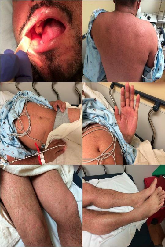
A series of clinical photographs documenting the exanthem and enanthem of measles (rubeola) in an adult male. The oral cavity image demonstrates pathognomonic Koplik spots: tiny, bluish-white or whitish-gray macules resembling grains of sand on an erythematous base, located on the buccal mucosa. The remaining images show a generalized, blanching, erythematous maculopapular rash. This exanthem exhibits a cephalocaudal progression, appearing as numerous discrete macules and papules that have coalesced into larger patches on the back, abdomen, and extremities, including the palms and feet. The presentation illustrates the classic clinical manifestations of a primary measles infection, highlighting the diagnostic importance of the enanthem during the prodromal phase and the characteristic distribution of the subsequent skin eruption for emergency and infectious disease medicine.
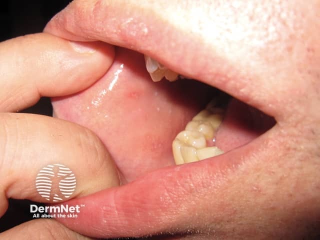
Close-up clinical photograph of the oral cavity focusing on the buccal mucosa reveals characteristic small bluish-white Koplik spots embedded in erythematous mucosa. The image corresponds to early measles (rubeola) infection on illness day 1, typically preceding the maculopapular rash. Koplik spots are pathognomonic for measles, reflecting cytopathic changes in mucosal epithelial cells with inflammatory exudate and adjacent hyperemia. In this view, the lips are gently everted to expose the inner cheek, with moist mucosa and subtle surrounding erythema near the molar region. These lesions may be transient and diminish as the exanthem develops over the next 1–3 days. The photograph supports the clinical diagnosis of measles and aids differentiation from other viral exanthems where such mucosal lesions are absent or non-specific. Clinically, identification of Koplik spots prompts immediate infection control measures and confirmatory testing (measles IgM serology or RT-PCR). For educational and research use, the image highlights oral cavity involvement, buccal mucosa localization, and the temporal relationship to fever and onset of rash. Differential considerations include rubella, enteroviral infections, and aphthous lesions, but Koplik morphology and distribution are highly characteristic of measles when present. This image serves as a teaching reference for clinicians and trainees. Facilitates rapid clinical decision-making.
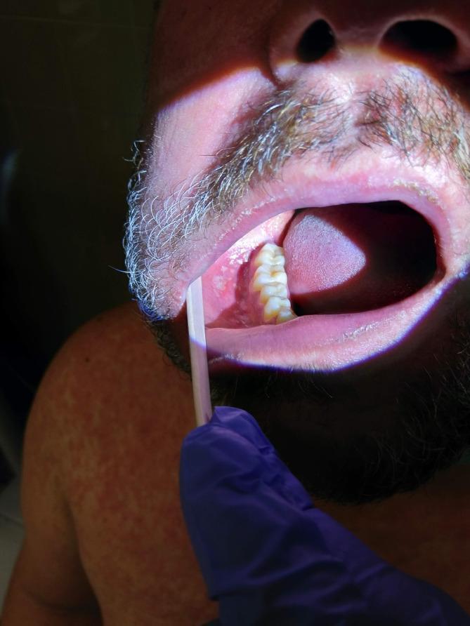
This clinical photograph shows the intraoral cavity and shoulder of an adult male, highlighting pathognomonic signs of a viral exanthem. The primary focus is the buccal mucosa, which is retracted to reveal Koplik's spots: multiple small, 1–2 mm, irregular, bluish-white or gray-white grain-like elevations on an erythematous base. These spots are located opposite the mandibular molars. In the background, the patient's shoulder and chest display a faint, widespread, red maculopapular rash. The image demonstrates key diagnostic features of the prodromal phase of measles (rubeola), where intraoral lesions precede the full systemic rash. This visual is suitable for medical education regarding infectious diseases, dermatology, and clinical diagnosis of childhood and adult exanthems.
measles epidemiology vaccination prevention
SAQ: Measles Vaccination (Park's Textbook, p. 161/pg. 168-170)
Measles Vaccination
Vaccine Type
- Monovalent (measles only)
- MR (Measles-Rubella)
- MMR (Measles-Mumps-Rubella)
- MMRV (Measles-Mumps-Rubella-Varicella)
Vaccine Properties
- Freeze-dried (lyophilized) product; reconstituted with sterile diluent before use
- Each dose = 0.5 ml containing ≥1000 viral infective units
- Contains sorbitol and hydrolysed gelatin as stabilizers; small amount of neomycin; no thiomersal
- Heat-labile: loses ~50% potency after 1 hour at 20°C; almost all potency after 1 hour at 37°C
- Sensitive to sunlight - stored in coloured glass vials
- Storage: 2-8°C (refrigerated); diluent must NOT be frozen but cooled before reconstitution
- After reconstitution: must be used within 4 hours, stored in dark at 2-8°C
Recommended Schedule (WHO)
| Setting | MCV1 | MCV2 |
|---|---|---|
| High-transmission / high-risk countries | 9 months | 15-18 months |
| Low-transmission / near-elimination countries | 12 months | At school entry (4-6 years) |
| Minimum interval between MCV1 and MCV2 | - | 4 weeks |
- MCV0: Supplementary dose given from 6 months of age in special situations (outbreaks, high-endemic areas, displaced populations, HIV-exposed infants, travellers to outbreak areas). Does not replace MCV1 and MCV2.
Immunity
- Immunity develops 11-12 days after vaccination
- Duration: probably lifelong
- One dose at 11-12 months: ~95% protection
- Two doses: ~98% protection
- Infants vaccinated at 9 months: seroconversion ~90%
Post-Exposure Prophylaxis
- Susceptible contacts >9-12 months: vaccine given within 3 days of exposure can protect (vaccine incubation ~7 days vs. natural measles incubation ~10 days)
Contraindications
- Severe immunosuppression (HIV with severe immunosuppression, congenital immunodeficiency, leukaemia, lymphoma, high-dose corticosteroids)
- MMRV: not approved for HIV-infected persons
- Moderate/severe acute illness: defer until recovery
- Receipt of antibody-containing blood products: delay by 3-11 months depending on product
Adverse Effects
- Toxic Shock Syndrome (TSS): occurs when vaccine is contaminated or same vial used >4 hours; presents as watery diarrhoea, vomiting, high fever within hours; cluster of cases; high CFR; totally preventable by good immunization practices
Passive Immunization (Immunoglobulin)
- Dose: 0.25 ml/kg IM, within 3-4 days of exposure
- Modifies or prevents disease
- Live vaccine should be given 8-12 weeks later
LAQ: Measles - Epidemiology, Control & Prevention
Koplik Spots (Pathognomonic Enanthem):
Measles Maculopapular Rash with Koplik spots:
Definition
1. EPIDEMIOLOGY
Global Disease Burden
- Endemic in virtually all parts of the world
- In 1980, before widespread vaccine use: ~2.6 million measles deaths per year
- By 2018: ~9.7 million cases and >140,000 measles-related deaths globally (73% reduction in deaths since 2000)
- Accounts for ~2% of under-five mortality worldwide
- Epidemics occur when susceptible children reach ~40% of population
- In virgin communities: >90% of the community infected when disease is introduced
- Pre-immunization programme: cyclical peaks every 3 years; ~2.47 lakh cases in 1987
- Post-UIP (2018): 20,895 cases and 34 deaths
- States most affected (2018): West Bengal, Assam, J&K, Maharashtra, Delhi, UP, Rajasthan
Epidemiological Determinants
A. Agent Factors
| Factor | Details |
|---|---|
| Agent | RNA Paramyxovirus; only one serotype |
| Reservoir | Humans only; no animal reservoir |
| Survival | Cannot survive outside human body for long; retains infectivity at sub-zero temperatures |
| Source | Only a case of measles; no carriers known |
| Infective material | Secretions of nose, throat, respiratory tract (prodromal + early rash stage) |
| Communicability | ~4 days before to 4 days after rash appearance; highly infectious during prodrome |
| Second attack | Rare; one serotype only; infection gives lifelong immunity |
B. Host Factors
| Factor | Details |
|---|---|
| Age | 6 months - 3 years (developing countries); >5 years (developed countries); post-vaccine era: older age groups affected |
| Sex | Equal incidence in males and females |
| Immunity | One attack gives lifelong immunity; maternal antibody protects infants up to 6-9 months |
| Nutrition | Measles hits malnourished children harder; precipitates kwashiorkor in borderline cases |
| Susceptibility | Universal in absence of immunity |
C. Environmental Factors
- Seasonal trend: Temperate zones - peaks in late winter/spring; tropics - during dry cooler months
- Overcrowding promotes spread
- Epidemic cycle: Every 2-3 years (pre-vaccine); longer cycles with higher vaccination coverage
- Virus spreads efficiently in conditions of poor ventilation and crowding
D. Transmission
- Airborne (primary route) - droplets and droplet nuclei
- Direct contact with nasal/throat secretions
- Indirect contact - freshly soiled articles with nasopharyngeal secretions
Incubation Period
- 10-14 days (range 7-18 days; typically about 10 days to fever, ~14 days to rash)
2. CLINICAL FEATURES
Prodromal Stage (3-5 days)
- High fever, severe malaise
- The 3 C's: Coryza, Cough, Conjunctivitis
- Koplik spots (pathognomonic): bluish-white/grayish spots on erythematous buccal mucosa, opposite lower molars; appear 1-2 days before rash
Eruptive Stage
- Maculopapular rash: starts on face and behind ears → spreads downward (centrifugal, cephalocaudal)
- Rash lasts ~5-6 days; may coalesce
- Fever rises with rash, subsides as rash fades
- Rash fades in order of appearance, leaving brownish discoloration
Complications
- Otitis media (most common complication)
- Pneumonia (most common cause of death)
- Laryngotracheobronchitis (Croup)
- Encephalitis (1 in 1000 cases)
- Subacute Sclerosing Panencephalitis (SSPE) - rare, fatal; years later
- Malnutrition/Kwashiorkor in developing countries
- Blindness (vitamin A deficiency + corneal ulceration)
3. PREVENTION
A. Active Immunization (Primary Prevention)
- Live attenuated measles vaccine (Oka/Edmonston-Zagreb strain)
- Schedule: 2-dose strategy (MCV1 + MCV2) - see SAQ above for full details
- Target: ≥95% coverage nationally; ≥80% in every district
- Achieves 95-98% protection (1 or 2 doses)
- Global target (WHO/UNICEF): reduce measles mortality by ≥95% vs. year 2000 levels
B. Passive Immunization
- Immunoglobulin (human): 0.25 ml/kg IM within 3-4 days of exposure
- For high-risk susceptible contacts (malnourished children, immunocompromised)
C. Vitamin A Supplementation
- WHO recommends vitamin A to all children with measles to reduce complications and mortality
- Dose: 200,000 IU orally for 2 days (children >12 months); 100,000 IU (<12 months)
- Reduces measles-related blindness and pneumonia mortality
D. Supplementary Immunization Activities (SIAs)
- Mass vaccination campaigns targeting unvaccinated/under-vaccinated children
- Conducted in areas with low routine coverage or during outbreaks
4. CONTROL MEASURES
A. Case Management
| Measure | Details |
|---|---|
| Notification | Notifiable disease - report promptly |
| Isolation | Isolate for 7 days from onset of rash (more than covers communicability period) |
| Treatment | Symptomatic; vitamin A; antibiotics for secondary bacterial complications |
| Hospitalize | Severe cases - pneumonia, encephalitis, malnutrition |
B. Contact Management
- Identify and vaccinate susceptible contacts within 3 days of exposure (post-exposure vaccination)
- High-risk susceptibles (infants <9 months, immunocompromised): administer immunoglobulin within 3-4 days
- Exclude unvaccinated susceptible children from school for 21 days from last exposure
C. Environmental Measures
- Improve ventilation in schools, hostels, hospitals
- Promote hand hygiene and respiratory etiquette
- Airborne + contact precautions in healthcare settings
D. Surveillance
- Case-based measles-rubella surveillance with adequate laboratory support (IgM serology, RT-PCR)
- Maintain vaccination records and track outbreaks
- Investigation and containment of clusters
5. ERADICATION
- Humans are the only reservoir
- No known carrier state
- Only one serotype
- An effective, safe, affordable vaccine exists
- Only 2 doses needed; more heat-stable vaccine developed
- Immunization coverage of ≥96% of children under 1 year
- Prevention of accumulation of immunity gaps across generations
Summary Table
| Parameter | Details |
|---|---|
| Causative agent | RNA Paramyxovirus (1 serotype) |
| Reservoir | Humans only; no carriers |
| Transmission | Airborne (droplets/nuclei), contact |
| Incubation | 10-14 days |
| Communicability | 4 days before to 4 days after rash |
| SAR | ~100% in susceptibles |
| Pathognomonic sign | Koplik spots |
| Vaccine | Live attenuated, 2-dose (MCV1 + MCV2) |
| Isolation | 7 days from rash onset |
| Immunity | Lifelong after natural infection |
Drug resistance tb in story format in simple language
drug resistant tuberculosis MDR TB chest xray
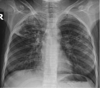
This diagnostic image is a posterior-anterior (PA) chest radiograph illustrating pulmonary manifestations of multi-drug resistant tuberculosis (MDR-TB). The radiograph reveals significant pathology characterized by bilateral, asymmetric opacities and infiltrates, predominantly involving the upper and mid-lung zones. Specifically, there are dense, patchy alveolar and interstitial infiltrates visible in both apical regions. Within these areas of consolidation, several lucent, thin-walled cavitary lesions are present, which are classic radiographic hallmarks of post-primary or secondary tuberculosis reactivation. The mediastinal shadow appears within normal limits, and there is no evidence of significant pleural effusion. The visible skeletal structures show no acute fractures, though the clinical context notes an underlying diagnosis of systemic lupus erythematosus (SLE), which may contribute to the patient's immunosuppression. This image serves as an educational example of advanced pulmonary infectious disease and the radiographic appearance of cavitation in the setting of drug-resistant mycobacterial infection.
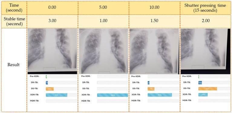
A comparison chart demonstrating a live image analysis interface for tuberculosis (TB) drug resistance diagnosis from chest X-rays (CXR). The graphic is divided into four chronological columns: 0.00 seconds, 5.00 seconds, 10.00 seconds, and shutter pressing time at 15 seconds. Each column displays a cropped diagnostic image focusing on the bilateral mid-to-lower lung fields and mediastinal borders. Below each image are AI-generated probability bars for five clinical classifications: Pre-XDR (pre-extensively drug-resistant TB), DR-TB (drug-resistant TB), DS-TB (drug-susceptible TB), XDR-TB (extensively drug-resistant TB), and MDR-TB (multidrug-resistant TB). The visual illustrates the real-time evolution of the deep learning model's diagnostic confidence. For instance, at 5.00 seconds, XDR-TB shows a 100% probability, while at the final shutter time (15 seconds), DS-TB becomes the most likely prediction at 66%. This infographic highlights the application of 'fast image analysis' in clinical informatics and infectious disease screening.
🫁 The Story of the Stubborn Bug
Drug-Resistant Tuberculosis - Told as a Story
Chapter 1: The Villain Arrives
Chapter 2: The Mutation Game
Chapter 3: The Big Mistake - Incomplete Treatment
Chapter 4: The Names of the Monster
| Name | What it means | In plain language |
|---|---|---|
| Mono-resistant TB | Resistant to ONE drug | The bug dodges one punch |
| Poly-resistant TB | Resistant to MORE than one drug (but not both Isoniazid + Rifampicin) | The bug is getting tougher |
| MDR-TB (Multi-Drug Resistant) | Resistant to at least Isoniazid AND Rifampicin - the two most powerful first-line drugs | The main weapons don't work anymore |
| Pre-XDR-TB | MDR-TB + resistant to a fluoroquinolone OR a second-line injectable | Almost impossible to treat |
| XDR-TB (Extensively Drug Resistant) | MDR-TB + resistant to a fluoroquinolone AND a second-line injectable (amikacin/streptomycin) | One of the hardest infections to treat |
Chapter 5: How Did We Get Here? - The Three Villains
Villain 1 - The Careless Patient 🧍
- Stopping medicines early ("I feel better")
- Skipping doses
- Not knowing why full treatment matters
- Poverty and social barriers making it hard to access medicines
Villain 2 - The Failing System 🏥
- Doctors prescribing the wrong drug combination or wrong dose
- No guidelines, or guidelines not followed
- Poor supervision and monitoring of patients
- Lack of trained health workers
Villain 3 - The Bad Medicine 💊
- Drug stockouts (medicines running out mid-treatment)
- Poor quality medicines
- Wrong storage (drugs damaged by heat or sunlight)
- Wrong dosage combinations in the packet
Chapter 6: The Battle Gets Harder - Treating MDR-TB
- Treatment duration: At least 18-24 months (compared to 6 months for normal TB)
- Number of drugs: 5 drugs in the intense phase, then 4 drugs in the continuation phase
- Medicines used: Bedaquiline, Linezolid, Moxifloxacin, Clofazimine, Cycloserine - names even doctors struggle to pronounce
- Cost: Second-line drugs cost 30 times more than normal TB drugs
- Side effects: These medicines can damage hearing, cause nerve pain, affect the liver, and disturb mental health
Chapter 7: The Even Scarier Monster - XDR-TB
Chapter 8: India's Story
- Primary MDR-TB in India: ~2.8% of new cases
- MDR-TB in retreatment cases: ~12%
- Though the percentage sounds small, because India has so many TB patients, this translates into very large absolute numbers of people suffering from MDR-TB
Chapter 9: Can We Win? - Prevention and Control
For the Patient
- Take ALL your medicines, EVERY day, for the FULL duration - no skipping, no stopping early
- DOTS (Directly Observed Treatment, Short-course) - a health worker watches you swallow your pill every day to make sure you don't miss a dose
- Inform your doctor immediately if you feel side effects rather than stopping on your own
For the Healthcare System
- Proper diagnosis with drug susceptibility testing before starting treatment
- Correct drug regimens, correct doses
- Regular monitoring of patient progress
- Adequate drug supply - no stockouts
- Rapid molecular tests (like GeneXpert/CB-NAAT) that detect resistance within hours, not weeks
For Society
- Improve ventilation and reduce overcrowding
- Address poverty and malnutrition that make people more vulnerable
- TB awareness campaigns
- Contact tracing - finding people who were exposed
Epilogue: What Raju Learned
"Six months of pills felt like too much. Two years of second-line treatment felt like forever. Please - finish your medicines the first time."