
vocal cord understand eassy
Reading File
The vocal cords (also called vocal folds) are two bands of mucous membrane in the larynx that vibrate to produce sound. They sit in the middle of the larynx, stretched across it, and open/close rapidly as air passes through.
Searching sources...
Finding Sources
Finding Sources
Finding Sources
Reading File
Finding Sources
Finding Sources
Reading File
Reading File
Reading File
Finding Sources
Now I have rich textbook content. Let me fetch a relevant image to include in the answer.
Reading File
Finding Sources
Reading File
Loading Image
Reading File
I now have all the information needed for a thorough, easy-to-understand essay on the vocal cords. Here is the complete answer:
The Vocal Cords - A Complete Easy Guide
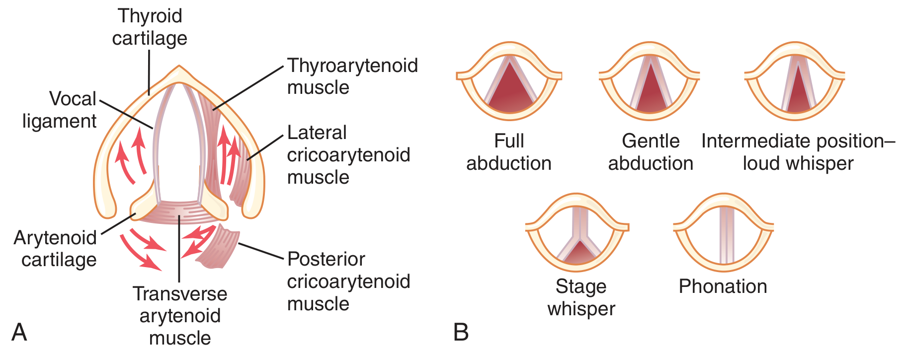
1. What Are the Vocal Cords?
The vocal cords (also called vocal folds) are two bands of specialized tissue stretching across the inside of the larynx (your "voice box"), which sits at the top of your windpipe (trachea). They are not cords at all in the rope sense - they look more like lips or folds of tissue.
There are actually two sets of folds:
- True vocal folds (lower) - these vibrate to make sound
- False vocal folds / vestibular folds (upper) - mainly protective, do not usually vibrate for normal speech
2. Where Are They Located? (The Larynx)
The larynx contains three unpaired cartilages (epiglottis, thyroid cartilage, cricoid cartilage) and three paired cartilages (arytenoid, corniculate, cuneiform). The vocal folds attach at the front to the thyroid cartilage and at the back to the arytenoid cartilages - and it is the movement of the arytenoids that opens and closes the vocal folds.
The space between the two vocal folds is called the glottis. The larynx has three regions:
- Supraglottis - above the folds (includes epiglottis)
- Glottis - the folds themselves and the 1 cm below
- Subglottis - below, down to the cricoid cartilage
(Sabiston Textbook of Surgery, p. 1361)
3. Layers of the Vocal Fold (Microarchitecture)
This is the key to understanding how the vocal cords work. From outside to inside, each fold has:
| Layer | Contents |
|---|---|
| Epithelium | Stratified squamous cells, ~50 microns thick |
| Superficial lamina propria (Reinke's space) | Loosely arranged collagen and elastin - acts as a shock absorber, allows the mucosal "wave" |
| Intermediate lamina propria | Predominantly elastic fibers |
| Deep lamina propria | Dense collagen fibers |
| Vocalis muscle (thyroarytenoid) | The muscular "body" of the fold |
The intermediate + deep layers together = the vocal ligament.
Hirano (a pioneer researcher) described this beautifully as a "body-cover" model: the vocalis muscle is the relatively stiff "body," while the epithelium + Reinke's space form the soft "cover" that ripples freely over it. This free rippling is what gives the voice its richness and range. If the Reinke's space is scarred (e.g., after surgery or injury), voice quality drops dramatically. (Cummings Otolaryngology, p. 1107-1108)
4. How Do the Vocal Cords Make Sound? (Phonation)
Sound production involves three steps: breath power, vibration, and resonance.
Step 1 - Air Power (Subglottic Pressure)
You breathe in, then begin to breathe out. The air below the vocal folds builds up pressure (subglottic pressure).
Step 2 - The Vibration Cycle (Myoelastic-Aerodynamic Theory)
This is the widely accepted explanation for how sound is made (van den Berg, 1950s):
- The vocal folds are held near the midline (adducted/closed)
- Air pressure from below pushes the folds apart
- As air rushes through the narrow gap, the Bernoulli effect (air moving fast = lower pressure) and the elastic recoil of the folds pull them back together
- The folds snap shut, pressure builds again - and the cycle repeats hundreds of times per second
The mucosal wave starts at the lower edge of the fold and travels upward - as the bottom is closing, the top is still opening. This is the "two-mass model" of vibration. (Cummings Otolaryngology, p. 1023-1024)
"Vocal fold structure determines whether the resulting vibration is periodic or chaotic." - Cummings Otolaryngology
Step 3 - Resonance
The raw buzzing sound is shaped into speech by the mouth, tongue, lips, soft palate, nose, and throat cavities. (Guyton & Hall, p. 502)
5. Controlling Pitch and Volume
| What changes | Effect |
|---|---|
| Increase tension in the fold (cricothyroid muscle stretches the fold) | Higher pitch |
| Shorten the fold | Higher pitch |
| Reduce mass of the fold | Higher pitch |
| Relax the vocalis muscle | Lower, richer "chest voice" |
| More air pressure (deeper breath, more effort) | Louder voice |
| Falsetto (very tight, thin folds, only top edges touch) | Very high pitch; mucosal wave disappears |
The cricothyroid muscle (the main pitch-control muscle) is the only intrinsic laryngeal muscle controlled by the external (superior) laryngeal nerve. All other intrinsic muscles are controlled by the recurrent laryngeal nerve (RLN). (Guyton & Hall, p. 501-502)
6. Positions of the Vocal Folds
As seen in the diagram above, the folds take different positions:
- Full abduction (wide open) - quiet breathing
- Gentle abduction - deep breathing
- Adduction/phonation (nearly closed) - speech and singing
- Stage whisper - folds apart but tightly tense, turbulent airflow
- Loud whisper - intermediate position
7. What Can Go Wrong? (Common Disorders)
| Disorder | What happens |
|---|---|
| Vocal nodules | Callus-like thickenings at the midpoint, from vocal overuse ("vocal overdoers") - e.g., teachers, singers |
| Vocal polyps | Soft, fluid-filled swellings, usually one-sided; from acute vocal injury |
| Vocal cord paralysis | One or both folds cannot move, usually from recurrent laryngeal nerve damage (e.g., thyroid surgery, lung cancer); causes breathy, weak voice |
| Reinke's edema | Fluid accumulation in Reinke's space; common in smokers; causes deep, rough voice |
| Laryngitis | Inflammation from infection or reflux; swelling prevents normal vibration |
| Vocal cord scar | Loss of the pliable Reinke's layer; permanent voice change |
| Papillomatosis | Wart-like growths from HPV infection |
(Scott-Brown's Otorhinolaryngology; Cummings Otolaryngology)
8. How Are Vocal Cord Problems Diagnosed?
- Laryngoscopy (flexible or rigid camera): the standard way to directly view the folds
- Videostroboscopy: uses a strobe light to slow down the apparent vibration and assess the mucosal wave - the most useful tool for diagnosing benign fold disorders
- High-speed video: true slow-motion capture of vibration (expensive, research use)
- Acoustic analysis: computerized measurement of voice quality
Quick Summary
VOCAL CORDS
    │
    ├── Location: Larynx (between thyroid & arytenoid cartilages)
    ├── Layers: Epithelium → Reinke's space → Vocal ligament → Vocalis muscle
    ├── How sound is made: Myoelastic-aerodynamic theory
    │       Air pressure pushes folds apart → Bernoulli + elastic recoil closes them
    │       → Vibration → Resonance in vocal tract → Speech
    ├── Pitch control: Tension (high pitch), relaxation (low pitch)
    └── Common problems: Nodules, polyps, paralysis, polyps, scar
The vocal folds are a marvel of biological engineering - two tiny tissue bands, each only about 15-20 mm long in an adult, yet capable of producing the full range of human speech, singing, and expression by vibrating up to 1000 times per second.
This is a shared conversation. Sign in to Orris to start your own chat.