
EMBRYOLOGY OF THE AUDITORY SYSTEM: WEEK-BY-WEEK DEVELOPMENT
Reading File
Now let me search the medical library for auditory system embryology content in parallel.
Finding Sources
Finding Sources
Finding Sources
Finding Sources
Excellent - I found the key sources. Let me now read the primary embryology content from "The Developing Human" and the otolaryngology references in parallel.
Reading File
Reading File
Great content found. Let me continue reading "The Developing Human" for more details on middle ear, external ear, and ossicle development, plus fetch the key images.
Reading File
Finding Sources
Let me read the section on middle and external ear development from The Developing Human, and also get content from Scott-Brown's.
Reading File
Reading File
I have rich content. Now let me fetch the key development diagram and read more of The Developing Human for middle/external ear content.
Loading Image
Loading Image
Excellent images retrieved. Now let me get the middle ear and external ear development content from The Developing Human.
Reading File
I now have comprehensive content from The Developing Human. Let me also get the middle ear ossicle development content and fetch one more key image.
Reading File
Loading Image
I now have all the content needed for a comprehensive week-by-week guide. Let me compile the full answer.
Embryology of the Auditory System: Week-by-Week Development
The ear is unique in human embryology because its three components - internal, middle, and external - arise from completely different embryonic origins yet must integrate into a single functional unit. The internal ear is the first to develop, beginning as early as week 3, and reaches adult size by week 20-22.
Overview of Embryonic Origins
| Component | Embryonic Tissue of Origin |
|---|---|
| Internal ear (membranous labyrinth) | Surface ectoderm (otic placode) |
| Bony labyrinth | Mesenchyme (cartilaginous otic capsule) |
| Middle ear cavity | Endoderm of 1st pharyngeal pouch |
| Ossicles (malleus, incus) | Cartilage of 1st pharyngeal arch (Meckel's) |
| Stapes crura/head | Neural crest cells (2nd pharyngeal arch) |
| Stapes footplate rim | Mesodermal cells |
| External acoustic meatus | Ectoderm of 1st pharyngeal groove |
| Tympanic membrane | Ectoderm + endoderm + mesenchyme |
| Auricle (pinna) | Mesenchyme of 1st and 2nd pharyngeal arches (auricular hillocks) |
Week 3 - The Otic Placode Appears
The internal ear is the first part of the ear to develop. Early in the fourth week (approximately day 22), a thickening of surface ectoderm called the otic placode appears bilaterally, adjacent to the myelencephalon (caudal hindbrain) (Fig. 18.13A).
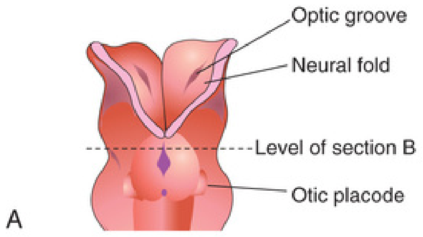
Molecular signals driving otic placode induction:
- Inductive signals from paraxial mesoderm and notochord
- FGF signaling initiates specification of otic-epibranchial progenitors from sensory precursors in the preplacodal region
- Transcription factors FoxL1/3, Wnt and Notch pathways, Pax2/8, and protein-encoding Dlx genes all regulate further development
- Pa2G4 protein-coding gene is involved
Week 4 - Otic Pit and Otic Vesicle Formation
The otic placode invaginates into the underlying mesenchyme, forming the otic pit (approximately day 24-26). The edges of the pit fuse to create the otic vesicle (otocyst) - the primordium of the entire membranous labyrinth (approximately day 28).
The otic vesicle then pinches off, losing all contact with the surface ectoderm.
Two regions are already distinguishable:
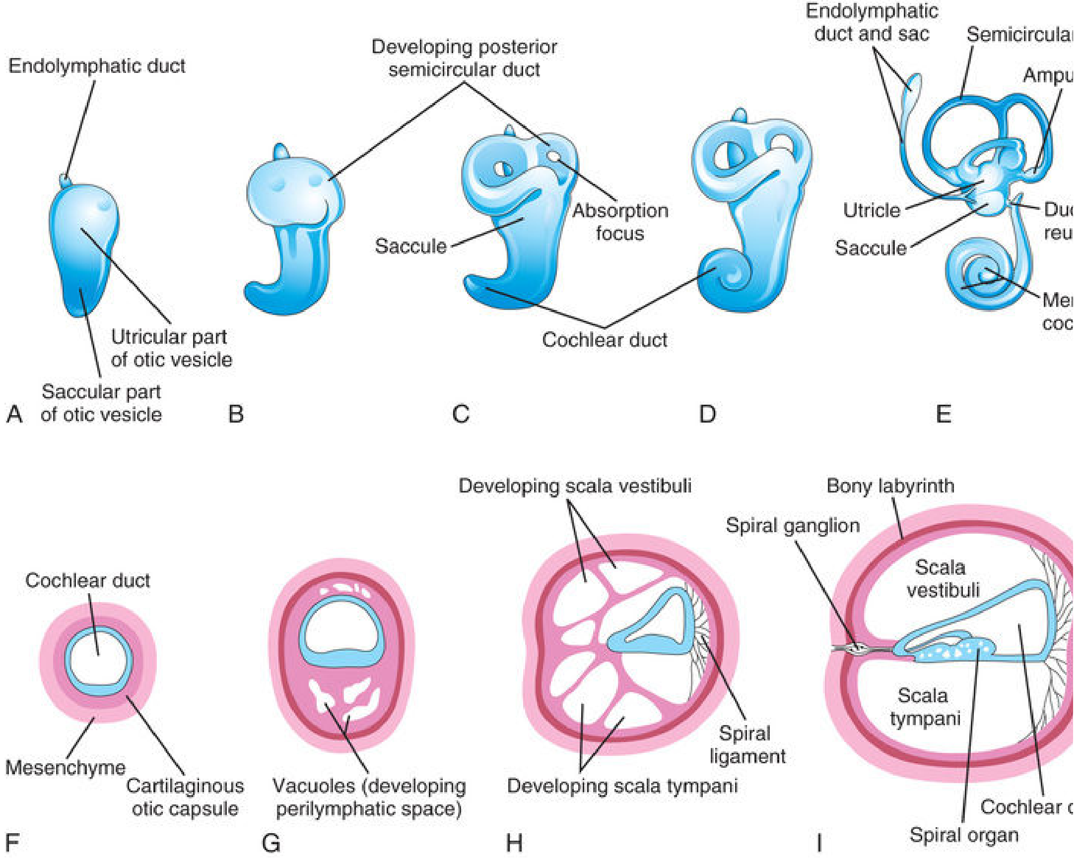
- Dorsal utricular part - will give rise to the endolymphatic duct, utricle, and semicircular ducts
- Ventral saccular part - will give rise to the saccule and cochlear duct
A diverticulum grows from the dorsal vesicle to form the endolymphatic duct and sac - this is the first structure to develop from the otocyst.
Simultaneously, the first pharyngeal pouch begins expanding toward the surface ectoderm - forming the future tubotympanic recess - the anlage of the middle ear cavity.
Week 5 - Cochlear Duct Emerges; Semicircular Canal Precursors Form
The cochlear duct (tubular diverticulum) grows from the ventral saccular part of the otic vesicle. The three disc-like diverticula from the utricular part represent the semicircular duct precursors. Their central portions begin to fuse and disappear, leaving only the peripheral arcs.
The otocyst now has two functional divisions:
- Pars superior - will become ampullae and semicircular canals (develop in order: superior, then posterior, then lateral)
- Pars inferior - will become saccule and cochlea
The tubotympanic recess continues expanding from the first pharyngeal pouch. Its proximal portion will become the pharyngotympanic (Eustachian) tube; its distal portion will become the tympanic cavity.
Week 6 - Cochlear Coiling Begins; Auricular Hillocks Appear
The cochlear duct begins its characteristic spiral coiling from the saccule (connected via the ductus reuniens). TBX1 expression in surrounding mesenchyme regulates cochlear duct formation via retinoic acid activity.
Auricle (pinna) formation begins: Six mesenchymal proliferations - auricular hillocks - develop around the first pharyngeal groove:
- Three from the 1st pharyngeal arch (Hillocks 1-3) - contribute mainly to the tragus
- Three from the 2nd pharyngeal arch (Hillocks 4-6) - contribute the remainder
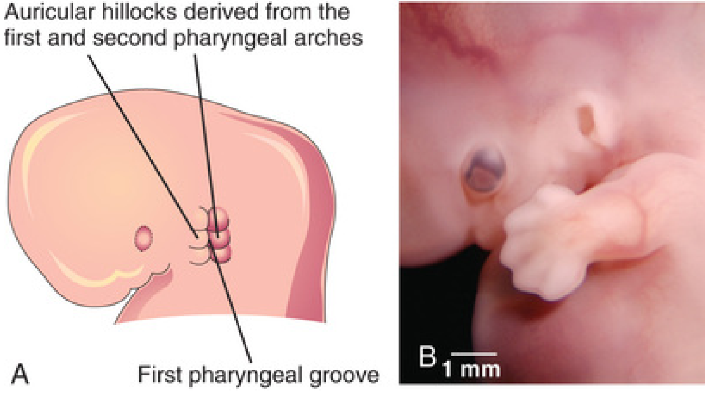
The auricles form low on the neck initially, migrating upward as the mandible develops.
Week 7 - Ossicle Condensation; Meatal Plug Forms
The malleus and incus condense from Meckel's cartilage (first pharyngeal arch). The stapes develops mainly from the second arch (Reichert's cartilage) and neural crest cells. Specifically:
- Crus, base of foot plate, and head of stapes = neural crest cells
- Outer rim of stapes foot plate = mesodermal cells
The stapedius muscle (attached to stapes) is derived from the second pharyngeal arch and is supplied by the facial nerve (CN VII). The tensor tympani (attached to malleus) is from the first arch, supplied by CN V3 (mandibular nerve).
The external acoustic meatus ectoderm proliferates to form a solid meatal plug - this temporarily blocks the canal.
Week 8 - Cochlea Reaches 2.5 Turns; Semicircular Canals Defined
The cochlear duct completes its 2.5 spiral turns by week 8. The membranous semicircular ducts are now defined as the central areas of the disc-like diverticula have been absorbed. Localized dilatations (ampullae) form at one end of each semicircular duct.
Specialized receptor areas differentiate:
- Cristae ampullares - in the ampullae (vestibular equilibrium receptors)
- Maculae utriculi and sacculi - in the utricle and saccule (linear acceleration/gravity receptors)
Inductive signals from the otic vesicle drive the surrounding mesenchyme to condense into a cartilaginous otic capsule - the precursor of the bony labyrinth.
Weeks 8-12 - Perilymphatic Spaces Form; Middle Ear Cavitation
Vacuoles appear in the cartilaginous otic capsule and coalesce to form the perilymphatic space (periotic space). This space divides into two scalae around the cochlear duct:
- Scala vestibuli (superior)
- Scala tympani (inferior)
Middle ear cavitation begins in the 3rd month (week ~9-12). The tubotympanic recess expands to gradually envelop the auditory ossicles, their tendons, ligaments, and the chorda tympani nerve. The entire lining of the tympanic cavity - including the ossicles - receives an epithelial investment derived from neural crest cell-origin endoderm via epithelial-mesenchymal transformation.
PAX2 gene expression is required for formation of the spiral organ of Corti and the spiral ganglion. Retinoic acid and TGF-β1 direct the formation of the otic capsule.
Weeks 10-12 - Spiral Ganglion; Cochlear Duct Nears Full Growth
Ganglion cells of CN VIII (vestibulocochlear nerve) migrate along the coils of the membranous cochlea to form the spiral ganglion. Nerve processes extend from the ganglion to terminate on hair cells in the spiral organ. Crucially, spiral ganglion cells retain their embryonic bipolar condition into adult life.
The cochlear duct reaches full 2.5 turns and begins to approach adult size.
Week 20 - Internal Ear Reaches Adult Size
The internal ear achieves adult size and shape by the middle of the fetal period (weeks 20-22). Cavitation of the middle ear is completed by week 8 of the fetal period (about month 8 of gestation).
The auricle reaches its adult structure by week 22. It migrates from its initial low-cervical position to the lateral side of the head as the mandible develops.
Week 21 - Organ of Corti Becomes Functional
The organ of Corti (spiral organ) becomes functionally active at approximately 21 weeks, although the cochlear duct already reached full adult size by ~20 weeks.
Week 26 - Functional Hearing
Functional hearing begins at approximately 26 weeks of gestation. This is the point at which the neural pathways are sufficiently mature for auditory processing.
Late Fetal Period - Meatal Plug Dissolves; Bony Labyrinth Complete
The central cells of the meatal plug degenerate, forming the inner portion of the external acoustic meatus. The meatus remains short at birth and reaches adult length at approximately 9 years of age.
The cartilaginous otic capsule ossifies to form the complete bony labyrinth.
Postnatal Milestones
- Endolymphatic sac and duct continue growing until age 3-4 years
- External acoustic meatus reaches adult length at ~9 years
Week-by-Week Summary Table
| Week | Event |
|---|---|
| 3 | Otic placode appears (surface ectoderm thickening) |
| 4 | Otic pit forms → otic vesicle (otocyst); endolymphatic duct first to develop; 1st pharyngeal pouch → tubotympanic recess begins |
| 5 | Cochlear duct emerges; semicircular canal disc-diverticula form; pars superior/inferior division of otocyst |
| 6 | Cochlear duct begins coiling; auricular hillocks appear (6 hillocks from arches 1+2); external auditory meatus forms from 1st groove |
| 7 | Ossicle condensation (malleus/incus from arch 1; stapes from arch 2/neural crest); meatal plug forms |
| 8 | Cochlea completes 2.5 turns; semicircular canals defined; ampullae and cristae differentiate; cartilaginous otic capsule condenses |
| 9-12 | Middle ear cavitation begins (month 3); perilymphatic space (scala vestibuli + tympani) forms |
| 10-12 | Spiral ganglion formed by CN VIII migration; spiral organ of Corti differentiates |
| 20 | Internal ear reaches adult size; cochlear duct full adult size |
| 21 | Organ of Corti becomes functional |
| 22 | Auricle reaches adult structure |
| 26 | Functional hearing present |
| Month 8 | Middle ear cavitation complete |
Key Molecular Players
| Gene/Molecule | Role |
|---|---|
| FGF signaling | Initiates otic-epibranchial progenitor specification |
| Pax2/8 | Otic placode induction; spiral organ/spiral ganglion formation |
| FoxL1/3 | Otic placode development |
| Wnt / Notch | Otic placode development |
| TBX1 | Cochlear duct formation (via retinoic acid regulation) |
| HoxA2 | Auricle development |
| Retinoic acid + TGF-β1 | Otic capsule formation; epithelial-mesenchymal interaction |
| FGF8 / EDN1 / TBX1 | Middle ear development signaling |
Common Congenital Anomalies and Their Developmental Basis
| Anomaly | Developmental Disruption |
|---|---|
| Microtia | Failure of auricular hillock fusion/growth |
| Atresia of external meatus | Failure of meatal plug recanalization |
| Preauricular tags/pits | Abnormal auricular hillock fusion |
| Low-set ears (Potter facies) | Failure of auricle to ascend with mandibular growth |
| Congenital sensorineural hearing loss | Otic vesicle malformation; PAX2 mutation; hair cell failure |
| Stapes fixation | Neural crest cell migration/differentiation defect |
| Semicircular canal aplasia | Failure of disc-diverticulum formation/absorption |
Sources: The Developing Human: Clinically Oriented Embryology (Moore & Persaud), Ch. 18; K.J. Lee's Essential Otolaryngology, Developmental Anatomy section; Cummings Otolaryngology Head and Neck Surgery.
This is a shared conversation. Sign in to Orris to start your own chat.