
.. i provide me possible short note questions and essay questions from thorax in anatomy in MGR University exa.
Reading File
Searching the Web
MGR University anatomy thorax short note essay questions exam
Reading Web Page
https://www.studocu.com/in/document/tamil-nadu-dr-mgr-medica…
Finding Sources
Finding Sources
Searching the Web
TNMGR university MBBS first year anatomy thorax previous year question papers essay short notes
Finding Sources
Reading Web Page
https://medical.iqcity.in/wp-content/uploads/2021/03/1st-Pro…
Reading File
Searching the Web
site:tnmgrmu.ac.in anatomy question paper thorax 2022 2023 2024
Now I have enough information to compile a thorough answer. Let me put together the complete question bank.
Here is a well-organized list of possible Short Note and Essay Questions from Thorax in Anatomy for MGR University (TNMGRMU) MBBS First Year examinations, compiled from previous year papers and standard exam patterns:
Thorax - Anatomy Question Bank (MGR University)
ESSAY QUESTIONS (Long Answer - 10-12 Marks)
Chest Wall & Bony Thorax
- Describe a typical intercostal space - its contents, arrangement of structures, and clinical importance. Add a note on the site for pleural tapping.
- Describe the sternum with its parts, ossification, and clinical importance.
- Describe the diaphragm - its attachments, openings with structures passing through each, nerve supply, blood supply, and applied anatomy. Explain why infant respiration is abdominal.
Pleura & Lungs
- Give an account of the pleura - its parts, reflections, recesses, nerve supply. What is the site of choice for inserting a needle to drain pleural fluid, and why?
- Describe the root and hilum of the lung with the arrangement of structures.
- Describe bronchopulmonary segments of the right and left lung. What is their clinical importance?
- A patient diagnosed with pleural effusion - give a brief account of pleura with its recesses, nerve supply, and the anatomical basis for pleural tapping.
Heart
- Describe the pericardium - its layers, sinuses (transverse and oblique), blood supply, nerve supply, and clinical importance.
- Describe the interior of the right atrium - its features and the development of the interatrial septum. What are its anomalies?
- Describe the interior of the left ventricle and compare it with the right ventricle.
- Describe the coronary arteries (arterial supply of the heart) under the following headings: origin, course, branches, area supplied, and clinical importance.
- Describe the conducting system of the heart with its clinical importance.
- Describe the fibrous skeleton of the heart and its significance.
- Describe the venous drainage of the heart.
- Write about Tetralogy of Fallot - anatomical defects, embryological basis, and clinical features.
- Describe the development and blood supply of the interventricular septum. Add a note on its anomalies.
Mediastinum
- Describe the superior mediastinum - its boundaries and contents.
- Describe the arch of the aorta - its relations, branches, and development. Add a note on the ligamentum arteriosum.
- Describe the superior vena cava - its formation, tributaries, relations, and clinical importance.
- Describe the thoracic duct - its origin, course, tributaries, termination, and clinical importance.
- Describe the oesophagus - its parts, relations at each part, blood supply, nerve supply, and constrictions. A patient has dysphagia after corrosive ingestion - name the possible sites of stricture.
- Describe the trachea - its extent, relations, blood supply, and clinical importance.
- Describe the azygos vein - its formation, tributaries, and clinical importance.
SHORT NOTES (5-6 Marks)
Chest Wall
- Inlet (superior aperture) of thorax
- Typical intercostal nerve
- 1st intercostal nerve (atypical)
- Internal thoracic (internal mammary) artery - branches and applied anatomy
- Costodiaphragmatic recess of pleura
- Nerve supply of the apical pleura
- Central tendon of diaphragm
- Subcostal nerve
Lungs & Airways
- Bronchopulmonary segments of the left lung
- Root of the lung
- Respiratory epithelium (histology)
- Microanatomy (histology) of the lung
- Lingula of the left lung
Heart & Pericardium
- Transverse pericardial sinus
- Oblique sinus of pericardium
- Left coronary artery
- Right coronary artery and its branches
- Coronary sinus - tributaries and termination
- Conducting system (SA node / AV node)
- Ligamentum arteriosum (and its developmental significance)
- Fossa ovalis - development and clinical importance
Mediastinum & Great Vessels
- Mediastinal surface of the left lung
- Arch of the aorta - branches
- Thoracic duct
- Azygos vein
- Superior vena cava
- Constrictions of the oesophagus
- Histology of the oesophagus
- Vagus nerve in the thorax
- Sympathetic chain in the thorax
Embryology / Development
- Development of the interatrial septum and its anomalies
- Tetralogy of Fallot
- Development of the interventricular septum
- Tracheoesophageal fistula (embryological basis)
- Coarctation of the aorta
"EXPLAIN WHY" / REASONING QUESTIONS (3 Marks)
- Why is the central tendon of the diaphragm blended with the base of the fibrous pericardium?
- Why does tracheoesophageal fistula occur?
- Why is respiration in infants abdominal, thoracic in adult females, and thoracoabdominal in adult males?
- Why are segments 2 and 6 (apical and apical-basal) most susceptible to tuberculosis?
- Why does referred pain from the diaphragm appear at the shoulder tip?
- Why is a pleural tap done at the 9th intercostal space in the midaxillary line?
- Why does Horner's syndrome occur in apical lung tumors (Pancoast tumor)?
DIAGRAMS FREQUENTLY ASKED
- Transverse section of thorax at T4 vertebra level (labeled)
- Bronchopulmonary segments of both lungs
- Interior of right atrium
- Interior of right ventricle
- Conducting system of the heart
- Diaphragm (inferior view)
- Transverse pericardial sinus
- Mediastinal surface of right/left lung
HIGH-PRIORITY TOPICS (Repeat Offenders in MGR Papers)
| Rank | Topic |
|---|---|
| ⭐⭐⭐ | Bronchopulmonary segments |
| ⭐⭐⭐ | Pleura - parts, recesses, nerve supply |
| ⭐⭐⭐ | Pericardium and its sinuses |
| ⭐⭐⭐ | Coronary arteries |
| ⭐⭐⭐ | Diaphragm |
| ⭐⭐ | Tetralogy of Fallot |
| ⭐⭐ | Development of interatrial septum |
| ⭐⭐ | Oesophagus and its constrictions |
| ⭐⭐ | Thoracic duct |
| ⭐⭐ | Ligamentum arteriosum |
| ⭐⭐ | Interior of right atrium |
| ⭐⭐ | Intercostal nerve / space |
Tips for MGR University exam pattern:
- Essay questions usually follow a clinical/applied format - e.g., "A patient presents with pleural effusion... describe the pleura."
- Short notes on embryology (interatrial septum, coarctation, TEF) are very common.
- Always add applied anatomy / clinical significance for extra marks.
- Labeled diagrams often fetch 2-3 extra marks even when not explicitly asked.
Sources: TNMGRMU official question bank | 10-year chapter-wise question bank | Gray's Anatomy for Students | Studocu MGR formative assessment papers
Give me best diagram to study pleura.
Searching sources...
Finding Sources
Reading File
Loading Image
Reading File
Loading Image
Here are the two best diagrams from Gray's Anatomy for Students to study the pleura:
Diagram 1 - Pleural Cavities & Parts of Parietal Pleura
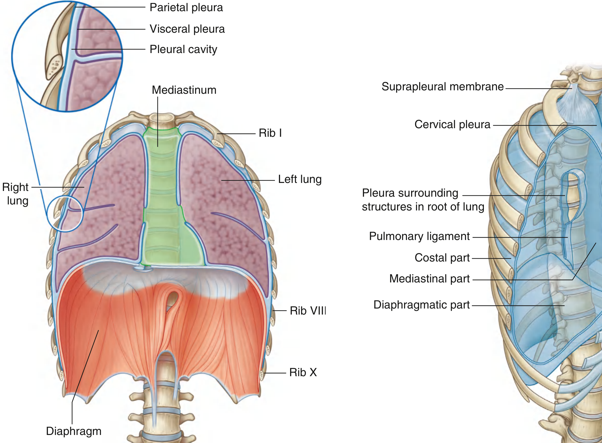
What this diagram shows:
- The difference between parietal pleura and visceral pleura (inset magnified view, top-left)
- The pleural cavity as the potential space between them
- The four named parts of parietal pleura: Costal, Diaphragmatic, Mediastinal, and Cervical (cupola)
- The suprapleural membrane at the apex
- The pulmonary ligament below the hilum
- Overall relationship of pleura to both lungs and the diaphragm
Diagram 2 - Pleural Recesses & Surface Markings (Exam Favourite!)
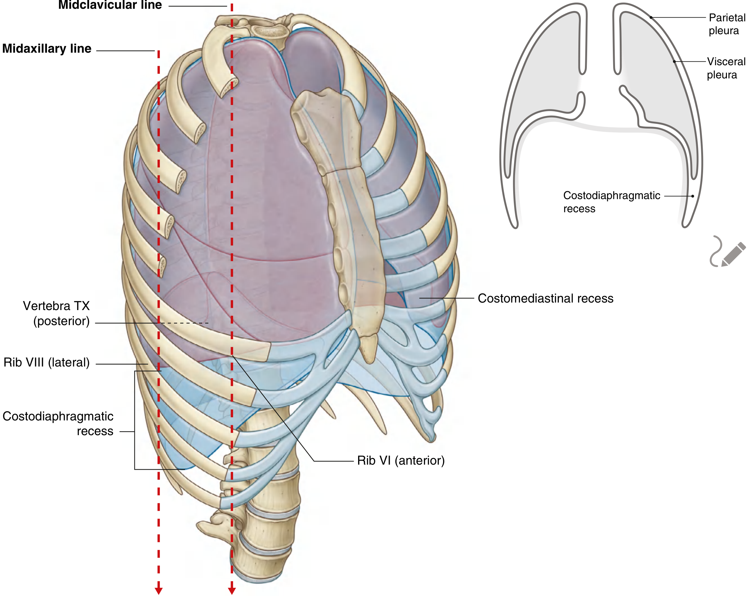
What this diagram shows:
- The costodiaphragmatic recess - the largest and most clinically important recess (site for pleural tapping)
- The costomediastinal recess anteriorly
- Surface marking lines you MUST memorize for exams:
| Line | Lung margin | Pleural cavity margin |
|---|---|---|
| Midclavicular | Rib VI | Rib VIII |
| Midaxillary | Rib VIII | Rib X |
| Posterior (vertebral) | T X | T XII |
Key Points to Write in Your Exam Answer
- Parietal pleura - somatic innervation (intercostal nerves for costal part; phrenic nerve for diaphragmatic and mediastinal parts) - PAIN SENSITIVE
- Visceral pleura - visceral afferent (bronchial vessels) - NOT pain sensitive
- Pleural tapping site - 9th intercostal space, midaxillary line (within costodiaphragmatic recess, above the rib to avoid neurovascular bundle)
- Cervical pleura / cupola - projects 2.5 cm above the medial one-third of the clavicle - can be injured in stab wounds to the root of the neck
Source: Gray's Anatomy for Students, Thorax chapter - Pleura section (pp. 201-203)
This is a shared conversation. Sign in to Orris to start your own chat.