
Tests to know do u have active hairfall
tests to diagnose active hair fall alopecia investigation
hair loss investigation pull test scalp examination alopecia
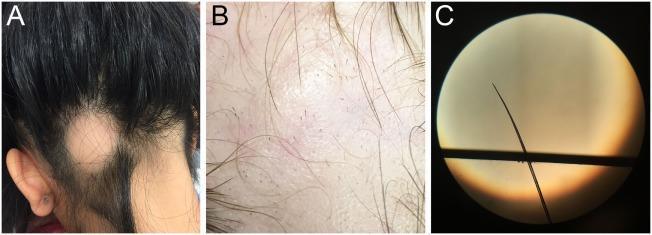
This composite educational image illustrates the clinical and microscopic features of alopecia areata (AA). Image A is a clinical photograph of a patient's posterior scalp, showing a single, well-demarcated, smooth patch of non-scarring hair loss located near the lower occiput and nape of the neck (ophiasis-like distribution). Image B provides a high-magnification view of the affected scalp, highlighting pathognomonic 'exclamation point hairs'—short, broken hairs that are narrower at the base near the scalp than at the distal end. Image C is a light microscopy frame of a hair shaft obtained via a pull test, demonstrating a classic tapered hair bulb characteristic of rapidly progressive AA. These visual findings are essential for distinguishing non-scarring autoimmune alopecia from other pediatric conditions like tinea capitis or trichotillomania. The educational focus is on identifying clinical morphology, trichoscopic markers, and microscopic hair shaft anomalies used in dermatologic diagnosis.
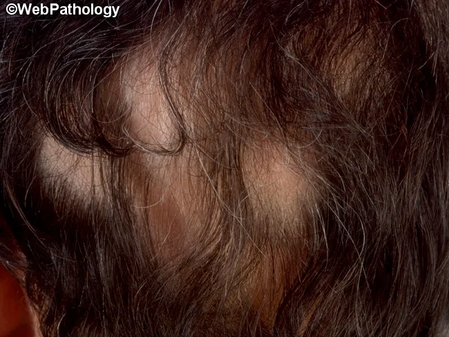
Clinical photography of the scalp reveals a single, well-demarcated patch of alopecia approximately X cm in diameter, with overlying mild to moderate scaling and marginal hair follicle disruption. The patch is hypo-hyperpigmented relative to surrounding skin. Hair within the patch is sparse with broken hair shafts. The lesion is round/oval, with a slightly raised, scaly border and no obvious crusting or pustulation. The surrounding scalp shows normal hair density and no additional patches. The appearance is classic for inflammatory hair loss due to dermatophyte infection (tinea capitis) or, less commonly, alopecia areata with background scale. The presence of scale and patchy hair loss increases suspicion for tinea capitis, particularly in children, and warrants mycological confirmation via KOH prep or fungal culture, as well as possibly wood lamp examination for fluorescing species. Differential diagnosis includes alopecia areata, seborrheic dermatitis, psoriasis, lupus-related scalp involvement, and traction alopecia. In clinical practice, the finding would prompt dermatologic examination, fungal testing, and initiation of antifungal therapy if confirmed. Clinically relevant features include perifollicular scaling, endothrix or ectothrix hair invasion not visible on gross inspection, and potential secondary bacterial infection if excoriation is present. This image demonstrates essential gross morphology for educational references and diagnostic pattern recognition.
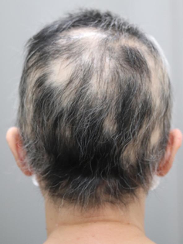
Clinical photograph showing the posterior view of a human scalp and neck, primarily illustrating Alopecia areata (AA). The image captures the crown, occipital region, and nape. Multiple non-scarring, circumscribed patches of hair loss are visible, varying in size from 1 to 3 centimeters. The affected scalp within these patches appears smooth, pale, and lacks visible inflammation, scaling, or scarring. Short, fine hairs are visible within some of the patches, consistent with the clinical stage of hair regrowth. The surrounding terminal hair is salt-and-pepper in color and appears normal in texture. This visual evidence supports the diagnosis of multifocal alopecia areata and demonstrates the progress of hair follicle regeneration following treatment, specifically 6 months post-thymectomy in the context of paraneoplastic autoimmune phenomena. The image serves as a clinical reference for dermatological examination and monitoring of autoimmune-related hair loss.
Tests to Know if You Have Active Hair Fall
1. Pull Test (Most Important Bedside Test)
2. Blood Tests (To Find the Cause)
| Test | What It Checks |
|---|---|
| CBC (Complete Blood Count) | Anaemia, infections |
| Serum Ferritin + Iron | Iron deficiency - one of the most common causes of hair fall |
| TSH, T3, T4 | Thyroid disorders (hypo/hyperthyroidism) both cause shedding |
| ANA (Antinuclear Antibody) | Lupus and autoimmune causes |
| Sex Hormones (Testosterone, LH, FSH) | Hormonal imbalance, PCOS in women |
| Prolactin | Elevated prolactin causes hair loss |
| Zinc + Total Protein/Albumin | Nutritional deficiencies |
| Blood Sugar / HbA1c | Diabetes-related hair loss |
| ESR + CRP | Systemic inflammation |
| VDRL (Syphilis) + HIV | Infectious causes of diffuse hair loss |
| Liver/Kidney function (AST, ALT, BUN/Cr) | Systemic disease |
3. Trichoscopy (Dermoscopy of the Scalp)
- Exclamation mark hairs - hallmark of alopecia areata
- Yellow dots - empty follicles
- Hair diameter diversity - sign of androgenetic (pattern) hair loss
- Peripilar signs - inflammation around follicles
4. Trichogram (Hair Root Microscopy)
5. Scalp Biopsy
- The diagnosis remains unclear after all other tests
- Scarring alopecia is suspected (irreversible hair loss)
- Terminal to vellus-like hair ratio needs to be measured
6. KOH (Potassium Hydroxide) Test + Fungal Culture
7. Urine Analysis
Clinical Image: Alopecia Areata Features
Summary: Which Tests Are Most Useful?
| Situation | Key Tests to Start With |
|---|---|
| General hair fall / shedding | Pull test + Ferritin + TSH + CBC |
| Patchy hair loss | Pull test + Trichoscopy + ANA |
| Female with diffuse thinning | Sex hormones + Prolactin + Ferritin + TSH |
| Scalp scaling + broken hairs | KOH test + Fungal culture |
| Unclear diagnosis | Scalp biopsy |