
differentiate cardiac and skeletal muscle properties of cardiac muscle and WBW syndrome
Wolff-Parkinson-White syndrome
Wolff Parkinson White syndrome ECG management guidelines accessory pathway
https://www.escardio.org/communities/councils/cardiology-pra…
1. Cardiac muscle versus skeletal muscle
| Property | Cardiac muscle | Skeletal muscle |
|---|---|---|
| Control | Involuntary | Voluntary |
| Site | Myocardium of heart | Attached to bones |
| Cell shape | Short, cylindrical, branched cells | Long, cylindrical, unbranched fibers |
| Nuclei | One, occasionally two, central nuclei | Many nuclei at the periphery |
| Striations / sarcomeres | Present | Present |
| Cell connection | Intercalated discs with desmosomes and gap junctions | Fibers are electrically separate; no intercalated discs |
| Functional organization | Functional syncytium: atria and ventricles act as separate syncytia | Each fiber is an anatomical syncytium, but fibers act independently |
| Initiation of contraction | Myogenic: SA node generates impulse automatically | Neurogenic: requires somatic motor-neuron stimulation |
| Rhythmicity | Automaticity and intrinsic rhythmic contraction present | No automaticity in normal muscle |
| Autonomic nerves | Modify rate, conduction, and force, but do not normally initiate beat | Motor nerves initiate contraction |
| Action potential | Long, with plateau due mainly to L-type Ca²⁺ current | Very short, no plateau |
| Refractory period | Long, nearly equal to contraction | Short |
| Tetanus / fatigue | Cannot be tetanized because of long refractory period | Can undergo summation and tetanus |
| Ca²⁺ source for contraction | Requires extracellular Ca²⁺ entry, which triggers SR Ca²⁺ release: calcium-induced calcium release | Mainly SR Ca²⁺ release; extracellular Ca²⁺ is not essential for each contraction |
| T-tubule and SR arrangement | T-tubules at Z line; dyads: one T-tubule + one terminal cisterna; SR less developed | T-tubules at A-I junction; triads: one T-tubule + two terminal cisternae; SR well developed |
| Force regulation | Depends strongly on initial fiber length and extracellular Ca²⁺, Frank-Starling mechanism | Mainly by motor-unit recruitment and frequency of stimulation |
| Fatigue | Highly fatigue-resistant, aerobic metabolism, many mitochondria | Variable, depending on fiber type |
2. WPW syndrome
Definition
- WPW pattern: ECG evidence of pre-excitation only, without symptomatic tachyarrhythmia.
- WPW syndrome: pre-excitation plus symptomatic arrhythmia, commonly AV re-entrant tachycardia (AVRT).
ECG findings in sinus rhythm
- Short PR interval: ≤120 ms
- Delta wave: slurred initial upstroke of QRS
- Wide QRS complex
Mechanism of tachycardia
- Orthodromic AVRT: impulse travels down through AV node and returns to atria via accessory pathway. Usually a regular narrow-complex SVT.
- Antidromic AVRT: impulse travels down the accessory pathway and returns via AV node. Usually a regular wide-complex tachycardia.
- Pre-excited atrial fibrillation: very rapid, irregular, often wide-complex rhythm. This can deteriorate into ventricular fibrillation and is an emergency.
Symptoms
- Palpitations
- Sudden episodes of rapid heartbeat
- Dizziness or syncope
- Chest discomfort
- Breathlessness
- Rarely, cardiac arrest
Management principles
- Unstable tachyarrhythmia: immediate synchronized cardioversion.
- Regular narrow-complex orthodromic AVRT: vagal maneuvers, then adenosine may be used under monitored acute-care conditions.
- Pre-excited AF or irregular wide-complex tachycardia: do not give AV-node blockers such as adenosine, verapamil/diltiazem, beta-blockers, digoxin, or IV amiodarone. They may preferentially direct conduction through the accessory pathway and worsen ventricular rate. Urgent cardioversion is needed if unstable; procainamide is commonly used when stable and appropriate.
- Definitive therapy for recurrent symptomatic WPW: electrophysiologic study and catheter ablation of the accessory pathway. The ESC summary lists symptomatic or recurrent AVRT as a Class I ablation indication.
Explain WPW ECG changes with a labeled diagram
Wolff Parkinson White syndrome ECG short PR interval delta wave wide QRS labeled diagram
Wolff-Parkinson-White syndrome ECG
Wolff Parkinson White ECG short PR delta wave wide QRS reputable cardiology
A clear educational labeled ECG diagram comparing normal sinus rhythm to Wolff-Parkinson-White pre-excitation. Focus on one enlarged WPW ECG trace with P wave, short PR interval, slurred delta wave at the start of the QRS, wide QRS complex, and normal ST-T segment labeled. Include a small simplified conduction schematic showing an accessory atrioventricular pathway bypassing the AV node and causing early ventricular activation. Labels must be medically accurate and easy to read.
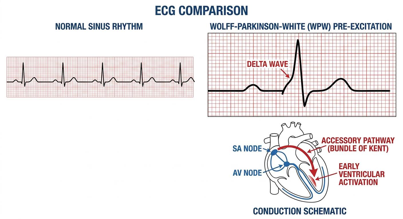
Why the ECG changes occur in WPW
1. Short PR interval: <120 ms
- The PR interval represents conduction from atrial depolarization to onset of ventricular depolarization.
- The AV node normally delays the impulse.
- In WPW, the impulse uses the accessory pathway and bypasses this delay.
- Therefore, ventricular activation begins earlier, giving a short PR interval.
2. Delta wave
- The earliest portion of ventricular muscle is activated slowly through the accessory pathway, not rapidly through the His-Purkinje system.
- This produces a slurred initial upstroke of the QRS complex, the delta wave.
- The delta wave is the ECG sign of early ventricular activation.
3. Wide QRS complex
- The QRS is widened because ventricular activation occurs by two routes at the same time:
- Early, slower activation through the accessory pathway
- Normal, fast activation via the AV node and His-Purkinje system
- This combined ventricular depolarization is called a fusion beat.
4. Secondary ST-T changes
- Because ventricular depolarization is abnormal, repolarization may also be abnormal.
- Therefore, there may be secondary ST-segment and T-wave changes. These should not automatically be interpreted as myocardial ischemia.
Exam summary
WPW ECG triad: short PR interval + delta wave + wide QRS complex.
| ECG feature | Mechanism |
|---|---|
| Short PR | Accessory pathway bypasses normal AV-nodal delay |
| Delta wave | Slow early activation of a portion of ventricle |
| Wide QRS | Fusion of pre-excited and normally conducted ventricular activation |
| ST-T change | Secondary repolarization abnormality |