
Coombs test
Reading File
The Coombs test (antiglobulin test) detects antibodies or complement proteins bound to red blood cells. There are two main types: the direct Coombs test (DAT), which detects antibodies already on RBCs, and the indirect Coombs test (IAT), which detects antibodies in serum.
Searching sources...
Finding Sources
Finding Sources
Good results. Let me read the key sections from Harrison's and Janeway's (which has the diagram), plus Goldman-Cecil.
Reading File
Reading File
Reading File
Excellent content. Now let me fetch the Coombs test diagrams from Janeway's and Harrison's, and also get the Robbins section on immunohemolytic anemia for clinical contexts/drug-induced causes.
Loading Image
Loading Image
Reading File
I now have comprehensive content from multiple authoritative textbooks and two excellent diagrams. Here is a thorough answer:
Coombs Test (Antiglobulin Test)
The Coombs test - also called the antiglobulin test (AGT) - detects antibodies (immunoglobulins) or complement components bound to red blood cell (RBC) surfaces. It was devised because certain anti-RBC antibodies (particularly IgG anti-Rh) are "incomplete" - they bind to RBCs but do not directly agglutinate them in vitro, making them undetectable by simple agglutination. The solution was to use a secondary anti-human immunoglobulin antibody (Coombs reagent) which bridges antibody-coated RBCs and causes visible agglutination.
Two Types
1. Direct Coombs Test (DAT - Direct Antiglobulin Test)
Principle: Detects antibody or complement already coating the patient's own RBCs in vivo.
Procedure:
- Take patient's RBCs from blood sample
- Wash them thoroughly (to remove unbound serum immunoglobulins)
- Add anti-human immunoglobulin reagent (Coombs serum - contains anti-IgG and anti-complement such as anti-C3/C4)
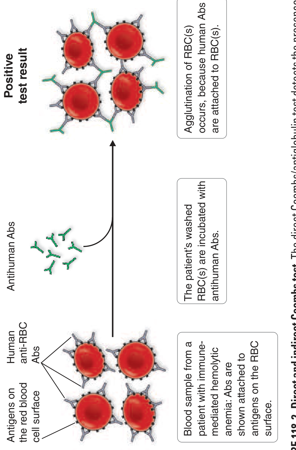
- Positive result = agglutination (the Coombs antibody bridges IgG/complement-coated RBCs)
2. Indirect Coombs Test (IAT - Indirect Antiglobulin Test)
Principle: Detects free antibodies in the patient's serum that would bind to RBCs.
Procedure:
- Take patient's serum
- Incubate with test RBCs of known antigen profile (e.g., Rh-positive cells)
- Wash away unbound antibody
- Add Coombs reagent (anti-human immunoglobulin)
- Positive result = agglutination (the test RBCs are now coated with patient's antibody, which is then bridged)
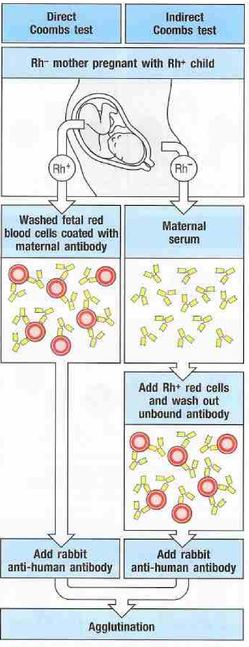
The diagram below (from Janeway's Immunobiology) shows both tests in the clinical context of Rh incompatibility:
The Coombs Reagent
The standard modern antiglobulin reagent (polyspecific) contains antibodies against:
- All four classes of IgG
- Complement components C3 and C4
Monospecific reagents targeting only IgG or only C3 can be used to identify which component is coating the RBCs - this has clinical significance (see table below).
Clinical Applications
| Situation | Test Used | Why |
|---|---|---|
| Autoimmune hemolytic anemia (AIHA) | DAT | Confirms IgG/complement on RBCs |
| Hemolytic disease of the fetus/newborn (HDN) | DAT (on cord blood) | Maternal IgG coating fetal RBCs |
| Pre-transfusion compatibility testing | IAT (crossmatch) | Detects recipient antibodies against donor RBCs |
| Prenatal antibody screening | IAT | Identifies anti-Rh or other alloantibodies in maternal serum |
| Drug-induced hemolytic anemia | DAT | Drug-antibody complexes or drug-induced autoantibodies on RBCs |
| Hemolytic transfusion reactions | DAT | Recipient antibodies coating transfused RBCs |
Immunohemolytic Anemia: DAT Patterns
Warm AIHA (70-80% of AIHA):
- Mediated by IgG (usually IgG1), active at 37°C
- DAT pattern: IgG-positive alone, or IgG + C3-positive
- Hemolysis is predominantly extravascular (splenic macrophages)
- Spherocytes visible on peripheral smear
- Causes: idiopathic (>60%), SLE, B-cell lymphomas/CLL, drugs (α-methyldopa)
Cold AIHA (15-20% of AIHA):
- Mediated by IgM cold agglutinins, active at <37°C (optimally 0-4°C)
- DAT pattern: C3-positive only (IgM itself elutes off at 37°C, leaving complement behind)
- Hemolysis is both intra- and extravascular
- Causes: Mycoplasma pneumoniae, infectious mononucleosis, lymphoplasmacytic lymphoma
Drug-Induced Mechanisms (DAT-Positive)
Three distinct mechanisms can produce a positive DAT with drugs:
- Hapten mechanism (e.g., penicillin): Drug binds covalently to RBC membrane proteins creating neoantigens; IgG anti-drug antibody forms and coats RBCs.
- Immune complex ("innocent bystander") (e.g., quinidine): Drug-antibody immune complexes deposit on RBC surface, fixing complement -> hemolysis.
- True autoantibody induction (e.g., α-methyldopa): Drug alters immunogenicity of native RBC epitopes (particularly Rh antigens), inducing genuine autoantibodies that persist even after stopping the drug.
Interpreting Results
False positives (DAT): Not uncommon. Incidence in normal blood donors: 1 in 100 to 1 in 15,000 depending on technique. Causes include:
- Nonspecific low-avidity IgG adherence to RBCs
- SLE or B-cell malignancies producing true auto-antibodies without active hemolysis
False negatives (DAT): Rare (0-5% of true AIHA). Causes:
- Low-affinity autoantibodies that elute off RBCs in vitro
- IgA antibodies (standard reagents primarily detect IgG)
- Antibody levels below the detection threshold of the test
A positive DAT alone does not diagnose AIHA - it must be interpreted alongside clinical and other lab findings (reticulocytosis, elevated LDH, low haptoglobin, indirect hyperbilirubinemia, spherocytes on smear).
Sources:
- Harrison's Principles of Internal Medicine, 22nd Ed. (2025), p. 954
- Goldman-Cecil Medicine, p. 1688
- Robbins & Kumar Basic Pathology, p. 391
- Janeway's Immunobiology, 10th Ed., Appendix A
- Washington Manual of Medical Therapeutics, Diagnosis of hemolysis
Coombs test
Reading File
The Coombs test detects antibodies or complement on RBCs. Two main types: the Direct Antiglobulin Test (DAT) finds antibody already on RBCs; the Indirect Antiglobulin Test (IAT) finds antibody in serum.
Searching sources...
Finding Sources
Finding Sources
Reading File
Reading File
Reading File
Now let me fetch the key diagrams in parallel:
Loading Image
Loading Image
Coombs Test (Antiglobulin Test)
Background & Why It's Needed
Certain anti-RBC antibodies - especially IgG anti-Rh antibodies - are "incomplete": they bind to RBCs but do not directly agglutinate them because their Fab arms cannot bridge the wide spacing of Rh antigens on adjacent cells. This made them invisible to simple agglutination tests. The Coombs test solves this by adding a secondary anti-human immunoglobulin antibody (the Coombs reagent) that bridges IgG-coated RBCs together, producing visible agglutination.
The modern standard Coombs reagent contains antibodies against all four IgG subclasses plus complement components C3 and C4.
Two Types
1. Direct Coombs Test (DAT - Direct Antiglobulin Test)
Detects antibody or complement already coating the patient's RBCs in vivo.
Steps:
- Collect patient's blood and isolate RBCs
- Wash thoroughly - removes free (unbound) immunoglobulin from serum
- Add Coombs reagent (anti-human IgG + anti-complement)
- Positive = agglutination (antibody-coated RBCs get cross-linked)
The Harrison's diagram below shows the DAT mechanism - antibodies on the RBC surface get bridged by antihuman antibodies, causing clumping:
2. Indirect Coombs Test (IAT - Indirect Antiglobulin Test)
Detects free antibodies in the patient's serum that would coat RBCs if present.
Steps:
- Take patient's serum
- Incubate with test RBCs of known antigen profile (e.g., Rh+ cells)
- Wash to remove unbound antibody
- Add Coombs reagent
- Positive = agglutination (serum antibody has now coated test RBCs, which are then bridged)
The Janeway's diagram below shows both tests side by side in the clinical context of Rh incompatibility:
Key difference in one line: DAT = antibody on the patient's cells; IAT = antibody in the patient's serum.
Clinical Applications
| Clinical Situation | Test | Purpose |
|---|---|---|
| Autoimmune hemolytic anemia (AIHA) | DAT | Confirms IgG/complement on patient's RBCs |
| Hemolytic disease of newborn (HDN) | DAT on cord blood | Detects maternal IgG coating fetal RBCs |
| Hemolytic transfusion reaction | DAT | Recipient antibodies coating transfused donor RBCs |
| Drug-induced hemolytic anemia | DAT | Drug-antibody complexes or autoantibodies on RBCs |
| Pre-transfusion crossmatch | IAT | Screens recipient serum for alloantibodies vs. donor RBCs |
| Prenatal Rh antibody screening | IAT | Detects anti-Rh (or other) alloantibodies in maternal serum |
DAT Pattern in Immunohemolytic Anemia
| Type | Antibody | Temperature | DAT Pattern | Hemolysis Site |
|---|---|---|---|---|
| Warm AIHA (70-80%) | IgG (usually IgG1), rarely IgA | 37°C | IgG+ alone, or IgG+ + C3+ | Extravascular (spleen) |
| Cold AIHA (15-20%) | IgM (cold agglutinins) | <37°C (best 0-4°C) | C3+ only (IgM elutes off at 37°C) | Intra- and extravascular |
| Mixed AIHA (~5%) | Both IgG and IgM | Both temperatures | IgG+ + C3+ | Mixed |
| PCH (rare) | IgG Donath-Landsteiner Ab | Cold (binds), warm (lyses) | C3+ | Intravascular |
Causes of Positive DAT
Warm AIHA:
- Primary/idiopathic (>60%)
- Systemic lupus erythematosus
- CLL and other B-cell neoplasms
- Drugs (α-methyldopa, penicillin, quinidine)
Cold AIHA:
- Acute: Mycoplasma pneumoniae infection, infectious mononucleosis
- Chronic: idiopathic, lymphoplasmacytic lymphoma
Drug-Induced Mechanisms (Three Distinct Pathways)
-
True autoantibody induction (e.g., α-methyldopa): The drug alters the immunogenicity of native RBC epitopes (especially Rh antigens), bypassing T-cell tolerance and inducing genuine autoantibodies - which persist even after stopping the drug.
-
Hapten mechanism (e.g., penicillin): Drug binds covalently to RBC membrane proteins, creating neoantigens. IgG antibodies form against the drug-protein complex and coat the RBCs.
-
Immune complex ("innocent bystander") (e.g., quinidine): Drug-antibody immune complexes form in circulation and deposit onto RBC surfaces, fixing complement or acting as opsonins - causing hemolysis.
Interpreting Results: Pitfalls
False-positive DAT - not uncommon:
- Incidence in normal blood donors: 1 in 100 to 1 in 15,000 (varies with technique)
- Most common cause: nonspecific low-avidity IgG adhering to RBCs
- SLE and B-cell malignancies can produce true anti-RBC antibodies without active hemolysis
- A positive DAT alone does not diagnose AIHA - must correlate with clinical and lab findings (reticulocytosis, elevated LDH, low haptoglobin, indirect hyperbilirubinemia, spherocytes on smear)
False-negative DAT - rare (0-5% of true AIHA):
- Low-affinity autoantibodies that elute off RBCs at 37°C in vitro
- IgA antibodies (standard reagents primarily detect IgG)
- Antibody below the detection threshold (~200-500 IgG molecules per RBC needed)
Sources: Harrison's Principles of Internal Medicine, 22nd Ed. (2025); Goldman-Cecil Medicine, p. 1688; Robbins & Kumar Basic Pathology, p. 391; Janeway's Immunobiology, 10th Ed., Appendix A
What are incomplete antibodies
"Incomplete antibodies" are antibodies (typically IgG) that bind to RBC antigens but fail to cause direct agglutination in saline - they "coat" the cells without crosslinking them. The classic example is IgG anti-Rh antibody.
Searching sources...
Finding Sources
Finding Sources
Finding Sources
Finding Sources
Finding Sources
Finding Sources
Finding Sources
Finding Sources
Reading File
Reading File
Good textbook content found. I have enough to give a thorough, accurate answer directly from the sources.
Incomplete Antibodies
Definition
"Incomplete antibodies" is an older serological term for antibodies - most typically IgG - that bind to antigens on red blood cell surfaces but fail to cause direct agglutination in saline. They "coat" the cells without crosslinking them, so they are invisible to a simple agglutination test.
The term contrasts with "complete antibodies" (typically IgM), which do directly agglutinate RBCs in saline.
Why IgG Fails to Agglutinate (The Mechanism)
There are two interrelated reasons:
1. Antigen Spacing on the RBC Surface
Rh antigens (the classic example) are widely spaced on the RBC surface. For agglutination to occur, one antibody molecule must simultaneously bind antigens on two different cells, physically bridging them. IgG, being relatively small (monomer, ~150 kDa with a short Fc-Fab span of ~12-14 nm), cannot reach across the gap between two adjacent RBCs to bridge them.
2. The Zeta Potential Barrier
Red blood cells carry a net negative surface charge (from sialic acid residues on glycophorins), which generates an electrostatic repulsion force - called the zeta potential - that keeps cells ~25 nm apart in saline suspension. IgM, being a large pentamer (~900 kDa) with long reach, can overcome this gap and bridge two cells. IgG cannot.
In contrast, ABO blood group antigens are densely expressed and more accessible, so even IgM antibodies (and sometimes IgG) can agglutinate ABO-incompatible cells directly.
As Janeway's Immunobiology states: "Rh antigens are widely spaced on the red blood cell surface, and so the IgG anti-Rh antibodies do not bind in the correct conformation... [and] do not agglutinate red blood cells, unlike antibodies against the ABO blood-group antigens."
IgG vs. IgM: Complete vs. Incomplete
| Feature | IgM ("Complete") | IgG ("Incomplete") |
|---|---|---|
| Structure | Pentamer (10 antigen-binding sites) | Monomer (2 antigen-binding sites) |
| Size | ~900 kDa, large reach | ~150 kDa, short reach |
| Overcomes zeta potential? | Yes | No |
| Direct agglutination in saline | Yes | No |
| Crosses placenta? | No | Yes |
| Example | Anti-A, anti-B (ABO system) | Anti-D (Rh system) |
| Detection | Direct agglutination test | Needs Coombs test |
Clinical Significance
Because incomplete (IgG) antibodies do not agglutinate, they were historically undetectable by simple tube agglutination. This was a serious problem because:
- Anti-Rh (anti-D) IgG from a sensitized Rh- mother crosses the placenta, coats fetal Rh+ RBCs, and causes hemolytic disease of the newborn (HDN) - even though the antibody cannot be seen on a standard agglutination test.
- Alloantibodies from prior transfusions (IgG class) can destroy transfused cells without triggering obvious saline agglutination.
The Coombs test was specifically devised to detect incomplete antibodies:
- The direct Coombs test reveals IgG coating the patient's own RBCs
- The indirect Coombs test detects free IgG in serum that would coat target RBCs
Adding anti-human immunoglobulin (Coombs reagent) bridges the IgG-coated RBCs together, finally producing visible agglutination even though the original IgG antibody was "incomplete."
Ways to Make Incomplete Antibodies Complete (Convert to Agglutination)
Several techniques can artificially promote agglutination by IgG antibodies, by either reducing the zeta potential barrier or bringing cells closer together:
- Albumin (22% bovine albumin): Reduces the zeta potential by lowering the dielectric constant of the medium, allowing cells to come closer together
- Enzyme treatment (papain, ficin, bromelin): Cleaves sialic acid-bearing glycoproteins from the RBC surface, reducing the negative charge and zeta potential
- Low ionic strength saline (LISS): Reduces electrostatic repulsion, increasing antibody uptake
- Polyethylene glycol (PEG): Concentrates antibody and promotes binding
- Coombs/antiglobulin technique: The gold standard - adds anti-IgG to bridge already-bound antibodies
Summary
Incomplete antibodies = IgG antibodies that bind RBC antigens but cannot directly agglutinate cells due to the physical constraints of IgG size vs. the zeta potential gap between RBCs. They are clinically important because they cause hemolytic disease (HDN, transfusion reactions, AIHA) while being invisible to conventional agglutination testing - requiring the Coombs test for detection.
Sources: Janeway's Immunobiology, 10th Ed., pp. 826-828; Goldman-Cecil Medicine, p. 1688
How is HDN prevented
HDN (hemolytic disease of the newborn) is prevented primarily by giving Rh-negative mothers anti-D immunoglobulin (Rh immune globulin / RhoGAM) to prevent sensitization to fetal Rh+ RBCs.
Searching sources...
Finding Sources
Finding Sources
Finding Sources
Finding Sources
Reading File
Reading File
Reading File
Finding Sources
Excellent - I have comprehensive content from multiple textbooks. Here is the full answer:
Prevention of HDN (Hemolytic Disease of the Newborn)
Understanding the Problem First
HDN (also called erythroblastosis fetalis) arises when an Rh-negative mother carries an Rh-positive fetus. Fetal Rh+ RBCs enter the maternal circulation - mainly at delivery - and trigger an immune response. The mother produces IgG anti-D antibodies, which cross the placenta in a subsequent pregnancy and attack the fetal RBCs, causing hemolysis.
Key point: the first Rh+ pregnancy usually does not cause harm (insufficient sensitization), but the risk rises progressively: ~3% of second babies, ~10% of third babies show signs of erythroblastosis fetalis.
The Cornerstone of Prevention: Rh Immunoglobulin (Anti-D Ig / RhoGAM)
The single most important preventive measure, developed in the 1970s, which dramatically reduced the incidence of HDN worldwide.
What it is: A preparation of exogenous IgG anti-D antibody (passive immunization) given to Rh-negative mothers.
When It Is Given
| Timing | Reason |
|---|---|
| 28 weeks of gestation (antenatal) | Prevents sensitization from any transplacental fetal cell leakage during late pregnancy - even during the first pregnancy |
| Within 72 hours of delivery (if baby is Rh+) | Clears fetal Rh+ RBCs from maternal circulation before the mother can mount an active immune response |
| After abortion (spontaneous or therapeutic) | Fetal cells may enter maternal circulation at any termination |
| After any sensitizing event | Amniocentesis, chorionic villus sampling, external cephalic version, antepartum hemorrhage, trauma |
How Anti-D Immunoglobulin Works (Mechanism)
The exact mechanism is not completely understood, but two effects are recognized:
-
Inhibition of B-lymphocyte activation: The administered anti-D antibody suppresses antigen-induced B-cell antibody production in the mother - a form of antibody-mediated immune suppression (AMIS). The pre-formed antibody signals that the antigen is already "handled," dampening the primary immune response.
-
Rapid clearance of fetal RBCs: Anti-D antibody attaches to D antigens on any Rh+ fetal RBCs that crossed into the maternal circulation. The opsonized cells are rapidly cleared by maternal macrophages before the immune system can recognize them and mount a response.
As Guyton & Hall explains: "The administered anti-D antibody... attaches to D antigen sites on Rh-positive fetal RBCs that may cross the placenta and enter the circulation of the expectant mother, thereby interfering with the immune response to the D antigen."
Antenatal Screening Protocol
- Early pregnancy: Test all women for blood group and Rh status
- If Rh-negative and partner is Rh-positive: Monitor anti-D antibody titers throughout pregnancy (at booking, 28 weeks, 34-36 weeks)
- If already sensitized (rising antibody titers): Refer to specialist centre - prevention is no longer possible; management shifts to monitoring and treatment
If Already Sensitized: Management of the Affected Fetus/Neonate
Once sensitization has occurred, anti-D immunoglobulin cannot help. Management then focuses on:
| Situation | Intervention |
|---|---|
| Severe fetal anemia in utero | Intrauterine transfusion (Rh-negative blood into fetal umbilical vein) |
| Neonate with HDN | Exchange transfusion with Rh-negative blood - replaces the baby's antibody-coated Rh+ cells; performed over 1.5+ hours, repeated as needed |
| Hyperbilirubinemia | Phototherapy (converts bilirubin to water-soluble isomers for excretion) |
| Goal of exchange transfusion | Keep bilirubin low to prevent kernicterus (bilirubin deposition in basal ganglia and brain causing permanent neurological damage) |
The maternal anti-Rh agglutinins circulate in the infant for 1-2 months after birth, continuing to destroy RBCs. The exchanged Rh-negative cells are gradually replaced by the infant's own Rh+ cells over ~6 weeks, by which time the maternal antibodies have been destroyed.
Why ABO Incompatibility Causes Milder HDN
Anti-A and anti-B antibodies are predominantly IgM - they cannot cross the placenta. This is why ABO incompatibility between mother and fetus rarely causes serious HDN, and why anti-D immunoglobulin specifically targets the Rh system.
Summary
| Step | Action |
|---|---|
| All pregnant women | Screen Rh status at booking |
| Rh-negative, unsensitized | Anti-D Ig at 28 weeks + within 72 hours of delivery if baby is Rh+ |
| Any sensitizing event | Anti-D Ig promptly after each event |
| Already sensitized | Specialist monitoring, intrauterine transfusion if needed, exchange transfusion at birth |
Sources: Guyton & Hall Textbook of Medical Physiology, pp. 477-478; Park's Textbook of Preventive and Social Medicine, p. 617; Janeway's Immunobiology, 10th Ed., p. 827
Blood grouping
Blood grouping classifies RBCs based on surface antigens. The two most clinically important systems are ABO and Rh. ABO is determined by carbohydrate antigens; Rh by protein antigens (mainly D).
Searching sources...
Finding Sources
Finding Sources
Finding Sources
Finding Sources
Reading File
Reading File
Reading File
Reading File
Now let me fetch the ABO antigen structure diagram and get the Guyton content on blood groups + the Quick Compendium on other blood group systems:
Loading Image
Loading Image
Finding Sources
Excellent - rich content and two great diagrams. I have everything needed for a comprehensive answer.
Blood Grouping
Blood grouping (blood typing) is the classification of blood based on the presence or absence of specific antigens on the surface of red blood cells. The two most clinically critical systems are ABO and Rh (Rhesus).
1. ABO Blood Group System
The Antigens
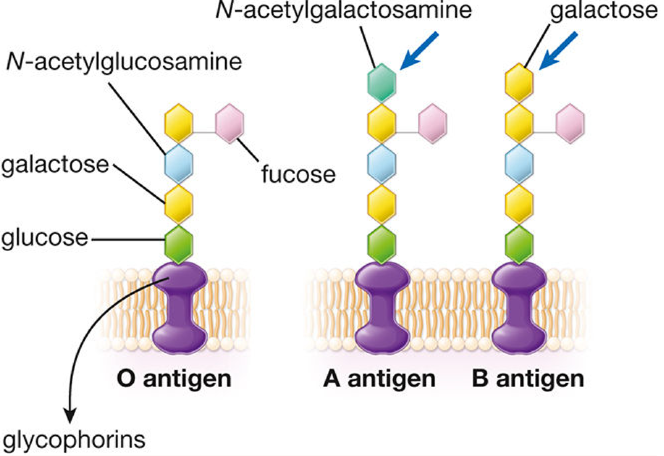
ABO antigens are not proteins - they are carbohydrate structures (oligosaccharide chains) attached to glycoproteins and glycolipids on the RBC surface (mainly on glycophorins and band 3 proteins). They are the products of glycosyltransferase enzymes, not direct gene products.
The biosynthetic pathway:
- All individuals make a core glycan attached to membrane proteins
- A fucosyltransferase (H-gene, chromosome 19) adds fucose to create the H antigen (the backbone)
- A single gene on chromosome 9 encodes a glycosyltransferase with three allele variants:
- A allele - adds N-acetylgalactosamine (GalNAc) to H antigen → A antigen
- B allele - adds galactose to H antigen → B antigen
- O allele - devoid of enzymatic activity → H antigen remains unmodified; no A or B antigen
The diagram below shows the molecular structures of the O, A, and B antigens attached to glycophorin in the RBC membrane:
Genetics: Codominance
The A and B alleles are codominant; O is recessive. This gives:
| Genotype | Blood Type |
|---|---|
| OO | O |
| AO or AA | A |
| BO or BB | B |
| AB | AB |
The Four Blood Groups + Their Antibodies
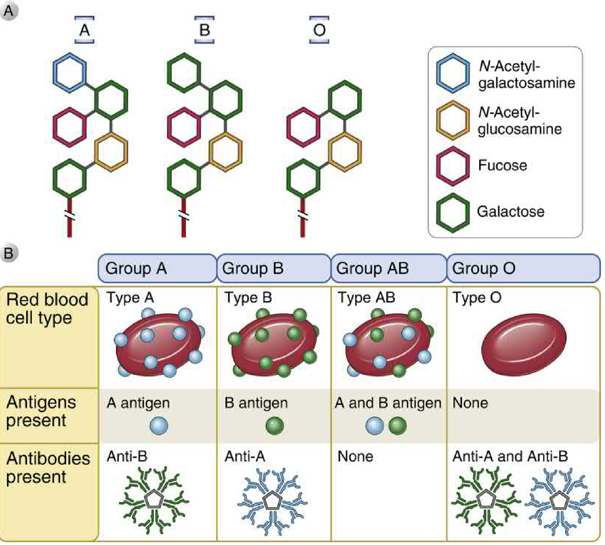
Individuals are tolerant to their own antigens and do not make antibodies against them. However, they have pre-formed "natural" IgM antibodies (likely raised against cross-reactive bacterial glycolipids in the gut) against the antigens they lack:
| Blood Group | Antigen on RBC | Antibody in Serum | Can Donate To | Can Receive From |
|---|---|---|---|---|
| A | A antigen | Anti-B (IgM) | A, AB | A, O |
| B | B antigen | Anti-A (IgM) | B, AB | B, O |
| AB | A and B antigens | None | AB only | A, B, AB, O (Universal recipient) |
| O | Neither A nor B (H antigen only) | Anti-A and Anti-B | A, B, AB, O (Universal donor) | O only |
Note: "Universal donor/recipient" is a simplification - other blood group antigens must still be considered in practice.
Why ABO Antibodies Are IgM (Not IgG)
ABO antibodies are predominantly IgM - this is clinically important because:
- IgM cannot cross the placenta → ABO incompatibility between mother and fetus generally does not cause serious HDN
- IgM is large and efficiently activates complement → ABO-incompatible transfusions cause acute intravascular hemolysis with complement activation, potentially leading to acute renal tubular necrosis, DIC, shock, and death
2. Rh Blood Group System
The Antigen
The Rh system is based on a 40 kDa non-glycosylated transmembrane polypeptide (Rh30), which is part of a larger 90 kDa complex that includes Rh50 glycoprotein. Unlike ABO, Rh antigens are protein-based.
Five clinically significant Rh antigens exist: D, C, c, E, e
- The D antigen is the most immunogenic and clinically dominant
- Rh-positive (Rh+): expresses the D antigen
- Rh-negative (Rh-): lacks the D antigen
Key Difference from ABO
| Feature | ABO | Rh |
|---|---|---|
| Antigen chemistry | Carbohydrate | Protein |
| Natural antibodies? | Yes (pre-formed IgM) | No - only form after exposure (transfusion/pregnancy) |
| Antibody class | IgM | IgG |
| Crosses placenta? | No (IgM) | Yes (IgG) → causes HDN |
| Reaction to first exposure | Immediate (pre-formed Ab) | No immediate reaction; sensitization occurs |
3. Blood Typing Procedure
Blood grouping involves two complementary tests:
Forward (Cell) Typing
- Patient's RBCs are mixed with known anti-A and anti-B sera
- Agglutination pattern identifies which antigens are on the cells
Reverse (Serum/Back) Typing
- Patient's serum is mixed with known group A and group B RBCs
- Confirms the expected antibodies in serum
- Results should be complementary to forward typing
Crossmatch (Pre-Transfusion)
- Recipient's serum is tested against donor's RBCs
- No reaction = compatible; reaction = incompatible - do not transfuse
4. Other Clinically Relevant Blood Group Systems
Over 30 blood group systems are recognized, but most cause only mild reactions (usually only after repeated transfusions triggering a secondary IgG response):
| System | Key Antigens | Notes |
|---|---|---|
| Kell | K, k | Highly immunogenic; K is second only to D in importance for transfusion |
| Duffy | Fy^a, Fy^b | Duffy-null (common in African populations) protects against Plasmodium vivax malaria |
| Kidd | Jk^a, Jk^b | Notorious for causing delayed hemolytic transfusion reactions |
| MNS | M, N, S, s | Generally weak; rarely cause severe reactions |
| Lewis | Le^a, Le^b | Carbohydrate antigens; also act as selectin ligands (E/P-selectin) in leukocyte migration; rarely cause strong transfusion reactions |
5. Clinical Importance of Blood Grouping
- Transfusion safety: ABO/Rh compatibility is mandatory; mismatched ABO transfusion can be rapidly fatal
- HDN prevention: Rh typing + anti-D immunoglobulin administration
- Organ transplantation: ABO antigens are expressed on endothelial cells; ABO incompatibility causes hyperacute rejection
- Forensics: ABO typing used in paternity testing and forensic identification
Sources: Cellular and Molecular Immunology (Abbas), pp. 1156-1160; Histology: A Text and Atlas (Ross), pp. 681-683; Guyton & Hall Textbook of Medical Physiology, p. 477
This is a shared conversation. Sign in to Orris to start your own chat.