
DIC
"disseminated intravascular coagulation" AND management
Disseminated Intravascular Coagulation (DIC)
Definition
Pathophysiology
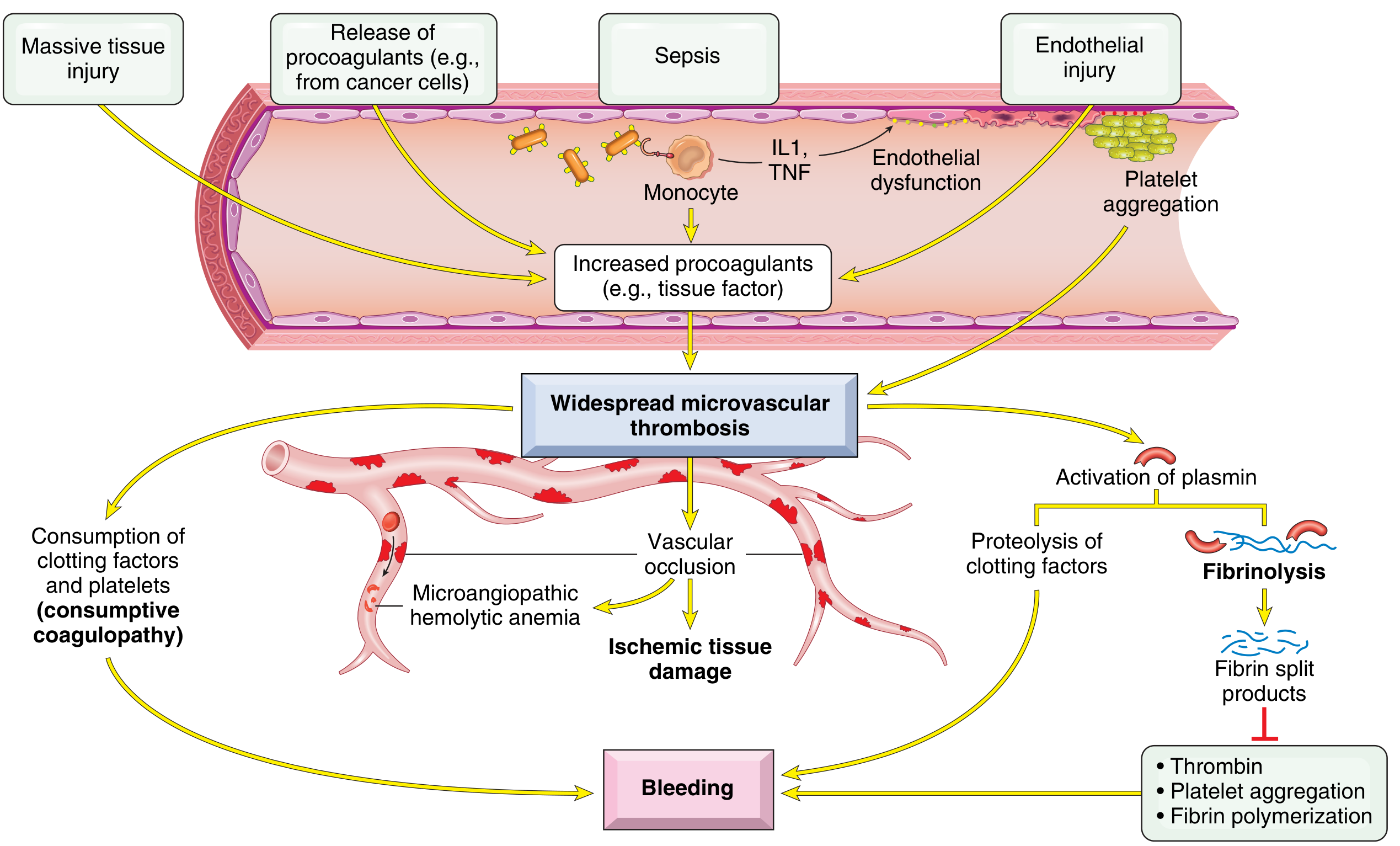
- Obstetric complications (placenta, amniotic fluid)
- Cancer cells (especially acute promyelocytic leukemia, adenocarcinoma)
- Massive tissue injury releasing thromboplastic material
- In bacterial sepsis: endotoxins/exotoxins stimulate tissue factor expression on monocytes. Activated monocytes release IL-1 and TNF, which simultaneously stimulate tissue factor expression on endothelial cells AND downregulate thrombomodulin (which activates protein C, an anticoagulant)
- Antigen-antibody complex deposition (e.g., SLE)
- Temperature extremes (heat stroke, burns)
- Meningococci, rickettsiae
- Deposits fibrin in small vessels of multiple organs
- Consumes platelets and clotting factors (especially fibrinogen, factors V, VIII, XIII) - consumptive coagulopathy
- Activates plasminogen activators - secondary fibrinolysis
- Cleaves fibrin and factors V and VIII
- Produces fibrin degradation products (FDPs/D-dimers) that: inhibit platelet aggregation, exert anti-thrombin activity, and impair fibrin polymerization - all worsening bleeding
Causes / Triggers
| Category | Examples |
|---|---|
| Obstetric | Abruptio placentae, amniotic fluid embolism, retained dead fetus (prothrombotic DIC), septic abortion, eclampsia, HELLP syndrome |
| Infections | Sepsis (gram-negative > gram-positive), meningococcemia, Rocky Mountain spotted fever, fungemia, malaria |
| Malignancy | Acute promyelocytic leukemia (APL), pancreatic/prostate/lung/gastric carcinoma |
| Massive tissue injury | Trauma (especially brain), burns, extensive surgery, aortic surgery with hypothermia |
| Miscellaneous | Acute intravascular hemolysis, snakebite, giant hemangioma, heat stroke, shock, liver disease, vasculitis, aortic aneurysm |
Note: Retained dead fetus causes a prothrombotic (not hemorrhagic) DIC - normal to shortened APTT (elevated FVIII as acute-phase reactant), mildly reduced platelets, normal/elevated fibrinogen, but elevated D-dimers.
Clinical Features
- Oozing from venipuncture sites, wounds, surgical incisions
- Mucosal bleeding (gums, GI tract)
- Petechiae and ecchymoses
- In severe cases: major hemorrhage
- Purpura fulminans (skin necrosis from dermal vessel thrombosis)
- Acral ischemia / digit gangrene
- Renal failure (glomerular fibrin thrombi)
- Altered mental status, focal neurological deficits
- Adrenal hemorrhage (Waterhouse-Friderichsen in meningococcemia)
Morphology (Autopsy Findings)
- Kidneys - glomerular fibrin thrombi, cortical necrosis in severe cases
- Adrenals - bilateral hemorrhagic infarction
- Brain - microinfarcts
- Heart - subendocardial microthrombi
- No organ is spared
Laboratory Diagnosis
| Test | Findings | Score |
|---|---|---|
| Platelet count | >100k = 0; <100k = 1; <50k = 2 | 0-2 |
| D-dimer (fibrin marker) | <0.4 µg/mL = 0; 0.4-4.0 = 2; >4.0 = 3 | 0-3 |
| PT prolongation | <3 sec = 0; 3-6 sec = 1; >6 sec = 2 | 0-2 |
| Fibrinogen | >100 mg/dL = 0; <100 mg/dL = 1 | 0-1 |
- Score ≥5 = compatible with overt DIC (repeat daily)
- Score <5 = suggestive of non-overt DIC (repeat in 1-2 days)
| Test | Finding | Mechanism |
|---|---|---|
| Peripheral smear | Low platelets, schistocytes, RBC fragments | RBC fragmentation on fibrin strands |
| Platelet count | Low (usually <100,000/mm³) | Consumed in clotting |
| PT | Prolonged | Factors II and IV consumed |
| PTT | Prolonged | Factors II, V, VIII consumed |
| Thrombin time | Prolonged | Decreased fibrinogen; in vivo fibrinolysis |
| Fibrinogen | Low (but may be deceptively normal as acute-phase reactant) | Consumed |
| D-dimer/FDPs | Elevated | Secondary fibrinolysis |
| Creatinine/UA | May be abnormal | Renal microvascular fibrin deposition |
Differential Diagnosis
| Condition | Key Distinguishing Features |
|---|---|
| Severe liver disease | Clinical jaundice, splenomegaly; factor VIII is normal (not synthesized by liver, so not low in liver failure); no fibrinolysis |
| Primary fibrinolysis | Rare; affects fibrinogen and fibrin but generally preserves platelets, factor V, and factor VIII in low-normal range |
| TTP/HUS | Thrombocytopenia + MAHA but coagulation tests (PT, PTT) usually normal |
| HELLP syndrome | Overlaps with DIC; if not resolved by day 3 postpartum, consider TTP |
Management
Step 1: Treat the underlying cause (always first)
Step 2: Specific intervention based on clinical picture
- Fresh frozen plasma (FFP) - corrects multiple factor deficiencies
- Cryoprecipitate - maintain fibrinogen >100 mg/dL
- Platelet transfusion - maintain count >20 × 10⁹/L
- Monitor: slowing of bleeding, falling FDPs, rising platelet count and fibrinogen
- Note: In non-bleeding patients with only lab DIC, do not give blood products - this can "add fuel to the fire" by exacerbating microthrombus formation
- Heparin (unfractionated or LMWH) is indicated
- Specific indications: purpura fulminans, retained dead fetus, giant hemangioma, APL
- Low-dose UFH (3-5 IU/kg/hour) in non-bleeding patients with lab DIC can help neutralize thrombin
- Heparin is not beneficial in: meningococcemia, abruptio placentae, severe liver disease, trauma
- Antithrombin III concentrate
- Prothrombin complex concentrate (PCC)
- Recombinant factor VIIa
- Activated protein C
Monitoring
- Repeat ISTH DIC score daily (in overt DIC)
- Track: fibrinogen, platelet count, PT/aPTT, D-dimers
- Normalization of clotting times occurs later in the course - less useful as an early monitor
- Also track renal function (common early organ impact)
Recent Evidence (2025)
- Robbins & Kumar Basic Pathology, p. 427
- Henry's Clinical Diagnosis and Management by Laboratory Methods, p. 954
- Rosen's Emergency Medicine, Chapter 111
Can you help to memorise the above easily by mind mapping or picturisation method?
What format would you like for the memory aid?
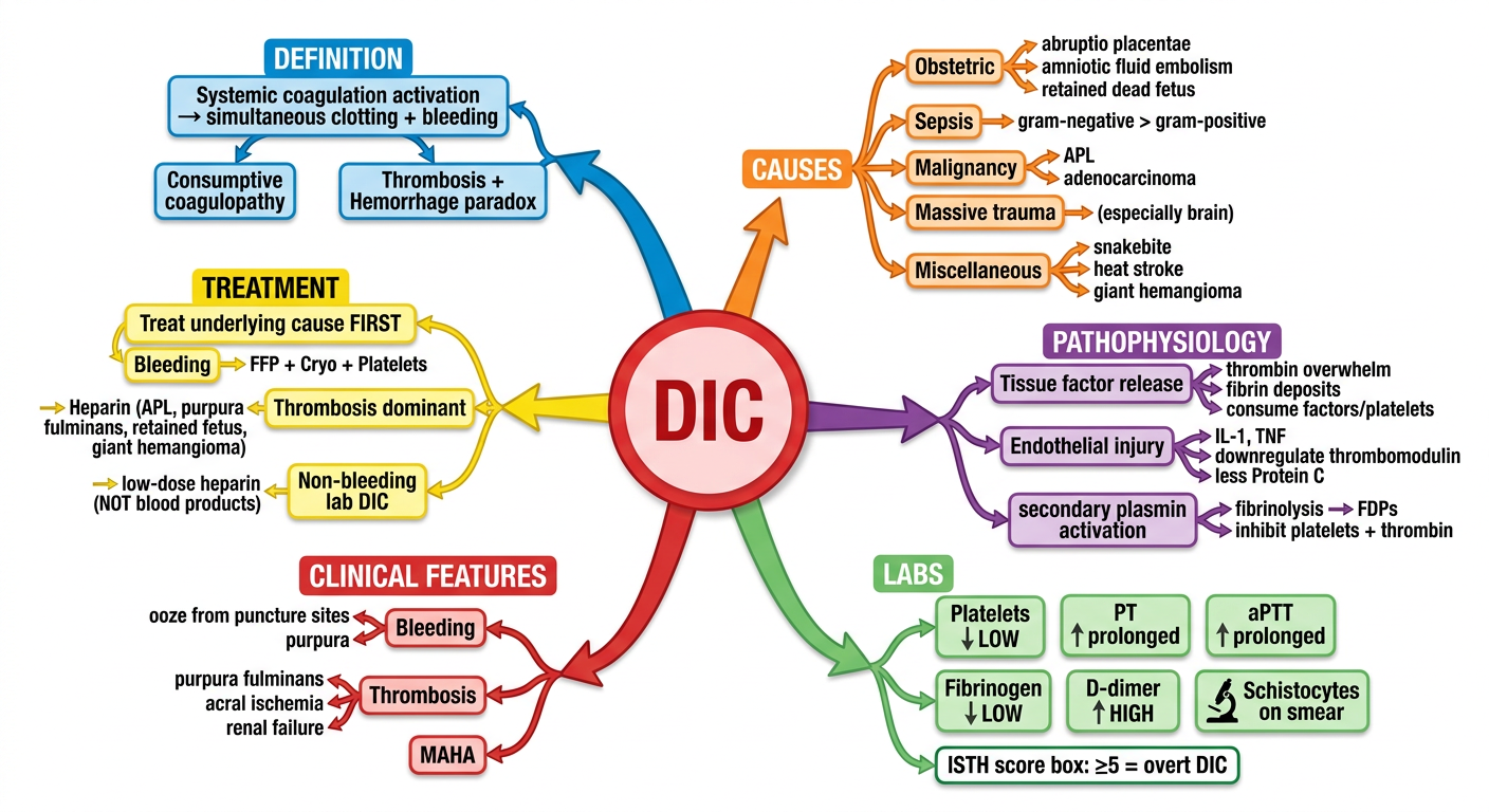
A detailed, colorful medical mind map for DIC (Disseminated Intravascular Coagulation) for medical students. Central node labeled "DIC" in bold red. Six main branches radiating outward: 1. DEFINITION branch (blue): "Systemic coagulation activation → simultaneous clotting + bleeding" with sub-nodes "Consumptive coagulopathy" and "Thrombosis + Hemorrhage paradox" 2. CAUSES branch (orange): main sub-nodes: Obstetric (abruptio placentae, amniotic fluid embolism, retained dead fetus), Sepsis (gram-negative > gram-positive), Malignancy (APL, adenocarcinoma), Massive trauma (especially brain), Miscellaneous (snakebite, heat stroke, giant hemangioma) 3. PATHOPHYSIOLOGY branch (purple): two triggers: "Tissue factor release" → thrombin overwhelm → fibrin deposits → consume factors/platelets; "Endothelial injury" → IL-1, TNF → downregulate thrombomodulin → less Protein C; secondary plasmin activation → fibrinolysis → FDPs → inhibit platelets + thrombin 4. LABS branch (green): boxes for: Platelets LOW, PT prolonged, aPTT prolonged, Fibrinogen LOW, D-dimer HIGH, Schistocytes on smear; ISTH score box: ≥5 = overt DIC 5. CLINICAL FEATURES branch (red): Bleeding (ooze from puncture sites, purpura), Thrombosis (purpura fulminans, acral ischemia, renal failure), MAHA 6. TREATMENT branch (yellow): "Treat underlying cause FIRST"; Bleeding → FFP + Cryo + Platelets; Thrombosis dominant → Heparin (APL, purpura fulminans, retained fetus, giant hemangioma); Non-bleeding lab DIC → low-dose heparin (NOT blood products) Clean white background, bright colors per branch, arrows showing connections, medical illustration style.
🗺️ Mind Map
🎬 Picturisation Story: "The DICTATORSHIP of the Blood Kingdom"
Scene 1 - The Dictator Arrives (Causes)
- 🤰 Gate 1 - The Pregnancy Palace: A pregnant queen has a abrupted placenta (torn off). Thromboplastin floods out. The amniotic fluid (like toxic oil) spills into the river. A dead baby left inside releases slow poison.
- 🦠 Gate 2 - The Germ Fortress: Gram-negative bacteria (picture black beetles) send endotoxin bombs that explode on monocyte guards. The guards panic and release IL-1 and TNF flares - lighting up tissue factor everywhere.
- 🦀 Gate 3 - The Cancer Castle: The leukemia prince (APL wearing a white crown) and pancreas/prostate cancer lords pour thromboplastin oil into the river.
- 💥 Gate 4 - The Trauma Tower: A massive brain explosion releases negatively charged membranes, activating the intrinsic pathway soldiers.
Sepsis, Obstetric, Acute leukemia (APL), Pancreas/Prostate/other cancers, Massive trauma
Scene 2 - The Dictator's First Act (Pathophysiology)
- Trap red blood cells like fish - the RBCs are sliced into fragments (schistocytes - picture half-moon shapes) → MAHA
- Block flow to kidneys, brain, adrenals, heart → ischemic damage
- Use up ALL the soldiers (factors V, VIII, XIII, fibrinogen) and ALL the workers (platelets) → the kingdom is now EMPTY of defenders
Scene 3 - The Rebellion (Secondary Fibrinolysis)
- Destroys factors V and VIII further
- Creates FDP/D-dimer rubble - this rubble blocks platelet aggregation, jams thrombin, and prevents fibrin from forming properly
The paradox: the blood that tried too hard to clot is now unable to clot at all.
Scene 4 - The Detective's Test (Lab Findings)
| Mnemonic | Test | Result |
|---|---|---|
| Please | Platelets | ↓ LOW |
| Don't | D-dimer | ↑ HIGH |
| Fire | Fibrinogen | ↓ LOW |
| Tommy | Time (PT, aPTT) | ↑ PROLONGED |
Scene 5 - Defeating the Dictator (Management)
🥇 Kill the dictator's CAUSE first - remove the trigger (deliver the baby, treat the sepsis, give chemo for APL). Many battles end here without needing anything else.
- Send fresh frozen plasma (restores all the soldiers)
- Send cryoprecipitate (restore fibrinogen > 100)
- Send platelet reinforcements (keep count > 20k)
- Send Heparin the Neutralizer to disarm thrombin
- Especially needed in: Purpura fulminans, APL, retained dead fetus, giant hemangioma
- Mnemonic: "PARG" → Purpura fulminans, APL, Retained dead fetus, Giant hemangioma
Quick Recall Chain
DICTATOR arrives → 4 gates (SOAP-M)
↓
Commands CLOT EVERYTHING → fibrin nets everywhere
↓
Consumes all factors + platelets (CONSUMPTIVE)
↓
Plasmin rebels → FDPs released → BLEEDING
↓
Paradox: Clot + Bleed simultaneously
↓
Labs: "Please Don't Fire Tommy" (Plt↓, D-dimer↑, Fibrinogen↓, Times↑)
↓
ISTH score ≥5 = overt DIC
↓
Treatment: KILL THE CAUSE FIRST → then FFP/Cryo/Plt for bleeding
                                  → Heparin for PARG conditions