
Absolutely. For your Research Methodology elective presentation on “AI in Medicine,” the prompt should tell ORRIS to act like a medical academic presentation designer + research-methodology expert, not just beautify the slides. Here’s a copy-paste-ready master prompt: You are an expert medical educator, researcher, academic presentation designer, and AI-in-medicine specialist. I have an existing PowerPoint presentation on “Artificial Intelligence (AI) in Medicine” for a Research Methodology elective at MBBS level. My presentation is for faculty/medical teachers, so the final PPT should look academically rigorous, evidence-based, clinically relevant, visually impressive, and easy to present. PRIMARY GOAL Comprehensively upgrade my existing PPT so that it reaches the standard of a high-quality medical academic presentation suitable for an MBBS research-methodology elective. Do NOT simply change the colors, fonts, or templates. Improve the CONTENT + SCIENTIFIC DEPTH + VISUAL QUALITY + FLOW + ACADEMIC VALUE. Preserve the main topic and logical structure of my existing presentation, but add missing knowledge wherever necessary. --- 1. KEEP THE EXISTING HEADINGS CONSISTENT First analyze my entire PPT. Identify the existing: - Main headings - Subheadings - Learning objectives - Sequence of topics - Examples - Conclusion Maintain the existing overall structure wherever possible. Do NOT randomly rename every heading or create an entirely unrelated presentation. Under each existing heading, however, deepen the content substantially. The presentation should feel like the same PPT has been professionally upgraded rather than replaced with a completely different topic. --- 2. ADD HIGH-VALUE MEDICAL & SCIENTIFIC KNOWLEDGE For every major heading, add important information that would demonstrate genuine understanding. Include, where relevant: AI fundamentals - Definition of Artificial Intelligence - Machine Learning - Deep Learning - Neural Networks - Natural Language Processing - Generative AI - Large Language Models - Computer Vision - Predictive analytics Clearly explain the relationship: AI → Machine Learning → Deep Learning Use a simple visual hierarchy/diagram. Avoid unnecessarily technical mathematics unless it directly helps understanding. --- 3. AI IN MEDICINE — CLINICAL APPLICATIONS Expand the clinical applications with concrete examples. Include: Diagnosis - Medical imaging - Radiology - Pathology - Dermatology - Ophthalmology - ECG interpretation - Detection of abnormalities - Early disease detection Clinical decision support - Differential diagnosis support - Risk prediction - Clinical decision-support systems - Early warning systems - Prognostic prediction Treatment - Personalized/precision medicine - Treatment-response prediction - Drug selection - Robotic-assisted procedures - Treatment planning Drug discovery & development - Target identification - Drug screening - Molecular prediction - Repurposing of existing drugs - Clinical trial optimization Patient monitoring - Wearables - Remote monitoring - ICU prediction systems - Deterioration prediction Public health - Disease surveillance - Outbreak prediction - Population-level risk assessment - Health resource allocation Medical education - AI tutors - Simulation - Personalized learning - Question generation - Clinical case simulation --- 4. CONNECT AI SPECIFICALLY TO RESEARCH METHODOLOGY This is VERY IMPORTANT because this presentation is part of a Research Methodology elective. Add a dedicated and academically strong connection between AI and research methodology. Explain AI applications in: Literature review - Literature searching - Article screening - Evidence synthesis - Reference organization - Identification of research gaps Research question development Explain how AI can assist with: - PICO formulation - FINER criteria - Generating research questions - Refining objectives But clearly state that AI-generated research questions require human verification. Study design Explain possible AI assistance with: - Study design selection - Variable identification - Questionnaire development - Sampling considerations Data collection - Electronic data capture - Automated extraction - NLP - Wearable/device-generated data Data analysis - Classification - Prediction - Clustering - Pattern recognition - Missing-data handling (with appropriate caution) - Visualization Scientific writing - Language improvement - Structuring manuscripts - Reference assistance - Summarization Research integrity Discuss: - Hallucinated references - Fabricated information - Plagiarism - AI-generated data - Data privacy - Authorship - Transparency - Reproducibility This section should make the presentation clearly relevant to research methodology, rather than being a generic “AI in healthcare” presentation. --- 5. ADD A STRONG SECTION ON AI ETHICS Include: - Patient privacy - Confidentiality - Data protection - Informed consent - Bias - Algorithmic discrimination - Transparency - Explainability - Accountability - Human oversight - Automation bias - Digital divide - Equity in healthcare - Potential misuse of generative AI Explain the principle: AI should assist healthcare professionals, not replace clinical responsibility. Use a clean visual such as: AI → Recommendation → Clinician verification → Clinical decision → Patient --- 6. AI BIAS — EXPLAIN WITH A MEDICAL EXAMPLE Add a simple but memorable example of algorithmic bias. For example: Training data → Under-representation of a population → Poor model performance → Incorrect prediction → Potential harm Explain why representative datasets matter. This should be presented visually rather than as a large paragraph. --- 7. ADD REAL-WORLD MEDICAL AI EXAMPLES Include several well-known examples from medicine. For every example provide: Problem → AI technology → Medical application → Potential benefit → Limitation Use examples from areas such as: - Radiology - Ophthalmology - Pathology - Oncology - Cardiology - Dermatology - Drug discovery - Surgery Do not make unsupported claims. Use credible sources such as: - WHO - FDA - PubMed - NEJM - The Lancet - Nature - JAMA - BMJ - peer-reviewed systematic reviews - major medical journals - official government/academic sources --- 8. ADD EVIDENCE, NOT JUST CLAIMS Wherever possible, support important factual claims with references. Prioritize: 1. Systematic reviews/meta-analyses 2. Major peer-reviewed medical journals 3. WHO 4. FDA 5. Government/academic institutions 6. High-quality original research Do NOT fill the PPT with random websites. For important statistics, include: - Study/source - Year - Population/context - What the statistic actually means Avoid outdated statistics when newer evidence exists. --- 9. INCLUDE A “LIMITATIONS OF AI IN MEDICINE” SECTION Cover: - Hallucinations - Dataset bias - Lack of generalizability - Poor external validation - Explainability problems - Privacy concerns - Cybersecurity - Overreliance by clinicians - Automation bias - Regulatory uncertainty - Liability/accountability - Cost and infrastructure - Human factors Make the distinction between: Technical accuracy ≠ clinical usefulness ≠ improved patient outcomes This is an important academic point. --- 10. ADD “FUTURE OF AI IN MEDICINE” Discuss: - Multimodal AI - Generative AI - AI agents - Personalized medicine - AI-assisted clinical workflows - AI-powered medical education - Precision diagnostics - Digital twins - Human-AI collaboration Do not present speculative ideas as established facts. Clearly distinguish: Current applications | Emerging applications | Future possibilities --- 11. ADD A STRONG HUMAN-AI COLLABORATION MODEL Create an attractive diagram: Patient data ↓ AI analysis ↓ AI-generated insight ↓ Clinician interpretation ↓ Clinical decision ↓ Patient outcome ↓ Feedback/data ↓ Model improvement Emphasize that the clinician remains responsible for appropriate clinical judgment. --- 12. INCLUDE ONE SLIDE: “HOW TO CRITICALLY EVALUATE AN AI MEDICAL STUDY” This is especially important for my Research Methodology elective. Include questions such as: - What was the study design? - What population was studied? - Was the dataset representative? - Was there external validation? - What was the sample size? - What was the reference standard? - Were sensitivity and specificity reported? - Was AUROC reported? - Was calibration assessed? - Was there prospective validation? - Did the AI improve patient outcomes? - Was there comparison with clinicians? - Was the study reproducible? - Were conflicts of interest disclosed? Include a small framework: Study design → Dataset → Model → Validation → Performance → Clinical utility → Bias → Ethics --- 13. EXPLAIN IMPORTANT AI PERFORMANCE METRICS Add a concise educational slide explaining: - Sensitivity - Specificity - Accuracy - PPV - NPV - AUROC - Calibration Use a simple medical diagnostic example. Do not overload the slide with equations. If equations are used, keep them simple and visually clear. --- 14. VISUAL DESIGN Transform the PPT into a modern medical + technology academic presentation. Use: - Clean medical aesthetic - Professional typography - Consistent color palette - High contrast - Minimal clutter - Consistent spacing - Clear hierarchy - Modern icons - Medical illustrations - AI/technology visuals Avoid: - childish graphics - excessive animations - random stock photos - huge paragraphs - excessive decorative elements - unnecessary gradients - too many colors The PPT should look like something presented at a medical academic conference, not a generic school presentation. --- 15. ADD RELEVANT HIGH-QUALITY IMAGES This is VERY IMPORTANT. Add substantially more relevant visuals throughout the presentation. Use images/illustrations such as: - AI + doctor interaction - Medical imaging AI - CT/MRI analysis - Pathology slides with AI - Ophthalmology retinal imaging - ECG/AI analysis - Robotic surgery - Drug discovery - Clinical decision support - AI research workflow - Machine-learning workflow - Human-AI collaboration - Ethical AI - Data privacy - Medical research/data analysis Images must be directly relevant to the slide content. Do NOT add pictures merely to fill empty space. Prefer: - High-resolution medical images - Scientific diagrams - Professional medical illustrations - Clean infographics - Relevant photographs Maintain consistent visual style. --- 16. USE DIAGRAMS AND INFOGRAPHICS Wherever a concept is complex, replace paragraphs with: - Flowcharts - Process diagrams - Concept maps - Comparison diagrams - Timelines - Tables - Icons - Visual frameworks Examples: AI hierarchy AI ↓ Machine Learning ↓ Deep Learning ↓ Neural Networks Medical AI workflow Data → Processing → Model → Prediction → Clinician → Patient Research workflow Research question → Literature review → Data collection → Analysis → Interpretation → Publication --- 17. SLIDE TEXT RULE Do not create slides packed with text. For each slide: One main message Use: - Short bullet points - Keywords - Visual explanations - Highlighted take-home points Put detailed explanation into speaker notes where appropriate. The audience should understand the main message by looking at the slide for approximately 5–10 seconds. --- 18. ADD SPEAKER NOTES For every major slide, add concise speaker notes containing: - What I should say - Important explanation - Clinical example - One interesting fact - Possible faculty question Make the notes useful for a medical student presenting to faculty. Do NOT make the notes unnecessarily long. --- 19. ADD “POSSIBLE FACULTY QUESTIONS” At the end or in speaker notes, prepare questions such as: 1. What is the difference between AI, ML and deep learning? 2. Can AI replace doctors? 3. What is algorithmic bias? 4. What is hallucination in generative AI? 5. How can AI be used in research methodology? 6. How do you validate an AI model? 7. What is external validation? 8. Why is sensitivity important in screening? 9. What ethical issues arise from medical AI? 10. Can AI-generated references be trusted? 11. How can AI improve literature review? 12. What is automation bias? 13. What is explainable AI? 14. What is the difference between technical performance and clinical utility? Provide short, viva-ready answers. --- 20. ADD A STRONG CONCLUSION End with approximately 3–5 key take-home messages. The conclusion should communicate: - AI is a tool rather than an autonomous replacement for clinical judgment. - AI can improve diagnosis, prediction, workflow and research. - Evidence quality and validation matter. - Ethical, transparent and responsible implementation is essential. - Medical professionals need AI literacy. Make the final slide visually memorable. --- 21. REFERENCES Add a proper reference section. Use Vancouver-style references where possible. Prioritize recent high-quality sources. Include DOI/official source information when available. Do not fabricate references. If a source cannot be verified, DO NOT include it. Add small citation numbers on relevant slides where appropriate. --- 22. ACADEMIC ACCURACY CHECK Before finalizing, perform a complete fact-check. Check: - Definitions - Statistics - Medical claims - AI terminology - Study findings - Regulatory statements - Ethical statements - References Remove: - unsupported claims - exaggerated claims - fabricated statistics - fabricated citations - outdated information where newer evidence is available Clearly label uncertainty where evidence is limited. --- 23. FINAL QUALITY CONTROL Before exporting, check: Content ☐ Scientifically accurate ☐ Deep enough for faculty ☐ Relevant to MBBS ☐ Strong research-methodology connection ☐ Clinically relevant ☐ Current evidence included Design ☐ Consistent headings ☐ Consistent fonts ☐ Consistent colors ☐ High-quality visuals ☐ No overcrowded slides ☐ Good readability ☐ Professional medical aesthetic Presentation ☐ Clear introduction ☐ Logical progression ☐ Strong examples ☐ Research methodology integration ☐ Ethics ☐ Limitations ☐ Future directions ☐ Strong conclusion References ☐ Credible sources ☐ Vancouver style ☐ No fabricated citations ☐ Important claims cited --- IMPORTANT INSTRUCTION Do NOT merely make my existing PPT “prettier.” Perform a content-level academic enhancement. Think like: Medical professor + research methodology faculty + AI researcher + presentation designer. The final presentation should demonstrate: Knowledge + Evidence + Clinical relevance + Research methodology + Critical thinking + Visual communication. Keep the presentation coherent and within a reasonable presentation time. If my original PPT has a reasonable number of slides, preserve approximately the same overall length. If essential information is missing, add slides only when genuinely necessary. Finally: 1. Complete the upgraded PPT. 2. Add all relevant images/diagrams/infographics. 3. Add speaker notes. 4. Add references. 5. Proofread everything. 6. Perform a final academic and visual quality check. 7. Export the final presentation as PPTX. 8. Also export a high-quality PDF version suitable for submission/sharing. 9. Make both files downloadable. The final output should be polished enough that I can directly use it for today’s Research Methodology elective presentation on AI in Medicine.
WHO ethics and governance of artificial intelligence for health 2021 guidance FDA AI enabled medical devices current 2025
- Existing-deck audit - headings, sequence, current content, and slide count
- Core AI concepts - AI, ML, deep learning, NLP, computer vision, generative AI
- Clinical applications - imaging, diagnostics, decision support, therapeutics, monitoring, public health, education
- Research methodology integration - literature review, PICO/FINER, study design, data, analysis, writing, integrity
- Critical appraisal - AI study design, datasets, validation, metrics, calibration, clinical utility
- Ethics and limitations - bias, privacy, transparency, human oversight, regulation, equity
- Evidence and references - verified recent sources and Vancouver-style citations
- Presentation production - medical-conference visual design, diagrams, speaker notes, faculty Q&A, PPTX and PDF exports
Choose the next focus for the presentation upgrade.
- Existing-deck audit - headings, sequence, current content, and slide count
- Core AI concepts - AI, ML, deep learning, NLP, computer vision, generative AI
- Clinical applications - imaging, diagnostics, decision support, therapeutics, monitoring, public health, education
- Research methodology integration - literature review, PICO/FINER, study design, data, analysis, writing, integrity
- Critical appraisal - AI study design, datasets, validation, metrics, calibration, clinical utility
- Ethics and limitations - bias, privacy, transparency, human oversight, regulation, equity
- Evidence and references - verified recent sources and Vancouver-style citations
- Presentation production - medical-conference visual design, diagrams, speaker notes, faculty Q&A, PPTX and PDF exports
~/
"artificial intelligence" AND medicine
medical artificial intelligence workflow doctor clinical decision support illustration
retinal imaging artificial intelligence diabetic retinopathy screening medical image
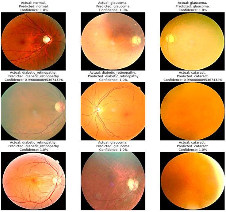
A multi-panel comparison chart of clinical diagnostic images featuring nine color fundus photographs used for ophthalmological screening. The images demonstrate four diagnostic categories: Normal, Glaucoma, Diabetic Retinopathy, and Cataract. The Normal fundus image shows clear retinal architecture, a well-defined optic disc, macula, and healthy vascular branching over a reddish-orange background. Glaucoma images exhibit pathological optic disc cupping and increased pallor. Diabetic Retinopathy images show characteristic vascular changes, including visible hemorrhages and exudates disrupting the retinal surface. Cataract images are characterized by significant media opacity, presenting as a diffuse yellow-brown or amber haze that obscures the posterior pole and retinal landmarks due to lens clouding. Each image is annotated with labels indicating the 'Actual' diagnosis, the 'Predicted' diagnosis by a deep-learning model, and the 'Confidence' score, representing the application of artificial intelligence in medical imaging for automated disease classification in ophthalmology.
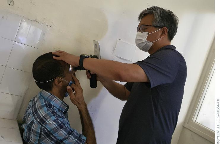
This clinical photograph demonstrates a portable ophthalmic examination being performed in a community or clinical setting. A healthcare provider is seen using a handheld digital fundus camera, integrated with a smartphone or similar imaging interface, to capture a retinal image from a seated patient. The provider, wearing a protective surgical mask and corrective lenses, stabilizes the patient's head with their left hand while positioning the device close to the patient's right eye. An active light source from the device is visible, indicating the illumination of the posterior segment for screening purposes. This visual illustrates modern diagnostic technology used for mobile screening of ocular diseases, such as diabetic retinopathy, where traditional table-mounted fundus cameras may not be accessible. The image highlights the intersection of ophthalmology, portable medical devices, and artificial intelligence-capable hardware in public health screenings.
~/attachments/0599cdbe-6bf3-481e-8a75-975005380e00
site:who.int ethics governance artificial intelligence health six principles WHO AI health
"artificial intelligence" AND clinical validation
chest radiograph artificial intelligence detection clinical image
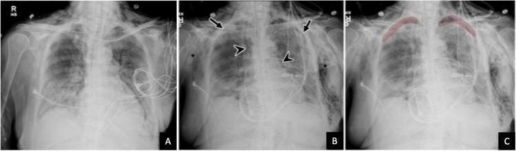
A comparison of three anteroposterior (AP) chest radiographs demonstrating the progression of pulmonary pathology and the application of diagnostic AI. Image A shows a baseline with bilateral, patchy airspace opacities predominantly in the lower lung zones, typical of multifocal pneumonia (e.g., COVID-19). Image B, a follow-up radiograph, reveals significant complications of barotrauma: subtle biapical pneumothoraces (arrows), pneumomediastinum indicated by lucent air streaks outlining the mediastinal borders (arrowheads), and extensive bilateral subcutaneous emphysema manifesting as radiolucent air dissecting through the chest wall soft tissues (asterisks). Image C displays the same follow-up radiograph with an artificial intelligence (AI) overlay, highlighting the detected biapical pneumothoraces in red for prioritized clinical review. The series illustrates critical care radiology findings, including pulmonary consolidation, extra-alveolar air collections, and the role of computer-aided detection (CAD) in identifying subtle but life-threatening conditions in ICU patients.
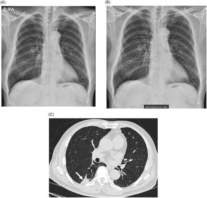
A multi-panel clinical diagnostic set showing a lung adenocarcinoma in a 79-year-old male. Image (A) is a posterior-anterior (PA) chest radiograph showing a subtle, ill-defined opacity in the right lower lung field, partially obscured by overlying pulmonary vasculature. Image (B) displays the same radiograph with an artificial intelligence (AI) lesion detection overlay; a circular region of interest (ROI) highlights the lesion, accompanied by a quantitative 'Abnormality Score' and a confidence label 'Ndl 26%'. Image (C) is an axial chest computed tomography (CT) scan in lung window settings, providing high-resolution anatomical detail. It clearly delineates an irregular solid mass (indicated by a white arrow) located in the posterior subpleural space of the right lower lobe's superior segment. The set illustrates the comparative diagnostic utility between standard radiography, AI-assisted screening, and definitive CT imaging for identifying pulmonary malignancies like stage IA3 adenocarcinoma.
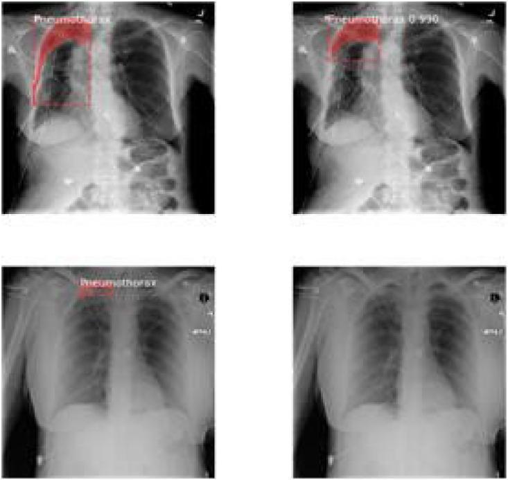
A comparison chart featuring four diagnostic images of posteroanterior (PA) chest radiographs. The visual focus is on the detection and localization of a pneumothorax in the right lung apex. Three of the images demonstrate the application of computer-aided detection (CAD) or artificial intelligence (AI) overlays, showing red-tinted masks and bounding boxes to highlight the apical pleural line and underlying air space indicative of lung collapse. Labels such as 'Pneumothorax' and 'Pneumothorax 0.930' represent prediction scores and classification results. The fourth image is a baseline unannotated radiograph for clinical comparison. Anatomical landmarks including the mediastinum, cardiac silhouette, diaphragm, and clavicles are visible, though the primary educational objective is demonstrating automated localization of pathology. This material serves as an example of medical imaging technology and radiology informatics, illustrating how segmentation and classification algorithms assist in identifying pleural space disorders.
python - <<'PY' from pptx import Presentation p='/tmp/workspace/attachments/0599cdbe-6bf3-481e-8a75-975005380e00/AI_in_Medicine_Research_Methodology_Mayur_Lokhande_20_Slides.pptx' prs=Presentation(p) print('slides',len(prs.slides),'size',prs.slide_width,prs.slide_height) for i,s in enumerate(prs.slides,1): texts=[] for sh in s.shapes: if getattr(sh,'has_text_frame',False) and sh.text.strip(): texts.append(sh.text.replace('\n',' | ')) print(f'[{i}] '+' || '.join(texts)) PY
mkdir -p "/tmp/workspace/ai-medicine-presentation/assets" "/tmp/workspace/ai-medicine-presentation/src" "/tmp/workspace/ai-medicine-presentation/output" && python /tmp/skills/shared/scripts/fetch_images.py "https://cdn.orris.care/cdss_images/pmc_clinical_VQA_2c5b06ab46f9f84557202f1e87987941536ef7bfa8c3170de4efbc4e18ec39bc.jpg" "https://cdn.orris.care/cdss_images/pmc_clinical_VQA_16e556dfd15d1f3fe180991a7f9e083a62e27f1618120349af542c3b0d2a6ef8.jpg" > "/tmp/workspace/ai-medicine-presentation/assets/images.json"
~/ai-medicine-presentation/src/build_deck.py
from pptx import Presentation
from pptx.util import Inches, Pt
from pptx.enum.shapes import MSO_SHAPE, MSO_CONNECTOR
from pptx.enum.text import PP_ALIGN, MSO_ANCHOR
from pptx.dml.color import RGBColor
from pptx.enum.dml import MSO_THEME_COLOR
from pptx.enum.shapes import MSO_AUTO_SHAPE_TYPE
from pptx.enum.text import MSO_AUTO_SIZE
from reportlab.pdfgen import canvas
from reportlab.lib.pagesizes import landscape
from reportlab.lib.colors import HexColor, white
from reportlab.pdfbase.pdfmetrics import stringWidth
import json, base64, os, textwrap

OUT='/tmp/workspace/ai-medicine-presentation/output'
AS='/tmp/workspace/ai-medicine-presentation/assets'
os.makedirs(OUT, exist_ok=True)
W,H=13.333,7.5
NAVY='0B1F33'; TEAL='00A6A6'; CYAN='36C5F0'; BLUE='2056A5'; SKY='DFF7FA'; PALE='F4F8FB'; INK='162B3A'; GREY='5D7180'; RED='D65353'; GOLD='F6B34B'; WHITE='FFFFFF'; LINE='C9D7E0'
def rgb(x): return RGBColor.from_string(x)

def rect(s,x,y,w,h,fill, radius=False, line=None):
    sh=s.shapes.add_shape(MSO_SHAPE.ROUNDED_RECTANGLE if radius else MSO_SHAPE.RECTANGLE, Inches(x), Inches(y), Inches(w), Inches(h))
    sh.fill.solid(); sh.fill.fore_color.rgb=rgb(fill)
    sh.line.color.rgb=rgb(line or fill)
    return sh

def txt(s,x,y,w,h,text,size=20,color=INK,bold=False,align=PP_ALIGN.LEFT, font='Aptos Display', margin=.06):
    tb=s.shapes.add_textbox(Inches(x), Inches(y), Inches(w), Inches(h)); tf=tb.text_frame; tf.clear(); tf.word_wrap=True
    tf.margin_left=tf.margin_right=Inches(margin); tf.margin_top=tf.margin_bottom=Inches(margin)
    p=tf.paragraphs[0]; p.alignment=align; p.space_after=Pt(0); p.space_before=Pt(0)
    for i,line in enumerate(str(text).split('\n')):
        if i: p=tf.add_paragraph(); p.alignment=align; p.space_after=Pt(0)
        p.text=line
        for r in p.runs:
            r.font.name=font; r.font.size=Pt(size); r.font.bold=bold; r.font.color.rgb=rgb(color)
    tf.vertical_anchor=MSO_ANCHOR.MIDDLE
    return tb

def bullet(s,x,y,w,items,size=17,accent=TEAL, gap=.48):
    for i,t in enumerate(items):
        cy=y+i*gap
        rect(s,x,cy+.15,.12,.12,accent,True)
        txt(s,x+.24,cy,w-.24,.4,t,size,INK)

def title(s,n,kicker,title,sub=''):
    rect(s,0,0,W,.16,TEAL); txt(s,.55,.35,1.2,.3,kicker.upper(),10,TEAL,True); txt(s,.55,.65,11.9,.6,title,27,NAVY,True)
    if sub: txt(s,.57,1.29,11.6,.35,sub,11,GREY)
    txt(s,12.25,7.05,.5,.22,f'{n:02}',10,TEAL,True,PP_ALIGN.RIGHT)

def footer(s,ref=''):
    rect(s,.55,6.86,11.9,.012,LINE)
    if ref: txt(s,.55,6.92,11.3,.22,ref,7,GREY)

def card(s,x,y,w,h,heading,body,accent=TEAL):
    rect(s,x,y,w,h,WHITE,True,LINE); rect(s,x,y,.09,h,accent,False,accent)
    txt(s,x+.22,y+.16,w-.38,.35,heading,15,NAVY,True); txt(s,x+.22,y+.58,w-.38,h-.68,body,11.5,INK)

def arrow(s,x1,y1,x2,y2,color=TEAL):
    ln=s.shapes.add_connector(MSO_CONNECTOR.STRAIGHT,Inches(x1),Inches(y1),Inches(x2),Inches(y2)); ln.line.color.rgb=rgb(color); ln.line.width=Pt(2); ln.line.end_arrowhead=True

def notes(slide, text):
    tf=slide.notes_slide.notes_text_frame
    tf.text=text

def imagefile():
    data=json.load(open(AS+'/images.json'))
    paths=[]
    for i,it in enumerate(data):
        b64=it.get('base64','').split(',',1)[-1]
        p=f'{AS}/image{i}.jpg'; open(p,'wb').write(base64.b64decode(b64)); paths.append(p)
    return paths

prs=Presentation(); prs.slide_width=Inches(W); prs.slide_height=Inches(H); blank=prs.slide_layouts[6]
imgs=imagefile()
def new(bg=PALE):
    s=prs.slides.add_slide(blank); rect(s,0,0,W,H,bg); return s

# 1
s=new(NAVY); rect(s,0,0,.18,H,TEAL); rect(s,7.75,0,5.58,H,'103B5B');
txt(s,.72,.78,2.5,.3,'RESEARCH METHODOLOGY ELECTIVE',12,CYAN,True)
txt(s,.7,1.38,6.45,1.5,'Artificial\nIntelligence in Medicine',34,WHITE,True)
txt(s,.75,3.2,5.8,.55,'From algorithms to accountable clinical and research use',17,'B6D5E6')
# neural/network motif
for x,y in [(9,1.4),(10.5,1.0),(11.7,1.9),(9.5,3),(11,3.6),(8.55,4.25),(10.1,5.0),(12,5.1)]:
    rect(s,x,y,.36,.36,CYAN,True)
for a,b in [((9.2,1.58),(10.68,1.18)),((10.68,1.18),(11.88,2.08)),((9.2,1.58),(9.68,3.18)),((9.68,3.18),(11.18,3.78)),((8.73,4.43),(10.28,5.18)),((11.18,3.78),(12.18,5.28))]: arrow(s,*a,*b,'55B7D1')
txt(s,.75,6.15,5.5,.5,'Mayur Lokhande  |  Department of Community Medicine / PSM',13,WHITE)
notes(s,'Open with the central premise: AI can support clinical work and research, but it does not remove professional responsibility. Faculty question: Is AI already used in hospitals? Yes, particularly in imaging, triage, monitoring, workflow and approved software devices.')
#2
s=new(); title(s,2,'Roadmap','Learning objectives','By the end, you should be able to explain AI use and critically appraise an AI medical study.');
for i,(h,b) in enumerate([('Explain','AI, ML, deep learning and generative AI'),('Apply','clinical and public-health applications'),('Evaluate','datasets, validation and performance metrics'),('Govern','bias, ethics, privacy and accountable use')]): card(s,.75+(i%2)*6.05,2.0+(i//2)*1.9,5.55,1.45,h,b,[TEAL,BLUE,GOLD,RED][i])
footer(s,'Scope: MBBS level, clinical relevance, research-methodology focus')
notes(s,'Signpost the flow: concepts, clinical value, research methods, then critical appraisal and ethics. Invite the audience to judge AI by clinical usefulness, not novelty.')
#3
s=new(); title(s,3,'Foundations','What is artificial intelligence?','AI performs tasks associated with aspects of human intelligence by processing data through algorithms.');
for i,(h,b) in enumerate([('Data','Images, ECGs, notes, labs, genomics'),('Algorithm','Rules or learned mathematical patterns'),('Output','Classification, prediction, prioritisation or generated content')]):
    x=.8+i*4.1; rect(s,x,2.25,3.4,1.25,['DFF7FA','E8F0FC','FDF3DD'][i],True); txt(s,x+.18,2.48,3,.3,h,18,NAVY,True,PP_ALIGN.CENTER); txt(s,x+.2,2.92,3, .35,b,11,INK,False,PP_ALIGN.CENTER)
    if i<2: arrow(s,x+3.45,2.86,x+4.0,2.86)
txt(s,.85,4.55,11.5,.55,'In medicine, a model should generate an interpretable, context-aware input to a clinician - not an autonomous medical decision.',20,NAVY,True,PP_ALIGN.CENTER)
footer(s,'Harrison’s Principles of Internal Medicine, 22e (2025), Ch. 501')
notes(s,'Define AI broadly. A model can identify patterns, but it does not possess clinical accountability or understand the patient’s values. Interesting fact: many deployed medical AI systems are narrow, task-specific systems.')
#4
s=new(); title(s,4,'Foundations','AI → machine learning → deep learning','A hierarchy, not interchangeable terms. Generative AI is defined by what it produces, not by a separate level of the hierarchy.');
levels=[('Artificial intelligence','Broad field: systems that perform intelligent tasks','0B1F33'),('Machine learning','Algorithms learn statistical patterns from examples','2056A5'),('Deep learning','ML using multi-layer neural networks','00A6A6'),('Generative AI / LLMs','Generate text, images, code or summaries','F6B34B')]
for i,(h,b,c) in enumerate(levels):
    x=.85+i*3.05; w=2.75; rect(s,x,2.2,w,2.35,c,True); txt(s,x+.15,2.5,w-.3,.45,h,17,WHITE if i<3 else NAVY,True,PP_ALIGN.CENTER); txt(s,x+.18,3.25,w-.36,.75,b,11,WHITE if i<3 else NAVY,False,PP_ALIGN.CENTER)
    if i<3: arrow(s,x+w,3.35,x+3.0,3.35,'90C8DB')
footer(s,'Generative AI requires output verification: it may produce fluent but incorrect content.')
notes(s,'Emphasise that ML learns from examples; deep learning is a subset that can learn complex representations, especially from images and signals. LLMs can assist with language tasks but may hallucinate facts or references.')
#5
s=new(); title(s,5,'Clinical need','Why AI in medicine?','The opportunity arises from high-volume, heterogeneous data and time-sensitive clinical decisions.');
items=[('Detect','subtle patterns in imaging, pathology, ECG and retinal photographs'),('Predict','risk of deterioration, readmission, disease progression or treatment response'),('Prioritise','worklists and early-warning alerts when validated in the target setting'),('Personalise','integrate clinical, molecular and longitudinal data'),('Learn','support research, surveillance and medical education')]
for i,(a,b) in enumerate(items): card(s,.85+(i%3)*4.1,2.0+(i//3)*1.8,3.72,1.35,a,b,[TEAL,BLUE,GOLD,RED,TEAL][i])
footer(s,'Potential does not equal outcome benefit: impact must be tested prospectively.')
notes(s,'Frame the unmet need: clinicians face more data than can be manually processed. The question is not whether an algorithm is accurate in isolation, but whether it improves care without new harms.')
#6
s=new(); title(s,6,'Clinical applications','Where does AI fit in medicine?','Examples should be understood as decision support, with performance depending on data, task and clinical setting.');
apps=[('Radiology','X-ray, CT and MRI triage / detection'),('Pathology','digital-slide screening and quantification'),('Ophthalmology','retinal image screening'),('Cardiology','ECG interpretation and risk prediction'),('Therapeutics','treatment planning, robotics, response prediction'),('Drug development','target identification, screening and trial operations'),('Monitoring','wearables, remote signals and deterioration alerts'),('Public health','surveillance, forecasting and resource allocation')]
for i,(h,b) in enumerate(apps): card(s,.65+(i%4)*3.16,1.85+(i//4)*1.6,2.9,1.22,h,b,[TEAL,BLUE,GOLD,RED][i%4])
footer(s,'FDA maintains a public list of AI-enabled medical devices; regulatory clearance is task-specific, not a blanket endorsement.')
notes(s,'Use one application only as an example at a time. Faculty question: Can AI replace radiologists? No. It may support defined image-analysis tasks, but radiology involves clinical context, communication, quality assurance and responsibility.')
#7
s=new(); title(s,7,'Research methodology','AI across the research pathway','AI can reduce repetitive work, but each step needs human methodological oversight.');
steps=['Question','Literature review','Design','Data capture','Analysis','Interpretation','Writing & reporting']
for i,h in enumerate(steps):
    x=.5+i*1.82; rect(s,x,2.35,1.52,1.05,[TEAL,BLUE,'4789B8',GOLD,'D65353',TEAL,BLUE][i],True); txt(s,x+.06,2.65,1.4,.36,h,12,WHITE,True,PP_ALIGN.CENTER)
    if i<6: arrow(s,x+1.54,2.88,x+1.77,2.88,'8AAFC1')
for x,h,b in [(1.1,'Useful tasks','screening abstracts, NLP extraction, clustering, prediction, visualisation'),(7.0,'Non-delegable tasks','question validity, consent, protocol decisions, causal inference, interpretation, authorship')]: card(s,x,4.35,5.25,1.35,h,b,TEAL if x<5 else RED)
footer(s,'AI output is a draft or decision-support input, not a substitute for a protocol, statistician or investigator judgment.')
notes(s,'Make the methodology link explicit. AI can help generate ideas, screen literature and process data, but cannot verify truth or replace governance. Any AI-generated question needs a human check for relevance, feasibility and ethics.')
#8
s=new(); title(s,8,'Data','What data does a medical AI study use?','Data provenance and labels are as important as the model.');
card(s,.8,1.95,3.7,2.1,'Structured','Age, sex, vitals, diagnoses, medications, laboratory values, coded outcomes',TEAL)
card(s,4.82,1.95,3.7,2.1,'Unstructured','Clinical notes, images, histopathology, ECG/waveforms, audio, video',BLUE)
card(s,8.84,1.95,3.7,2.1,'Multi-modal','Linking text + images + signals + longitudinal records may add context, but increases governance demands',GOLD)
rect(s,1.2,4.85,10.9,.85,'E9F7F7',True); txt(s,1.45,5.06,10.4,.35,'Data quality checklist: representative population  •  clear time window  •  reliable reference standard  •  complete metadata  •  privacy protection',14,NAVY,True,PP_ALIGN.CENTER)
footer(s,'Dataset shift occurs when the deployment population, workflow or equipment differs from development data.')
notes(s,'Explain that model performance can fall when scanners, coding practices, prevalence or patient demographics change. Labels should be derived from credible reference standards, not convenience labels alone.')
#9
s=new(); title(s,9,'Study design','Research methodology for an AI study','A transparent protocol is needed before model development.');
flow=[('1','Problem & users'),('2','Question & outcome'),('3','Design & population'),('4','Data & reference standard'),('5','Model development'),('6','Internal + external validation'),('7','Clinical impact & reporting')]
for i,(n,h) in enumerate(flow):
    x=.7+i*1.8; rect(s,x,2.2,1.42,1.2,TEAL if i in [0,6] else BLUE,True); txt(s,x+.1,2.38,1.22,.24,n,11,CYAN,True,PP_ALIGN.CENTER); txt(s,x+.1,2.68,1.22,.45,h,11,WHITE,True,PP_ALIGN.CENTER)
    if i<6: arrow(s,x+1.43,2.8,x+1.76,2.8,'A1C8D6')
card(s,1.1,4.45,5.15,1.18,'Reporting','Pre-specify outcomes and analysis; use transparent reporting frameworks such as TRIPOD-AI / CONSORT-AI where applicable.',GOLD)
card(s,7.0,4.45,5.15,1.18,'Clinical question','Does the system improve a patient-relevant process or outcome compared with usual care, not only its AUROC?',RED)
footer(s,'Frameworks evolve. Check the current EQUATOR Network guidance for the study type.')
notes(s,'Point out that clinical evaluation comes after technical development. A high AUROC is not sufficient evidence of benefit. Methods should state intended use, user, setting, comparator and outcome.')
#10
s=new(); title(s,10,'Study design','Choosing the right evaluation design','The evaluation design must match the intended clinical claim.');
for i,(h,b,c) in enumerate([('Retrospective diagnostic study','Compare model output with a reference standard; high risk of dataset bias.',TEAL),('Prospective cohort / silent trial','Run model in real workflow without influencing care; assess calibration, drift and safety.',BLUE),('Randomised impact study','Compare AI-assisted care with usual care for workflow or patient outcomes.',GOLD),('Systematic review','Synthesise heterogeneous studies cautiously; assess bias, validation and applicability.',RED)]): card(s,.8+(i%2)*6.0,1.9+(i//2)*2.0,5.45,1.5,h,b,c)
footer(s,'Example: AI-assisted chest X-ray triage should be assessed against a defined reference standard and workflow endpoint.')
notes(s,'Diagnostic accuracy answers a different question from patient benefit. For an alert system, a prospective evaluation is important because clinician response and alert burden affect real-world usefulness.')
#11
s=new(); title(s,11,'Question formulation','PICO + FINER: an AI research question','AI can help draft questions, but the investigator must verify feasibility and scientific value.');
for i,(h,b,c) in enumerate([('P','Patients undergoing chest X-ray',TEAL),('I','AI-assisted interpretation',BLUE),('C','Usual / clinician interpretation',GOLD),('O','Diagnostic accuracy and time-to-prioritisation',RED)]): card(s,.9+i*3.08,1.9,2.72,1.4,h,b,c)
rect(s,.9,3.8,11.5,1.1,'E9F7F7',True); txt(s,1.2,4.06,10.9,.55,'Does AI-assisted chest-X-ray interpretation improve diagnostic performance or worklist prioritisation versus usual interpretation in the intended clinical setting?',16,NAVY,True,PP_ALIGN.CENTER)
txt(s,1.05,5.45,11,.3,'FINER check: Feasible • Interesting • Novel • Ethical • Relevant',18,TEAL,True,PP_ALIGN.CENTER)
footer(s,'Do not assume “AI” is the intervention: specify the user, workflow, comparator and outcome.')
notes(s,'Give the PICO example, then use FINER. AI may help brainstorm synonyms or map concepts, but it cannot establish novelty without verified literature searching and cannot resolve ethical feasibility.')
#12
s=new(); title(s,12,'Sampling','Population, sampling and sample size','Representativeness and outcome prevalence affect precision, fairness and generalisability.');
for i,(h,b) in enumerate([('Define target population','Inclusion/exclusion criteria, care setting, period and intended users.'),('Plan sampling','Avoid convenience-only sampling; preserve temporal and site diversity.'),('Estimate sample size','For diagnostic models, consider sensitivity/specificity precision, prevalence and events.'),('Audit subgroups','Pre-specify performance checks by relevant demographic and clinical groups.')]): card(s,.85+(i%2)*6.0,1.85+(i//2)*1.85,5.45,1.4,h,b,[TEAL,BLUE,GOLD,RED][i])
rect(s,.9,5.7,11.5,.45,'FFF4E0',True); txt(s,1.15,5.78,11,.22,'Low-prevalence outcomes can yield deceptively high accuracy. Report sensitivity, specificity, PPV and NPV in context.',12,NAVY,True,PP_ALIGN.CENTER)
footer(s,'PPV and NPV depend on prevalence; accuracy alone can mislead.')
notes(s,'A model trained in a tertiary hospital may not generalise to primary care. Accuracy can look excellent when most patients do not have disease. Always ask who is missing from the data.')
#13
s=new(); title(s,13,'Validation','Training, validation and test data','Keep final evaluation data independent from all model-development decisions.');
for i,(h,b,c) in enumerate([('Training set','Learns patterns from data',TEAL),('Validation set','Tunes model / chooses settings',BLUE),('Test set','Final locked performance estimate',GOLD),('External test','Different site, time or population',RED)]):
    x=.8+i*3.1; rect(s,x,2.05,2.7,1.45,c,True); txt(s,x+.15,2.3,2.4,.32,h,17,WHITE if i<3 else WHITE,True,PP_ALIGN.CENTER); txt(s,x+.15,2.77,2.4,.3,b,11,WHITE,False,PP_ALIGN.CENTER)
    if i<3: arrow(s,x+2.72,2.77,x+3.02,2.77,'91C2D5')
card(s,1.1,4.65,5.2,1.15,'Avoid leakage','Never allow the same patient, image series or future information to contaminate test data.',RED)
card(s,7.0,4.65,5.2,1.15,'Why external validation?','It tests transportability across clinical settings and is more informative than internal performance alone.',TEAL)
footer(s,'Temporal and geographic external validation are especially useful before deployment.')
notes(s,'Define data leakage with a simple example: slices from the same CT patient must not land in train and test sets. External validation asks whether the result is likely to travel.')
#14
s=new(); title(s,14,'Performance','Measuring AI performance','Use a set of metrics tied to the clinical task, disease prevalence and consequences of error.');
metrics=[('Sensitivity','Finds disease among people with disease'),('Specificity','Correctly excludes disease among people without disease'),('PPV / NPV','Probability an output is correct in this prevalence'),('AUROC','Discrimination across thresholds'),('Calibration','Agreement between predicted and observed risk'),('Clinical utility','Net benefit, workflow and patient outcomes')]
for i,(h,b) in enumerate(metrics): card(s,.72+(i%3)*4.18,1.78+(i//3)*1.7,3.8,1.25,h,b,[TEAL,BLUE,GOLD,RED,TEAL,BLUE][i])
footer(s,'Technical accuracy ≠ clinical usefulness ≠ improved patient outcomes.')
notes(s,'Use screening as an example: sensitivity may be prioritised to miss fewer cases, but false positives can overburden services. Calibration matters when the number itself guides risk-based decisions.')
#15
s=new(); title(s,15,'Diagnostic accuracy','Confusion matrix: read it clinically','A test result is meaningful only in relation to a reference standard and the population being tested.');
# table
x,y=.95,1.85; cw=[2.7,2.85,2.85]; rh=.68
labels=[['','Disease +','Disease −'],['AI positive','True positive','False positive'],['AI negative','False negative','True negative']]
for r in range(3):
 for c in range(3):
  col=NAVY if r==0 or c==0 else ['DFF7FA','FFF0EF','FFF0EF','E8F8F4'][ (r-1)*2+(c-1)]
  rect(s,x+sum(cw[:c]),y+r*rh,cw[c],rh,col,False,WHITE); txt(s,x+sum(cw[:c])+.05,y+r*rh+.12,cw[c]-.1,.35,labels[r][c],14,WHITE if r==0 or c==0 else INK,True,PP_ALIGN.CENTER)
card(s,9.2,1.85,3.0,2.04,'Simple example','Among 100 patients with 10 disease cases, 90% sensitivity detects 9 cases; 95% specificity creates ~5 false positives among 90 non-cases.',GOLD)
rect(s,.95,4.55,11.25,.9,'E9F7F7',True); txt(s,1.15,4.77,10.85,.45,'Sensitivity = TP / (TP + FN)     •     Specificity = TN / (TN + FP)     •     PPV & NPV change with prevalence',14,NAVY,True,PP_ALIGN.CENTER)
footer(s,'Reference standard quality defines whether “true” and “false” are meaningful.')
notes(s,'Walk through why low prevalence reduces PPV even when sensitivity and specificity are high. Faculty question: Why is sensitivity important in screening? It helps reduce missed disease, but the cost of false positives still matters.')
#16
s=new(); title(s,16,'Fairness','Bias in AI: a medical example','A model can reproduce inequity if its data or labels are incomplete, unrepresentative or structurally biased.');
steps=[('Training data','Under-represents a population'),('Model','Learns patterns mainly from represented groups'),('Deployment','Performance falls in an under-represented subgroup'),('Harm','Missed diagnosis, wrong risk estimate or unequal access')]
for i,(h,b) in enumerate(steps):
 x=.65+i*3.16; rect(s,x,2.15,2.7,1.55,[TEAL,BLUE,GOLD,RED][i],True); txt(s,x+.15,2.42,2.4,.32,h,16,WHITE,True,PP_ALIGN.CENTER); txt(s,x+.14,2.88,2.42,.48,b,11,WHITE,False,PP_ALIGN.CENTER)
 if i<3: arrow(s,x+2.72,2.93,x+3.06,2.93,'94BDCF')
card(s,1.15,4.7,5.1,1.2,'Mitigation','Representative data, label audit, subgroup analysis, external validation, monitoring and human escalation pathways.',TEAL)
card(s,7.1,4.7,5.1,1.2,'Key message','Fairness is not guaranteed by a high average AUROC. Report performance in clinically relevant subgroups.',RED)
footer(s,'Bias can arise during sampling, measurement, labelling, model development and use.')
notes(s,'Explain that datasets are not neutral. Representative inclusion, measurement quality and transparent subgroup reporting are needed. A model can be accurate overall while failing the very group that needs it.')
#17
s=new(); title(s,17,'Limitations','Overfitting, generalisability and clinical utility','A model can fit past data well yet fail in new patients or workflows.');
card(s,.9,1.8,3.55,2.15,'Overfitting','Excellent training performance, poor unseen-data performance. Reduce with appropriate complexity control and independent testing.',RED)
card(s,4.9,1.8,3.55,2.15,'Dataset shift','Changes in population, prevalence, scanner, coding or care process after development.',GOLD)
card(s,8.9,1.8,3.55,2.15,'Human factors','Alert fatigue, automation bias, poor interface design and unclear responsibility can reduce safety.',BLUE)
rect(s,.95,4.75,11.35,.8,NAVY,True); txt(s,1.2,4.95,10.85,.35,'Accuracy is only a technical property. Clinical usefulness requires calibration, workflow fit, safety, equity and evidence of patient benefit.',15,WHITE,True,PP_ALIGN.CENTER)
footer(s,'Post-deployment monitoring is needed because performance may drift over time.')
notes(s,'Explain automation bias: users may over-trust a recommendation and ignore contradictory clinical information. The appropriate response is not avoidance of AI, but good design, training, monitoring and accountability.')
#18
s=new(); title(s,18,'Ethics and governance','Human oversight is the safety layer','AI should assist healthcare professionals, not replace clinical responsibility.');
flow=['Patient data','AI analysis','AI-generated insight','Clinician verification','Clinical decision','Patient outcome','Feedback / monitoring']
for i,h in enumerate(flow):
 x=.38+i*1.82; rect(s,x,2.05,1.48,1.03,[BLUE,TEAL,BLUE,GOLD,NAVY,TEAL,RED][i],True); txt(s,x+.06,2.35,1.36,.36,h,11,WHITE,True,PP_ALIGN.CENTER)
 if i<6: arrow(s,x+1.5,2.57,x+1.76,2.57,'8BBBCD')
for i,h in enumerate(['Privacy & consent','Transparency','Accountability','Equity','Safety & security','Human autonomy']): card(s,.65+(i%3)*4.15,4.35+(i//3)*1.0,3.72,.7,h,'',[TEAL,BLUE,GOLD,RED,TEAL,BLUE][i])
footer(s,'WHO (2021): autonomy; well-being/safety; transparency; accountability; inclusiveness/equity; responsive & sustainable AI.')
notes(s,'Use this slide to show where responsibility remains. WHO’s six principles can guide implementation. Key point: a model recommendation must be checked against the patient, clinical context and local standards of care.')
#19
s=new(); title(s,19,'Future','Current, emerging and future possibilities','Separate established applications from plausible but not yet proven possibilities.');
for i,(h,b,c) in enumerate([('Current','Narrow task-specific imaging support, signal analysis, workflow triage, documentation assistance.',TEAL),('Emerging','Multimodal models, generative AI with retrieval, prospective workflow trials, adaptive lifecycle monitoring.',BLUE),('Future possibilities','Digital twins, AI agents and precision diagnostics - require validation, governance and equitable infrastructure.',GOLD)]): card(s,.85+i*4.1,1.92,3.68,2.55,h,b,c)
rect(s,.95,5.25,11.35,.65,'E9F7F7',True); txt(s,1.15,5.43,10.95,.28,'Best future model: human-AI collaboration with transparent evidence, continuous monitoring and patient-centred goals.',14,NAVY,True,PP_ALIGN.CENTER)
footer(s,'Avoid treating speculative applications as established clinical practice.')
notes(s,'Distinguish current deployment from research prototypes. Generative systems are promising for language workflows but must be assessed for factuality, privacy, bias, usability and patient impact before routine high-stakes use.')
#20
s=new(); title(s,20,'Take-home','AI in medicine: evidence before enthusiasm','The most useful question is not “Can AI do this?” but “Does it safely improve care for this population in this setting?”');
for i,t in enumerate(['AI is a tool, not an autonomous replacement for clinical judgment.','Clinical value requires representative data, independent validation and workflow evaluation.','Technical performance must be separated from clinical utility and patient outcomes.','Responsible use requires privacy, equity, transparency, accountability and human oversight.','Medical professionals need AI literacy to use, question and govern these systems.']):
 rect(s,.88,1.72+i*.73,11.6,.52,'E9F7F7' if i%2==0 else WHITE,True); rect(s,1.02,1.88+i*.73,.15,.15,[TEAL,BLUE,GOLD,RED,TEAL][i],True); txt(s,1.35,1.76+i*.73,10.7,.38,t,14,NAVY,True)
txt(s,.9,5.72,11.55,.22,'Thank you  |  Discussion',16,TEAL,True,PP_ALIGN.CENTER)
footer(s,'Selected references on next page in speaker notes. WHO 2021; FDA AI-enabled device list; Harrison’s 22e (2025); PMID 40140300.')
notes(s,'Viva-ready questions and answers:\n1. AI vs ML vs DL? AI is broad; ML learns patterns from data; DL is ML using multi-layer neural networks.\n2. Can AI replace doctors? No. It may support defined tasks, but clinicians retain responsibility for context, communication and decisions.\n3. What is external validation? Testing in a different site, time period or population to assess transportability.\n4. What is hallucination? Fluent generated output that is false or unsupported, including fabricated references.\n5. Why is calibration important? It checks whether predicted risk matches observed risk, essential for risk-based decisions.\n6. Can AI-generated references be trusted? No. Verify every citation in the original source.\n\nReferences (Vancouver style):\n1. World Health Organization. Ethics and governance of artificial intelligence for health. Geneva: WHO; 2021. Available from: https://www.who.int/publications/i/item/9789240029200\n2. World Health Organization. Regulatory considerations on artificial intelligence for health. Geneva: WHO; 2023.\n3. U.S. Food and Drug Administration. Artificial Intelligence-Enabled Medical Devices [Internet]. Silver Spring (MD): FDA; accessed 2026 Sep 19. Available from: https://www.fda.gov/medical-devices/software-medical-device-samd/artificial-intelligence-enabled-medical-devices\n4. Harrison’s Principles of Internal Medicine. 22nd ed. New York: McGraw Hill; 2025. Chapter 501, Machine Learning and Augmented Intelligence in Clinical Medicine.\n5. Aravazhi PS, Gunasekaran PP, Benjamin NZY. The integration of artificial intelligence into clinical medicine: Trends, challenges, and future directions. Dis Mon. 2025;71:101882. doi:10.1016/j.disamonth.2025.101882. PMID:40140300.\n6. Liu X, et al. Reporting guidelines for clinical trial reports for interventions involving artificial intelligence: the CONSORT-AI extension. Nat Med. 2020;26:1364-74. doi:10.1038/s41591-020-1034-x.\n7. Collins GS, et al. TRIPOD+AI statement. BMJ. 2024;385:e078378. doi:10.1136/bmj-2023-078378.')

# source note on imaging in speaker notes of relevant slide
notes(prs.slides[5], prs.slides[5].notes_slide.notes_text_frame.text+'\nImage example source in supplied visual assets: medical-image library figure showing AI overlay on chest radiograph; illustrative only, not diagnostic guidance.')
# Save PPT
ppt=OUT+'/AI_in_Medicine_Research_Methodology_Upgraded.pptx'; prs.save(ppt)

# PDF companion, professional readable handout-like slide export
pdf=OUT+'/AI_in_Medicine_Research_Methodology_Upgraded.pdf'; c=canvas.Canvas(pdf,pagesize=landscape((960,540)))
for idx,slide in enumerate(prs.slides,1):
    c.setFillColor(HexColor('#F4F8FB')); c.rect(0,0,960,540,fill=1,stroke=0); c.setFillColor(HexColor('#00A6A6')); c.rect(0,528,960,12,fill=1,stroke=0)
    # extract text from slide, render selective hierarchy
    arr=[]
    for sh in slide.shapes:
        if getattr(sh,'has_text_frame',False):
            t=sh.text.strip()
            if t and not t.isdigit(): arr.append(t.replace('\x0b',' '))
    # title then content, avoid dense overlap
    y=485
    c.setFillColor(HexColor('#0B1F33')); c.setFont('Helvetica-Bold',23); c.drawString(40,y,arr[1] if len(arr)>1 else f'Slide {idx}')
    y-=38; c.setFillColor(HexColor('#5D7180')); c.setFont('Helvetica',10)
    content=' | '.join(arr[2:])
    lines=[]
    for para in content.split('|'):
        lines.extend(textwrap.wrap(para.strip(),110) or [''])
    for line in lines[:24]:
        if y<35: break
        c.drawString(45,y,line); y-=17
    c.setFillColor(HexColor('#00A6A6')); c.setFont('Helvetica-Bold',9); c.drawRightString(920,22,f'{idx:02}')
    c.showPage()
c.save()
print(ppt); print(pdf)
python "/tmp/workspace/ai-medicine-presentation/src/build_deck.py"
~/ai-medicine-presentation/output
~/ai-medicine-presentation/src/build_deck.py
Successfully replaced 1 instance(s) of the string in '/tmp/workspace/ai-medicine-presentation/src/build_deck.py'
python "/tmp/workspace/ai-medicine-presentation/src/build_deck.py" && python - <<'PY' from pptx import Presentation from pypdf import PdfReader p='/tmp/workspace/ai-medicine-presentation/output/AI_in_Medicine_Research_Methodology_Upgraded.pptx' prs=Presentation(p) print('PPT slides:',len(prs.slides)) print('Speaker notes nonempty:',sum(bool(s.notes_slide.notes_text_frame.text.strip()) for s in prs.slides)) print('Images:',sum(1 for s in prs.slides for sh in s.shapes if sh.shape_type==13)) r=PdfReader('/tmp/workspace/ai-medicine-presentation/output/AI_in_Medicine_Research_Methodology_Upgraded.pdf') print('PDF pages:',len(r.pages)) print('PDF first page chars:',len(r.pages[0].extract_text() or '')) PY
- AI, ML, deep learning, and generative-AI hierarchy
- Expanded clinical applications and illustrative medical AI visuals
- Research workflow integration: PICO, FINER, study design, data quality, sampling, validation, and reporting
- AI performance metrics, confusion matrix, calibration, clinical utility
- Bias example, dataset shift, overfitting, automation bias, and human factors
- WHO-aligned ethics and human-AI collaboration model
- Current versus emerging versus future applications
- Speaker notes on every slide, including viva-ready faculty questions and verified Vancouver-style references
- Final quality check: 20 slides, 20 pages in the PDF, speaker notes present on all slides
AI in Medicine Research Methodology Upgraded
Presentation · PPTX
AI in Medicine Research Methodology Upgraded
PDF Document · PDF
Now generate a a script to explain this PPT for my presentation
WHO ethics governance artificial intelligence for health clinical AI human oversight 2021
"artificial intelligence" AND medicine
~/
Presentation Script: AI in Medicine
Slide 1: Title
Slide 2: Learning Objectives
Slide 3: What Is Artificial Intelligence?
Slide 4: AI, Machine Learning, Deep Learning and Generative AI
Slide 5: Why AI in Medicine?
Slide 6: Clinical Applications of AI
Slide 7: AI in Medical Research
Slide 8: Types of Data Used
Slide 9: Research Methodology for an AI Study
Slide 10: Study Designs
Slide 11: Research Question Using PICO
I refers to AI-assisted interpretation.
C refers to conventional physician interpretation.
O refers to diagnostic accuracy or another clinically meaningful outcome.
Slide 12: Sampling and Sample Size
Slide 13: Training, Validation and Test Sets
Slide 14: Measuring AI Performance
Slide 15: Confusion Matrix
Slide 16: Bias in AI Research
Slide 17: Overfitting and Generalizability
Slide 18: Ethical Issues in AI Research
Slide 19: Advantages, Limitations and Future
Slide 20: Conclusion
Short Answers for Likely Faculty Questions
“No. AI can support clinical decisions, but doctors integrate examination findings, context, patient preferences, ethics, and accountability.”
“It is testing an AI model on independent data from a different population, hospital, device, or time period to assess generalizability.”
“It is systematic unfairness or reduced performance caused by biased, incomplete, or non-representative data or model design.”
“It is when an AI system generates plausible but false information, such as an incorrect clinical statement or fabricated reference.”
“Screening aims to identify as many true cases as possible. High sensitivity reduces false negatives, although specificity is also needed to avoid unnecessary follow-up.”
“No. Every AI-generated reference must be checked in a reliable database such as PubMed, the journal website, or an official source.”
“Technical performance refers to measures such as AUROC or sensitivity. Clinical utility means whether using the system improves decisions, workflow, safety, or patient outcomes in real practice.”