
About 6 months
It is about 6 months ago
- Are you describing a symptom or health issue that started 6 months ago?
- Is this about a medication or treatment you started 6 months ago?
- Something else entirely?
Ulcer
What type of ulcer are you referring to? · What would you like help with?
Mouth
oral ulcer persistent non-healing
oral ulcer chronic diagnosis
aphthous mouth ulcer oral cavity

Type: Clinical Photographs. Anatomical Region: Oral cavity, specifically the tongue. Description: A series of three intraoral photographs showing a patient's open mouth with the tongue protruded. The primary finding is a well-circumscribed, painful-appearing aphthous ulcer (stomatitis) located on the left anterolateral aspect of the tongue. The lesion presents as a shallow mucosal defect with a slightly depressed center and an erythematous (reddish) peripheral halo, characteristic of recurrent aphthous stomatitis (RAS). The surrounding lingual mucosa appears moist and otherwise normal. Normal dentition and lips are partially visible. Clinical Significance: These images demonstrate the classic clinical presentation of an aphthous ulcer in a common non-keratinized mucosal site. Such lesions are frequently associated with various systemic conditions, including nutritional deficiencies or gastrointestinal disorders like H. pylori-associated gastritis, and serve as a key diagnostic visual for medical education in oral medicine and dermatology.
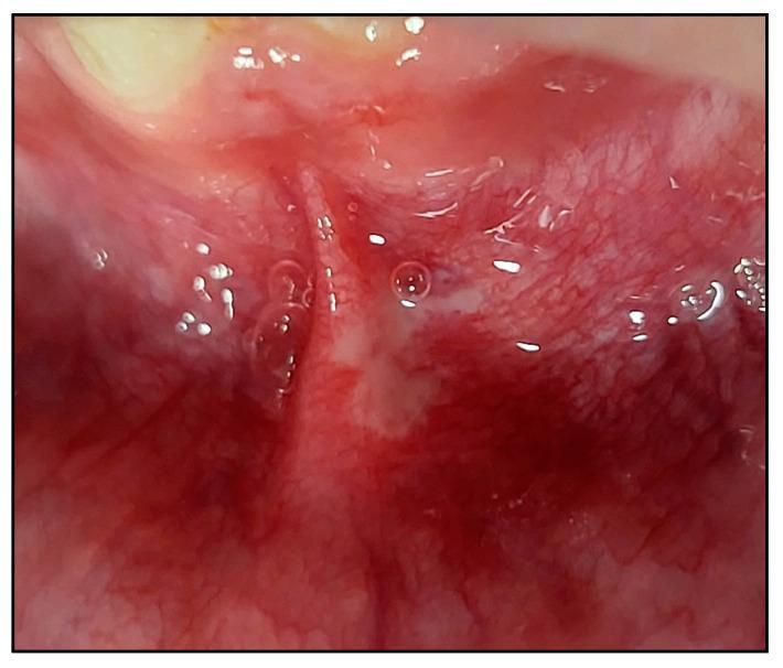
A close-up clinical photograph of the oral cavity showcasing a Major Recurrent Aphthous Stomatitis (MaRAS) lesion. The lesion is located on the non-keratinized labial mucosa, specifically involving the labial sulcus and extending across the lateral border of the lower midline frenum. Visually, the ulcer presents as a well-defined area covered by a white-to-yellowish pseudomembranous slough. This central ulceration is encircled by a prominent, diffuse erythematous halo, indicating acute inflammatory response. The surrounding mucosal tissue appears moist and highly vascularized. This image serves as a clinical example of the morphology and anatomical distribution typical of major aphthous ulcers, emphasizing their size and involvement of mucosal folds compared to minor variants.
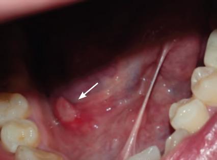
A close-up intraoral clinical photograph showing a minor recurrent aphthous ulcer on the right floor of the mouth. The lesion, indicated by a white arrow, is a small, well-circumscribed, ovoid ulcer characterized by a central pseudomembranous base that is yellowish-white in color. This central area is surrounded by a prominent, erythematous (reddened) halo, indicating localized inflammation. The surrounding oral mucosa of the floor of the mouth appears moist and pink with visible sublingual vasculature. Other visible structures include the lingual frenulum stretching vertically and several healthy molar and premolar teeth on the lateral borders of the image. This photograph serves as an educational example of aphthous stomatitis (canker sores), demonstrating the classic morphology and location within the non-keratinized oral mucosa for students and clinical professionals.
Mouth Ulcer Lasting 6 Months - What You Need to Know
Why This Is Concerning
Possible Causes of a Long-Standing Mouth Ulcer
| Category | Examples |
|---|---|
| Malignant (must rule out) | Squamous cell carcinoma, Lymphoma |
| Trauma | Ill-fitting dentures, sharp tooth, cheek biting - causing a chronic traumatic ulcer |
| Skin/mucosal disease | Erosive lichen planus, Pemphigus, Pemphigoid |
| Infection | Tuberculosis, Syphilis, HIV-related ulcer |
| Inflammatory bowel disease | Crohn's disease, Ulcerative colitis |
| Blood disorders | Leukaemia, Neutropenia, Vitamin B12/folate/iron deficiency |
| Autoimmune | Behçet's syndrome (mouth + genital ulcers) |
| Aphthous (major type) | Major aphthous ulcers can occasionally last weeks-months but 6 months is atypical |
Red Flag Features - Seek Urgent Care if You Have Any of These
- Ulcer has not healed in 3+ weeks
- Ulcer is painless (oral cancer is often painless)
- Hard or raised edges around the ulcer
- Ulcer is growing in size
- Unexplained bleeding from the ulcer
- Lump in the neck (swollen lymph node)
- Difficulty swallowing or speaking
- You use tobacco or alcohol (major risk factors for oral cancer)
What the Doctor Will Do
- Full history - Is this one continuous ulcer or recurrent episodes? Where exactly is it located?
- Examination - Size, shape, edge character, associated lymph nodes
- Biopsy - A small tissue sample is almost certainly needed after 6 months. This is the definitive test to rule out cancer
- Blood tests - Check for deficiencies (B12, folate, iron), blood disorders, inflammatory markers
- Referral - To oral medicine specialist, ENT, or oral surgeon
What You Should Do Right Now
Bottom line: A mouth ulcer lasting 6 months needs an urgent biopsy to rule out oral cancer. Please see a doctor or dentist as soon as possible.