
Intrauterine death
"intrauterine fetal death" OR "stillbirth" management
intrauterine fetal death stillbirth ultrasound signs
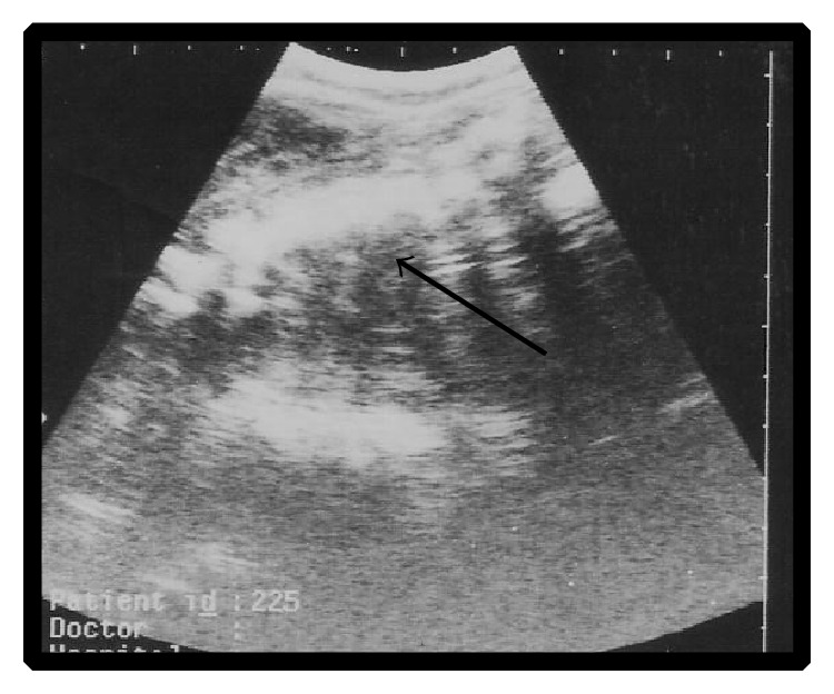
**Imaging Modality:** B-mode pelvic ultrasound (sonography). **Anatomical Region:** Intrauterine environment during the second trimester of gestation. **Observed Pathology:** Intrauterine fetal demise (IUFD) at approximately 120 days of gestation. **Characteristic Visual Features:** The image displays a sagittal or oblique view of a single fetus. Key sonographic findings include the loss of normal fetal anatomical definition and a disorganized echogenic mass within the uterine cavity. There is a notable absence of distinct cardiac activity or fetal movement. An arrow indicates the non-viable fetal remains, which appear heterogeneously echogenic compared to the surrounding hypoechoic amniotic fluid. **Key Diagnostic Features:** The presence of a collapsed fetal pole and fragmented structural echoes (consistent with maceration or early fetal resorption) are primary indicators of non-viability at this gestational stage. The image captures the diagnostic visualization of fetal death in a clinical obstetric setting.
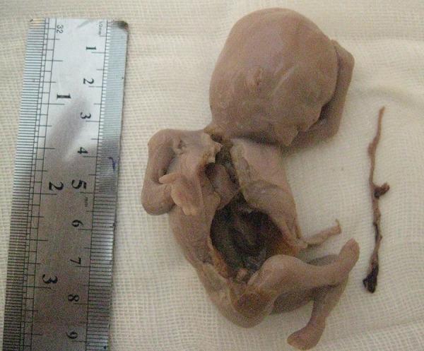
This clinical photograph shows a second-trimester human fetus alongside a metric ruler, documenting a case of intrauterine fetal death (IUFD) associated with Thin Cord Syndrome (TCS). The fetus exhibits signs of post-mortem maceration, including diffuse brownish skin discoloration and tissue softening. An open thoracic and abdominal cavity reveals internal organs undergoing ischemic necrosis. Positioned to the right of the fetus is a segment of the umbilical cord, which is notably pathological; it appears extremely thin, shriveled, and dark, measuring approximately 1 mm in diameter. This morphology indicates a severe deficiency of Wharton’s jelly, a condition that can lead to cord constriction, vascular compromise, and subsequent fetal demise. The presence of the ruler provides a scale for the approximately 9 cm long, filamentous cord. This image is an educational example for obstetrics and pathology, illustrating rare umbilical cord abnormalities and the macroscopic features of fetal death at 21 weeks of gestation.
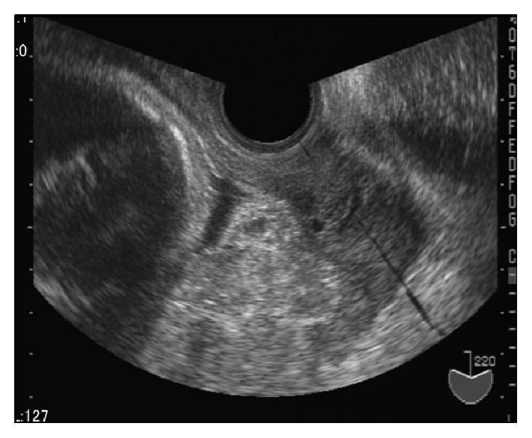
This diagnostic ultrasound image in the transverse plane demonstrates a placenta previa at approximately 33 weeks of gestation. The primary clinical focus is on the heterogeneous echotexture of the placental tissue following intrauterine fetal death (IUFD). The placenta occupies a significant portion of the lower uterine segment and is characterized by a granular, irregular appearance. Notable features include multiple scattered, irregular hyperechoic regions (bright white areas) embedded within the placental parenchyma. These high-echo regions are interspersed with areas of low-level echoes, indicating varied echogenicity across the tissue. This visual pattern is associated with reduced placental blood supply and changes occurring post-IUFD. The imaging modality is transvaginal or transabdominal ultrasonography, and the target audience includes obstetrics and gynecology professionals or medical students studying placental pathology and fetal growth restriction (FGR).
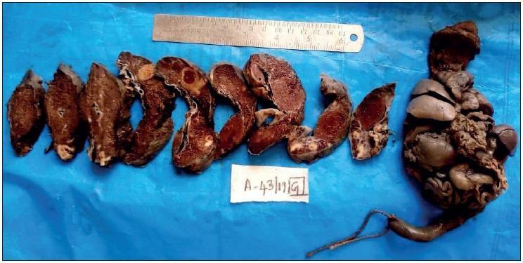
Gross clinical photograph of pathology specimens from a 32-week stillbirth, including cut sections of the placenta and an en bloc eviscerated fetus. The placental sections demonstrate diffuse parenchymal changes, including brownish-red thrombi and focal areas of pale, firm tissue indicative of chronic infarction, common in maternal eclampsia. The en bloc fetal specimen reveals multiple congenital anomalies: marked cardiomegaly, splenomegaly, a hypoplastic liver, and the absence of the left lung. The fetal tissue exhibits post-mortem discoloration and altered morphology consistent with intrauterine death. A ruler is included for scale against a blue background, along with a specimen identification tag. This image serves as a teaching tool for distinguishing between fetal death caused by hypertension-related placental pathology versus primary congenital malformations.
Intrauterine Death (IUD) / Intrauterine Fetal Demise (IUFD)
Definition
- WHO definition: Death before expulsion or extraction from the mother, at any gestational age, with no signs of life (no breathing, no heartbeat, no cord pulsation, no voluntary muscle movements)
- US definition: Fetal death at ≥20 weeks' gestation (23,595 such deaths reported in the US in 2013)
- Perinatal mortality includes late fetal death at ≥28 weeks + early neonatal death under 7 days - used as a quality-of-care indicator
Epidemiology
- Perinatal mortality rate in the US was stable at ~6/1,000 births (2014-2016)
- Rates are higher in women under 20 years (7.07-7.60/1,000)
- Single twin demise occurs in ~6% of all twin pregnancies
Causes / Risk Factors
Placental / Uteroplacental
- Uteroplacental vascular insufficiency (most common overall)
- Placental abruption
- Placental infarction, thrombosis, perivillous fibrin deposition
- Placenta previa
Maternal Medical Conditions
- Chronic hypertension
- Systemic lupus erythematosus (SLE)
- Antiphospholipid antibody syndrome (APAS)
- Thrombophilia
- Thyroid disease
- Intrahepatic cholestasis of pregnancy
- Diabetes mellitus
- Obesity
- Nutritional deficiencies
Fetal Causes
- Congenital anomalies / structural defects
- Chromosomal abnormalities (aneuploidy)
- Fetal growth restriction (FGR) - risk of stillbirth outweighs prematurity after 34 weeks
- Hydrops fetalis (immune and non-immune)
Infectious
- Parvovirus B19
- Cytomegalovirus (CMV)
- Syphilis - intrauterine death and perinatal death each occur in ~25% of untreated congenital syphilis
- Malaria
- Listeria monocytogenes
- Chorioamnionitis
Umbilical Cord Complications
- True knot
- Cord prolapse
- Thin cord syndrome (Wharton's jelly deficiency - severe cord constriction)
- Cord entanglement (especially in monoamniotic twins)
Other
- Multiple gestation (especially monochorionic)
- Smoking
- African American race (independent risk factor)
- Previous preterm birth with SGA neonate (7/1,000 risk in subsequent pregnancy)
Pathophysiology Shared with Preterm Birth
- LPS-induced preterm birth models show cytotoxic NK cell placental infiltration causing placental cell death - similar to inflammation-induced fetal demise
- Conditions like APAS are associated with shared placental histologic features (thrombosis, infarction, abruption, perivillous fibrin deposition)
Diagnosis
Symptoms / Clinical Presentation
- Absent fetal movements (primary complaint)
- Absent fetal heart sounds on auscultation
- Uterine size not increasing
Ultrasound Signs
- Absent fetal cardiac activity
- Absent fetal movement
- Spalding's sign: overlapping of fetal skull bones (after several days)
- Robert's sign: gas in the great vessels (sign of IUD on X-ray)
- Maceration pattern: heterogeneous echogenicity, collapsed fetal pole, fragmented structural echoes, loss of anatomical definition
X-ray Signs (historical, now largely replaced by ultrasound)
- Robert's sign: Gas in the great vessels or heart
- Spalding's sign: Overlapping of skull bones
- Hyperflexion of the spine
- Halo sign: Edema around fetal skull
Complications of Retained Dead Fetus
Disseminated Intravascular Coagulation (DIC)
- If a dead fetus is retained for >4 weeks, thromboplastin released from macerated fetal tissues can trigger DIC
- Fibrinogen levels fall (consumptive coagulopathy)
- Monitor fibrinogen levels weekly if expectant management is chosen
- Delivery should be expedited if fibrinogen falls below 150 mg/dL
Infection / Sepsis
- Risk of chorioamnionitis with retained fetus
- Maternal systemic infection can result
Psychological
- Grief, depression, post-traumatic stress - maternal psychological support is essential
Management
General Principles
- Confirm diagnosis with ultrasound
- Emotional support and counseling
- Investigate the cause
- Plan delivery
Investigation / Workup
- Maternal bloods: CBC, clotting studies (PT, APTT, fibrinogen), Kleihauer-Betke test (fetomaternal hemorrhage), blood group and antibody screen, glucose, thyroid function, ANA, anticardiolipin antibodies, lupus anticoagulant, TORCH serology
- Fetal/placental investigations: Placental histology, fetal karyotype (amniocentesis or fetal tissue), fetal autopsy (with consent)
- Cord blood culture, maternal blood cultures if infection suspected
Delivery
- Most women deliver spontaneously within 2 weeks of IUD
- Induction of labour is generally recommended:
- Cervical ripening with prostaglandins (misoprostol, dinoprostone)
- Oxytocin infusion
- In the second trimester, higher doses of misoprostol are used
- Caesarean section is rarely indicated and carries morbidity; reserved for specific obstetric indications
- Expectant management is possible but carries DIC risk; requires close monitoring
Twin Gestation - Special Considerations
- Chorionicity is the most important predictive factor for outcomes after one twin dies
- In monochorionic twins: vascular connections mean death of one fetus can cause profound neurologic injury or death of the co-twin via acute haemodynamic disturbance
- ~15% of co-twins also die after monochorionic single fetal death (vs ~3% in dichorionic)
- 34% show abnormal neonatal cranial imaging
- Injury can occur as early as 12 weeks
- Dichorionic: risk to co-twin is much lower
- "Vanishing twin": occurs in ~21% of twin pregnancies in the first trimester, usually without detriment to the remaining fetus
Post-mortem Features
Maceration
| Time after Death | Features |
|---|---|
| 12-24 hours | Skin desquamation, discoloration (brownish-red) |
| 2-7 days | Bullae formation, overlapping skull bones |
| >1 week | Mummification if oligohydramnios; widespread soft tissue changes |
Prevention
- Monitoring fetal movements (kick counts)
- Treatment of maternal medical conditions
- Fetal surveillance with ultrasound in high-risk pregnancies
- Avoidance of smoking
- Antenatal corticosteroids and timely delivery in FGR (after 34 weeks)