Vitamin d and skin

Your credits are exhausted. You can purchase a top-up or subscribe to a plan to continue using Orris. If you're already subscribed, your daily credits will reset tomorrow.

Vitamin d and skin

Reading File
Searching the Web

vitamin D skin synthesis roles dermatology 2025

Searching PubMed

"vitamin D" AND skin

Finding Sources
Reading Article

PMID: 39683522

Searching Images

vitamin D synthesis skin UVB pathway

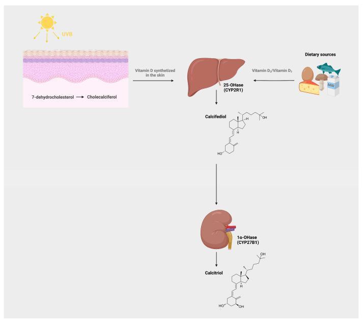
This pathophysiology diagram illustrates the metabolic pathway of Vitamin D in humans. The process begins with two sources: endogenous synthesis in the skin and exogenous intake from dietary sources (e.g., fish, eggs, mushrooms, milk). In the skin, UVB radiation facilitates the conversion of 7-dehydrocholesterol to cholecalciferol (Vitamin D3). Both synthesized and dietary Vitamin D travel to the liver, where the enzyme 25-hydroxylase (25-OHase/CYP2R1) converts it to calcifediol (25(OH)D3), shown with its chemical structure. Calcifediol then undergoes a second hydroxylation in the kidney by the enzyme 1α-hydroxylase (1α-OHase/CYP27B1) to form calcitriol (1α,25(OH)2D3), the biologically active form of Vitamin D, also depicted with its molecular structure. The diagram uses anatomical illustrations of the skin, liver, and kidney to delineate the multi-organ progression of this endocrine pathway, making it suitable for medical education regarding endocrinology, nutrition, and metabolic bone disease.
This pathophysiology diagram illustrates the metabolic pathway of Vitamin D in humans. The process begins with two sources: endogenous synthesis in the skin and exogenous intake from dietary sources (e.g., fish, eggs, mushrooms, milk). In the skin, UVB radiation facilitates the conversion of 7-dehydrocholesterol to cholecalciferol (Vitamin D3). Both synthesized and dietary Vitamin D travel to the liver, where the enzyme 25-hydroxylase (25-OHase/CYP2R1) converts it to calcifediol (25(OH)D3), shown with its chemical structure. Calcifediol then undergoes a second hydroxylation in the kidney by the enzyme 1α-hydroxylase (1α-OHase/CYP27B1) to form calcitriol (1α,25(OH)2D3), the biologically active form of Vitamin D, also depicted with its molecular structure. The diagram uses anatomical illustrations of the skin, liver, and kidney to delineate the multi-organ progression of this endocrine pathway, making it suitable for medical education regarding endocrinology, nutrition, and metabolic bone disease.

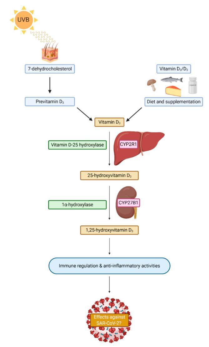
This pathophysiology diagram illustrates the metabolic pathway of Vitamin D synthesis and activation in humans, emphasizing its role in immune modulation. The flow begins with two primary sources: endogenous synthesis in the skin (UVB radiation converting 7-dehydrocholesterol to Previtamin D3 and subsequently Vitamin D3) and exogenous intake (dietary sources like fish, eggs, and mushrooms or supplementation). The pathway then details the two-step hydroxylation process: first in the liver, where Vitamin D-25 hydroxylase (CYP2R1) converts Vitamin D3 into 25-hydroxyvitamin D3; and second in the kidney, where 1α-hydroxylase (CYP27B1) converts it into the bioactive form, 1,25-dihydroxyvitamin D3. The diagram concludes by highlighting the clinical significance of active Vitamin D in promoting immune regulation and anti-inflammatory activities, specifically questioning its potential protective effects against SARS-CoV-2. This schematic serves as an educational tool for understanding endocrine metabolism and its systemic physiological impacts.
This pathophysiology diagram illustrates the metabolic pathway of Vitamin D synthesis and activation in humans, emphasizing its role in immune modulation. The flow begins with two primary sources: endogenous synthesis in the skin (UVB radiation converting 7-dehydrocholesterol to Previtamin D3 and subsequently Vitamin D3) and exogenous intake (dietary sources like fish, eggs, and mushrooms or supplementation). The pathway then details the two-step hydroxylation process: first in the liver, where Vitamin D-25 hydroxylase (CYP2R1) converts Vitamin D3 into 25-hydroxyvitamin D3; and second in the kidney, where 1α-hydroxylase (CYP27B1) converts it into the bioactive form, 1,25-dihydroxyvitamin D3. The diagram concludes by highlighting the clinical significance of active Vitamin D in promoting immune regulation and anti-inflammatory activities, specifically questioning its potential protective effects against SARS-CoV-2. This schematic serves as an educational tool for understanding endocrine metabolism and its systemic physiological impacts.

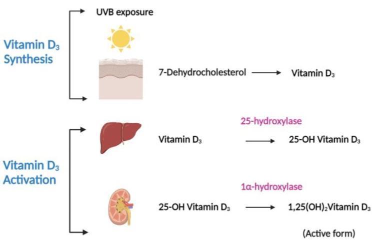
A pathophysiology diagram illustrating the multi-organ process of Vitamin D3 synthesis and activation. The diagram is divided into two primary stages: Synthesis and Activation. In the Synthesis stage, ultraviolet B (UVB) exposure acts on the skin, facilitating the conversion of the precursor 7-dehydrocholesterol into Vitamin D3 (cholecalciferol). The Activation stage depicts a sequential two-step hydroxylation process. First, within the liver, the enzyme 25-hydroxylase converts Vitamin D3 into 25-hydroxyvitamin D3 (calcifediol). Second, within the kidney, the enzyme 1̠-hydroxylase converts 25-OH Vitamin D3 into 1,25-dihydroxyvitamin D3 (calcitriol), which is labeled as the physiologically active form. The visual utilizes anatomical icons for the skin, liver, and kidney alongside biochemical flowcharts to demonstrate the endocrine pathway of Vitamin D metabolism, making it a key educational resource for understanding calcium homeostasis and metabolic bone disease foundations.
A pathophysiology diagram illustrating the multi-organ process of Vitamin D3 synthesis and activation. The diagram is divided into two primary stages: Synthesis and Activation. In the Synthesis stage, ultraviolet B (UVB) exposure acts on the skin, facilitating the conversion of the precursor 7-dehydrocholesterol into Vitamin D3 (cholecalciferol). The Activation stage depicts a sequential two-step hydroxylation process. First, within the liver, the enzyme 25-hydroxylase converts Vitamin D3 into 25-hydroxyvitamin D3 (calcifediol). Second, within the kidney, the enzyme 1̠-hydroxylase converts 25-OH Vitamin D3 into 1,25-dihydroxyvitamin D3 (calcitriol), which is labeled as the physiologically active form. The visual utilizes anatomical icons for the skin, liver, and kidney alongside biochemical flowcharts to demonstrate the endocrine pathway of Vitamin D metabolism, making it a key educational resource for understanding calcium homeostasis and metabolic bone disease foundations.

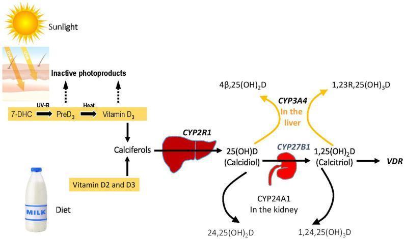
This pathophysiology diagram illustrates the metabolic pathway of Vitamin D homeostasis and its associated genetic enzymes. The process begins with two sources: endogenous synthesis in the skin via Sunlight (UV-B) converting 7-DHC to PreD3 and then to Vitamin D3 (Cholecalciferol) through heat, and dietary intake (Vitamin D2 and D3). These converge into 'Calciferols'. The primary activation pathway shows the liver enzyme CYP2R1 hydroxylating calciferols into 25(OH)D (Calcidiol). This metabolite then moves to the kidney, where CYP27B1 converts it to the active form, 1,25(OH)2D (Calcitriol), which binds to the Vitamin D Receptor (VDR). The diagram also highlights catabolic inactivation pathways: in the kidney, CYP24A1 converts 25(OH)D and 1,25(OH)2D into inactive products (24,25(OH)2D and 1,24,25(OH)3D respectively). In the liver, CYP3A4 facilitates an alternative inactivation route, producing 4β,25(OH)2D and 1,23R,25(OH)3D. Key genes involved in Vitamin D-Dependent Rickets (VDDR) are highlighted in bold, emphasizing the clinical significance of these enzymatic steps in mineral metabolism.

This pathophysiology diagram illustrates the metabolic pathway of Vitamin D homeostasis and its associated genetic enzymes. The process begins with two sources: endogenous synthesis in the skin via Sunlight (UV-B) converting 7-DHC to PreD3 and then to Vitamin D3 (Cholecalciferol) through heat, and dietary intake (Vitamin D2 and D3). These converge into 'Calciferols'. The primary activation pathway shows the liver enzyme CYP2R1 hydroxylating calciferols into 25(OH)D (Calcidiol). This metabolite then moves to the kidney, where CYP27B1 converts it to the active form, 1,25(OH)2D (Calcitriol), which binds to the Vitamin D Receptor (VDR). The diagram also highlights catabolic inactivation pathways: in the kidney, CYP24A1 converts 25(OH)D and 1,25(OH)2D into inactive products (24,25(OH)2D and 1,24,25(OH)3D respectively). In the liver, CYP3A4 facilitates an alternative inactivation route, producing 4β,25(OH)2D and 1,23R,25(OH)3D. Key genes involved in Vitamin D-Dependent Rickets (VDDR) are highlighted in bold, emphasizing the clinical significance of these enzymatic steps in mineral metabolism.

Reading File
Finding Sources
Reading File
Reading File
Reading File
I now have solid material from multiple authoritative sources. Let me compile the comprehensive answer.

Vitamin D and the Skin

1. The Skin as the Primary Source of Vitamin D

Vitamin D is unique among vitamins because under normal conditions the skin is its major source, not diet. The process begins when UVB radiation (290-315 nm) hits the epidermis and causes photolysis of 7-dehydrocholesterol (an intermediate in cholesterol synthesis that accumulates in skin) to form previtamin D3. Over the next few hours, previtamin D3 undergoes a temperature-dependent isomerization to the stable form cholecalciferol (vitamin D3), which then diffuses into the dermal vasculature.
  • Harper's Illustrated Biochemistry, 32nd Ed, p. 550
  • Harrison's Principles of Internal Medicine, 22E (2025)
In temperate climates, plasma vitamin D levels peak at end of summer and are lowest at end of winter. Beyond approximately 40° north or south latitude, UVB of the appropriate wavelength is nearly absent in winter months. Chronological skin aging substantially reduces the skin's capacity to photosynthesize vitamin D3.
Vitamin D synthesis pathway - skin to liver to kidney

2. Activation Pathway

Once absorbed into circulation, cholecalciferol undergoes two sequential hydroxylations:
StepOrganEnzymeProduct
1stLiver25-hydroxylase (CYP2R1)Calcidiol - 25(OH)D3 (main storage form, bound to vitamin D-binding globulin)
2ndKidney1α-hydroxylase (CYP27B1)Calcitriol - 1,25(OH)₂D3 (biologically active form)
Importantly, keratinocytes and other skin cells can also take up calcidiol from circulation and synthesize calcitriol locally - acting in an autocrine/paracrine manner within the skin itself.
  • Harrison's Principles of Internal Medicine, 22E

3. Roles of Vitamin D in Skin Biology

Keratinocyte Regulation

Vitamin D receptors (VDRs) are expressed on keratinocytes. Calcitriol:
  • Inhibits keratinocyte proliferation
  • Augments epidermal differentiation signaling
  • Supports skin barrier integrity
These effects are directly exploited therapeutically - topical vitamin D analogues (calcipotriol, calcitriol) are first-line treatments for psoriasis, where keratinocyte hyperproliferation is a hallmark.

Immune Modulation

VDRs are also expressed on T lymphocytes, B lymphocytes, and natural killer cells in the skin. Vitamin D promotes anti-inflammatory activity and helps regulate immune responses, which has implications for inflammatory dermatoses including atopic dermatitis, psoriasis, and other immune-mediated conditions.

Skin Barrier and Wound Healing

Recent evidence (2025) shows vitamin D accelerates wound healing by promoting macrophage polarization (M1→M2), enhancing angiogenesis, and stimulating keratinocyte migration via the Hippo pathway. It also plays a role in regulating tight junction proteins and barrier-related gene expression.

4. Vitamin D Deficiency and Skin Disorders

Conditions associated with vitamin D deficiency or impaired skin synthesis:
  • Congenital ichthyoses (lamellar ichthyosis, X-linked ichthyosis, epidermolytic hyperkeratosis) - defective vitamin D synthesis in affected skin
  • Xeroderma pigmentosum - sun avoidance leads to deficiency
  • Rickets (children) / Osteomalacia (adults) - the classic systemic consequence
  • Seborrheic dermatitis and atopic dermatitis - lower serum vitamin D levels observed
  • Dark skin tones - melanin competes with 7-dehydrocholesterol for UVB photons, significantly reducing synthesis efficiency; this is why exclusively breastfed infants with darker skin tones are at elevated risk for rickets
  • Fitzpatrick's Dermatology, Volumes 1-2

5. Vitamin D and Inflammatory Skin Disease - What the Evidence Shows

A 2024 systematic review and meta-analysis (Nielsen et al., PMID 39683522) pooled 11 RCTs with 686 patients and found that vitamin D supplementation significantly reduced atopic dermatitis severity (standardized mean difference = -0.41, 95% CI: -0.67 to -0.16, p < 0.01) compared to controls, in both children and adults. Larger-scale trials are still needed to confirm long-term benefit.

6. Vitamin D and Skin Cancer

  • Vitamin D has antiproliferative properties that may influence skin cancer development
  • Evidence links vitamin D deficiency to increased Breslow thickness in melanoma and possibly higher nevus count
  • Whether correcting deficiency causally reduces skin cancer risk remains uncertain
  • The sunscreen dilemma: theoretically, sunscreen could reduce vitamin D synthesis, but real-world use of insufficient amounts and increased sun-seeking behavior in sunscreen users means no significant impact on deficiency rates has been demonstrated
  • For Fitzpatrick skin Type II, as little as 5 minutes of summer midday sun, 2-3 times per week is sufficient to meet physiologic vitamin D requirements
  • Fitzpatrick's Dermatology, p. 2239

7. Therapeutic Applications in Dermatology

ApplicationAgentEvidence
PsoriasisTopical calcipotriol, calcitriolFirst-line topical therapy
Atopic dermatitisOral vitamin D supplementationMeta-analysis supports moderate benefit
Keloids / hypertrophic scarsIntralesional + systemic vitamin D2025 clinical trial data
Non-melanoma skin cancer preventionNicotinamide (vitamin B3, not D)Established
  • Dermatology 2-Volume Set 5e; Fitzpatrick's Dermatology

Key takeaway: The skin is both the factory and a major target organ for vitamin D. Its roles span keratinocyte differentiation, immune regulation, barrier integrity, and wound healing - with well-established therapeutic use in psoriasis and emerging evidence in atopic dermatitis and wound repair.
This is a shared conversation. Sign in to Orris to start your own chat.