
polycystic ovary syndrome diagnosis criteria ultrasound
polycystic ovary syndrome ultrasound multiple follicles
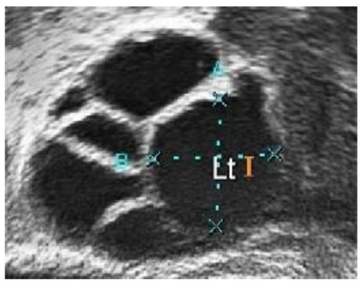
This diagnostic ultrasound image displays a close-up view of an ovary with characteristic features of Polycystic Ovary Syndrome (PCOS). The image shows multiple anechoic (dark), fluid-filled follicles of varying sizes and shapes, primarily circular and oval, distributed within the hyperechoic (bright) ovarian stroma. The largest follicle in the center-right is marked with calipers and annotations ('A', 'B', 'Et I') indicating manual measurement of its dimensions. The follicles exhibit well-defined borders against the surrounding light-pixel tissue, which has a grainy texture due to inherent speckle noise. Clinically, the presence of these numerous developing follicles is a primary diagnostic marker for polycystic morphology. This image is used in medical education to demonstrate the visualization of follicular accumulation and the process of diagnostic measurement during gynecological sonography.
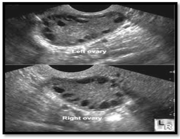
Diagnostic ultrasound imaging displaying a split-view of the left and right ovaries in a patient with Polycystic Ovary Syndrome (PCOS). Both ovaries exhibit classic sonographic features of polycystic morphology, characterized by an increased ovarian volume and a high count of small, peripherally arranged antral follicles. The follicles appear as multiple anechoic (dark), circular, or oval structures, typically measuring between 2 to 9 mm in diameter, creating a 'string of pearls' appearance. The central ovarian stroma is echogenic (brighter gray) and appears thickened or hypertrophied. Each ovary contains more than 12 visible follicles, meeting the Rotterdam diagnostic criteria for PCO. This clinical photograph is a primary tool in reproductive endocrinology and gynecology for evaluating menstrual irregularities, hyperandrogenism, and infertility. It serves as an educational reference for distinguishing normal follicular development from the pathological follicular arrest seen in PCOS.
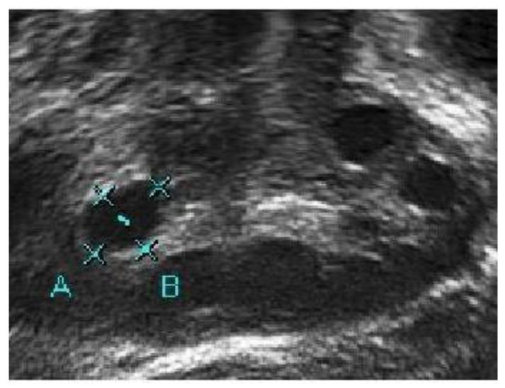
This diagnostic image is a B-mode ultrasound scan of a human ovary, demonstrating characteristic features of Polycystic Ovary Syndrome (PCOS). The image shows multiple hypoechoic (dark) regions representing ovarian follicles. Two specific regions are labeled 'A' and 'B'. Region A is a rounded, anechoic structure marked with measurement calipers to determine follicle size. Region B is a larger, more elongated hypoechoic area situated lower in the stroma. The image quality is characterized by low contrast and significant speckle noise, which is typical for raw clinical ultrasound data before digital enhancement. These factors result in poorly defined follicle boundaries that blend into the surrounding echogenic ovarian stroma. The educational focus of this image is on the visual identification and manual measurement of follicles for PCOS diagnosis, highlighting the technical challenges in distinguishing fluid-filled cysts from adjacent tissue and pelvic capillaries in low-light-intensity ultrasound captures.
USG Report Interpretation - Meema, 19F
What the Report Shows
- Liver, gallbladder, CBD, pancreas, spleen - all normal
- Both kidneys - normal size, shape, no hydronephrosis, no calculi
- Urinary bladder - normal
- Uterus - anteverted (normal position), size 39x86x28 mm (normal), endometrial thickness 5.0 mm (normal for proliferative phase)
- Cervix - normal, no intrauterine collection
The Key Finding - PCOS Morphology
| Finding | Right Ovary | Left Ovary |
|---|---|---|
| Volume | 10.4 cc | 11 cc |
| Follicle size | 2-3 mm multiple | 2-3 mm multiple |
| Follicle arrangement | Peripheral ("string of pearls") | Peripheral |
| FNPS | >10 | >10 |
What is PCOS Morphology on Ultrasound?
- Ovarian volume > 10 mL (this patient: R=10.4 mL, L=11 mL - both borderline to mildly elevated)
- Follicle number per ovary ≥ 12 follicles measuring 2-9 mm
- Multiple small peripheral follicles (2-3 mm) - the classic "string of pearls" sign
- Bilateral ovarian volumes slightly above 10 cc
- FNPS > 10 in both ovaries
Important: USG Alone Does NOT Diagnose PCOS
- Oligo/anovulation (irregular or absent periods)
- Clinical or biochemical hyperandrogenism (excess hair, acne, elevated androgens)
- Polycystic ovarian morphology on USG (which this patient has) ✓
Recommended Next Steps
- LH, FSH (LH:FSH ratio > 2:1 is suggestive of PCOS)
- Total and free Testosterone
- DHEA-S (to rule out adrenal source)
- Prolactin (to rule out hyperprolactinemia)
- TSH (thyroid disorders can mimic PCOS)
- AMH (anti-Mullerian hormone - typically elevated in PCOS)
- Fasting insulin and blood glucose (for insulin resistance assessment)
- Menstrual history (regularity, cycle length)
- Signs of hyperandrogenism (hirsutism, acne, hair loss)
- BMI and waist circumference
- Family history of PCOS or diabetes
Summary
| Status | |
|---|---|
| Ultrasound finding | Polycystic ovarian morphology (bilateral) |
| Ovarian volumes | Mildly increased (>10 cc both sides) |
| Other pelvic/abdominal organs | Normal |
| Diagnosis confirmed? | No - USG alone is insufficient |
| Action needed | Hormonal blood tests + clinical correlation |
Note: Having PCOM on USG alone - especially at 19 years - does not mean PCOS is confirmed. Around 20-30% of normal women of reproductive age can have polycystic-appearing ovaries without the syndrome. A full clinical and hormonal evaluation is needed before any treatment is started.
Find recent clinical guidelines for PCOS management in adolescents
polycystic ovary syndrome adolescents management guidelines
PCOS adolescent management guidelines 2023 2024 international evidence-based
PCOS adolescent treatment lifestyle metformin oral contraceptive
PMID: 37804097
PMID: 40069730
PMID: 38595887
PMID: 42105324
PCOS Management in Adolescents - 2023 International Guidelines
1. Diagnosis in Adolescents - Key Differences from Adults
Required for Diagnosis (BOTH must be present):
| Criterion | Details |
|---|---|
| Irregular menstrual cycles | Defined based on time since menarche (see below) |
| Clinical OR biochemical hyperandrogenism | Hirsutism (Ferriman-Gallwey score), elevated testosterone, elevated free androgen index |
Menstrual Irregularity Definitions by Pubertal Stage:
- < 1 year post-menarche: Cycles < 21 days or > 45 days
- 1-3 years post-menarche: Cycles < 21 days or > 35 days (or < 8 cycles/year)
- > 2-3 years post-menarche: Cycles < 21 days or > 35 days (same as adult criteria)
What is NOT Used for Diagnosis in Adolescents:
- Pelvic ultrasound (PCOM) - NOT recommended until 8 years post-menarche, because multifollicular ovaries are physiologically normal in teens
- AMH levels - NOT recommended as a diagnostic criterion due to poor specificity in adolescents
For this patient (19-year-old Meema): The ultrasound showing PCOM cannot confirm PCOS diagnosis on its own. Clinical features (cycle regularity, signs of hyperandrogenism) and hormonal profile are mandatory. If she is < 8 years post-menarche (likely, given age 19), the USG PCOM finding is NOT a valid diagnostic criterion.
"At Risk" Category (new 2023 concept):
2. Assessment - What to Screen For
| Domain | What to Screen |
|---|---|
| Metabolic | Fasting glucose, lipids, blood pressure, BMI/waist circumference |
| Psychological | Anxiety, depression (use validated tools - PHQ-9, GAD-7) |
| Sleep | Screen for obstructive sleep apnea if obese |
| Reproductive | Future fertility counseling |
| Thyroid / Prolactin | To exclude mimics |
3. Management
First Line - Lifestyle Modification (ALL adolescents)
- Healthy diet - no single best diet; low glycaemic index and Mediterranean diets have evidence
- Regular physical activity - 150 min/week moderate-intensity or 75 min/week vigorous activity
- Weight management - prevent weight gain in all; modest weight loss (5-10%) in those with overweight significantly improves menstrual regularity, hyperandrogenism, and metabolic markers
- Weight stigma must be minimized - healthcare providers should seek permission before discussing weight and frame it around health, not appearance
Second Line - Pharmacological Treatment
For Menstrual Irregularity + Hyperandrogenism:
- Regulates cycles, reduces androgens, treats acne and hirsutism
- No specific preparation is recommended - use lowest effective dose
- Prefer preparations with low androgenic progestins (e.g., norgestimate, desogestrel, drospirenone)
- Duration: minimum 3-6 months to assess hirsutism response
- Always discuss VTE risk, and counsel on contraception/STI prevention (Machado et al., JCEM 2026)
For Metabolic Features (insulin resistance, impaired glucose tolerance):
- Improves insulin sensitivity, supports weight management, helps cycle regulation
- Also considered when COCP is contraindicated or not desired
- Start low, titrate (500 mg OD → 1500-2000 mg/day with food) to minimize GI side effects
- Evidence supports its use as first-line for metabolic comorbidities in adolescents
Additional Options (evidence emerging):
| Drug | Use | Evidence Level |
|---|---|---|
| Anti-androgens (spironolactone, finasteride) | Hirsutism refractory to COCP | Limited in adolescents; use with contraception |
| Inositol (myo-inositol) | Insulin resistance, cycle regulation | Emerging, not yet guideline-endorsed |
| Vitamin D supplementation | If deficient | Some benefit on metabolic markers |
| GnRH antagonists | Investigational | Still in trials (SPIOMET4HEALTH trial [PMID: 37715279]) |
Note on SPIOMET (spironolactone + pioglitazone + metformin): A 2023 multicentre phase II RCT protocol is evaluating this combination vs. lifestyle alone in adolescent PCOS [PMID: 37715279] - results are awaited.
4. Psychological Care
- Screen routinely for anxiety, depression, and eating disorders
- PCOS adolescents have ~2x higher rates of anxiety and depression vs. peers
- Cognitive behavioural therapy (CBT) and mindfulness are recommended adjuncts
- Use non-stigmatizing language around weight and fertility
5. Long-Term Planning
- Develop a lifelong health plan starting in adolescence
- Ensure transition of care to adult endocrinology/gynecology at 18-21 years
- Counsel on long-term risks: type 2 diabetes, cardiovascular disease, endometrial hyperplasia (from chronic anovulation)
- Reassess diagnosis at transition to adult care - some adolescents diagnosed "at risk" may no longer meet criteria in adulthood
Summary Table
| Aspect | 2023 Guideline Recommendation |
|---|---|
| Minimum diagnostic criteria | Irregular cycles + hyperandrogenism (BOTH required) |
| Role of USG in adolescents | Not recommended for diagnosis until 8 yrs post-menarche |
| Role of AMH | Not recommended for diagnosis |
| First-line treatment | Lifestyle modification |
| First-line pharmacotherapy | COCP (for cycle/hyperandrogenism) + Metformin (for metabolic) |
| Psychological screening | Mandatory - screen for depression, anxiety |
| Anti-androgens | Second-line; use with contraception |
| Long-term | Transition to adult care, metabolic surveillance |
- Peña AS et al. International evidence-based recommendations for PCOS in adolescents. BMC Med. 2025 [PMID: 40069730]
- Machado IFR et al. Approach to diagnosis and treatment of PCOS in adolescent patients. JCEM. 2026 [PMID: 42105324]
- Vadan RL et al. Comparative management methods for adolescents with PCOS. Cureus. 2024 [PMID: 38595887]
- 2023 International PCOS Network Guideline (Teede HJ et al., Fertil Steril 2023;120:767-793)