
CRP test
CRP test C-reactive protein clinical uses reference range 2025
CRP C-reactive protein inflammation acute phase response diagram
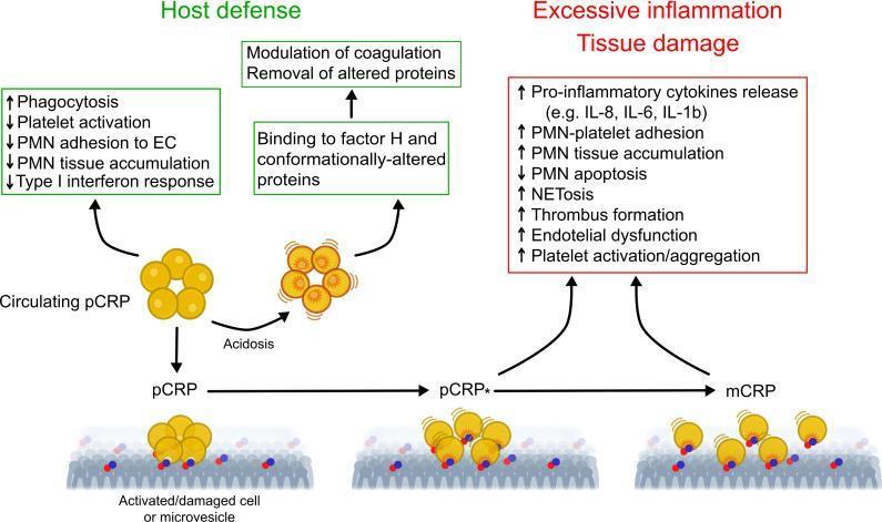
This pathophysiology diagram illustrates the conformational changes of C-reactive protein (CRP) and their divergent biological roles in the inflammatory response. The diagram depicts three distinct isomers: native pentameric CRP (pCRP), a partially dissociated pentamer (pCRP*), and monomeric CRP (mCRP). Circulating pCRP, shown as a yellow disc-shaped pentamer, is associated with 'Host defense' mechanisms, including increased phagocytosis and decreased platelet and PMN activation. Under conditions of acidosis or upon binding to activated/damaged cell membranes or microvesicles, pCRP undergoes structural transformation. The transition from membrane-bound pCRP to pCRP* and finally to individual mCRP subunits is shown along the bottom. These conformationally-altered forms (pCRP* and mCRP) are linked to 'Excessive inflammation / Tissue damage,' characterized by increased pro-inflammatory cytokine release (IL-8, IL-6, IL-1b), PMN-platelet adhesion, NETosis, thrombus formation, and endothelial dysfunction. The graphic emphasizes how the local microenvironment modulates CRP's function from a protective, anti-inflammatory circulating protein to a potent pro-inflammatory tissue-bound mediator.
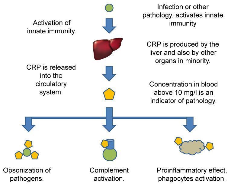
A pathophysiology flow diagram illustrating the synthesis, systemic release, and immunological functions of C-reactive protein (CRP). The pathway begins with an initial trigger, such as infection or other pathology, leading to the activation of innate immunity. The central vertical axis shows the liver as the primary site of CRP production, followed by the release of CRP (represented by a yellow pentagon) into the circulatory system. Text annotations specify that a blood concentration above 10 mg/L serves as a clinical indicator of pathology. The lower portion of the diagram branches into three distinct downstream effects: 1) Opsonization of pathogens, showing CRP bound to a green spherical target; 2) Complement activation, depicted by CRP interacting with a protein complex; and 3) Pro-inflammatory effects, specifically the activation of phagocytes (represented by a grey irregular cellular structure). This schematic summarizes CRP's role as an acute-phase reactant in human host defense and its diagnostic utility in clinical medicine.
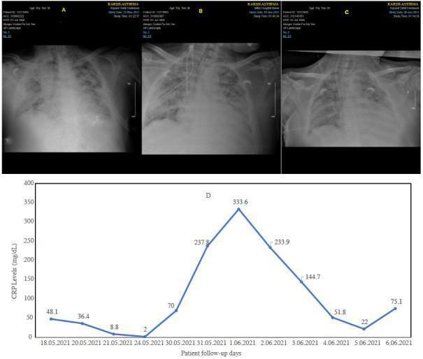
This composite educational graphic displays a serial progression of chest radiographs (A, B, C) and a corresponding line graph of C-reactive protein (CRP) levels (D) in a patient with Acute Respiratory Distress (ARD). Panel A shows initial bilateral diffuse pulmonary opacities and evidence of mechanical ventilation. Panel B, representing the third day of re-admission, demonstrates worsening bilateral alveolar and interstitial infiltrates with visible medical devices, including an endotracheal tube and central venous lines. Panel C, on the sixth day, reveals a slight improvement in lung aeration compared to B, though significant opacity remains. Graph D tracks the biochemical inflammatory response, showing a dramatic spike in CRP levels (reaching 333.6 mg/dL) coinciding with clinical deterioration and septic shock, followed by a decline. This clinical timeline illustrates the correlation between radiological findings of progressive lung injury and laboratory markers of systemic inflammation in critical care medicine.
C-Reactive Protein (CRP) Test
What Is CRP?
- Quick Compendium of Clinical Pathology, 5th ed.
Two Types of CRP Tests
| Feature | Standard CRP | High-Sensitivity CRP (hs-CRP) |
|---|---|---|
| Detection limit | ~5-10 mg/L | < 0.3 mg/L |
| Main use | Acute infection, inflammation monitoring | Cardiovascular risk assessment |
| Population screened | Symptomatic patients | Apparently healthy or intermediate-risk individuals |
- Tietz Textbook of Laboratory Medicine, 7th ed.
- Henry's Clinical Diagnosis and Management by Laboratory Methods
Reference Ranges
Standard CRP (reported in mg/dL by many US labs; mg/L by most others)
| CRP Level (mg/dL) | Classification | Possible Causes |
|---|---|---|
| < 0.3 | Normal | Normal |
| 0.3 - 1.0 | Minor elevation | Obesity, pregnancy, depression, diabetes, sedentary lifestyle |
| 1.0 - 10.0 | Moderate elevation | RA, autoimmune disease, MI, pancreatitis, bronchitis |
| > 10.0 | Marked elevation | Bacterial/viral infection, systemic vasculitis, major trauma |
| > 50.0 | Severe elevation | Acute bacterial infection (~90% of cases) |
Note: 1 mg/dL = 10 mg/L. Always check units on your lab report.
hs-CRP Population Distribution (mg/L)
| Population | 25th %ile | 50th %ile | 75th %ile | 95th %ile |
|---|---|---|---|---|
| American women | 0.6 | 1.5 | 3.5 | 9.1 |
| American men | 0.8 | 1.5 | 3.2 | 8.6 |
| European women | 0.9 | 1.7 | 3.4 | 8.8 |
| European men | 0.8 | 1.6 | 3.3 | 8.6 |
- Tietz Textbook of Laboratory Medicine, 7th ed., Table 36.26
hs-CRP Cardiovascular Risk Stratification (AHA/CDC)
| hs-CRP (mg/L) | Cardiovascular Risk |
|---|---|
| < 1.0 | Low |
| 1.0 - 3.0 | Average |
| > 3.0 | High |
Clinical Uses
1. Infection and Inflammation
- CRP > 50 mg/L is linked to bacterial infection in ~90% of cases.
- Guides antibiotic decisions (especially in neonates, sepsis workup, community-acquired pneumonia).
- Monitors treatment response - levels fall quickly when therapy is effective.
- Useful for distinguishing bacterial vs. viral infection (bacterial causes larger rises).
- Monitors flares in autoimmune/inflammatory diseases (RA, IBD, lupus, vasculitis).
2. Cardiovascular Risk Assessment (hs-CRP)
- Intermediate-risk patients (Framingham 10-year risk 10-20%) to help guide therapy decisions.
- NOT recommended for universal population screening.
- NOT useful for secondary prevention (management is aggressive regardless).
- Robbins, Cotran & Kumar Pathologic Basis of Disease
- Tietz Textbook of Laboratory Medicine, 7th ed.
3. Post-Surgical and Critical Care Monitoring
4. Neonatal Sepsis
Mechanism and Biological Role
Analytical Considerations
- Sample: Serum or plasma. No fasting required - CRP is unaffected by food intake and shows almost no circadian variation.
- Methods: Immunoturbidimetry, nephelometry, immunoassay; newer point-of-care devices (e.g., ProciseDx CRP Assay, FDA-approved 2022) give results in < 5 minutes.
- Serial testing: For cardiovascular risk, take two measurements ≥ 2 weeks apart for reliability (same protocol as lipid panels).
- Intraindividual variability: Relatively low in clinically stable patients - biological variability is comparable to serum cholesterol.
- Interfering factors: Obesity, HRT (oral estrogen raises CRP), pregnancy, and metabolic syndrome all elevate CRP independently of disease.
CRP vs. ESR
| Feature | CRP | ESR |
|---|---|---|
| Response to inflammation | Hours (6-12h) | Days (24-48h) |
| Fall after recovery | Rapid | Slow |
| Influenced by age/sex | Minimally | Significantly |
| Best use | Monitoring acute changes, treatment response | Chronic inflammatory conditions, myeloma |
| Current preference | Preferred by most guidelines | Supplementary |