
/generateimage 3dcutaway heart
3D cutaway diagram of the human heart, showing internal chambers (left and right atria, left and right ventricles), valves (mitral, tricuspid, aortic, pulmonary), major vessels (aorta, pulmonary artery, vena cava), myocardium wall thickness, chordae tendineae, and septum. Medical illustration style, detailed anatomical labels, cross-section view revealing interior structure, realistic rendering with depth and shading.
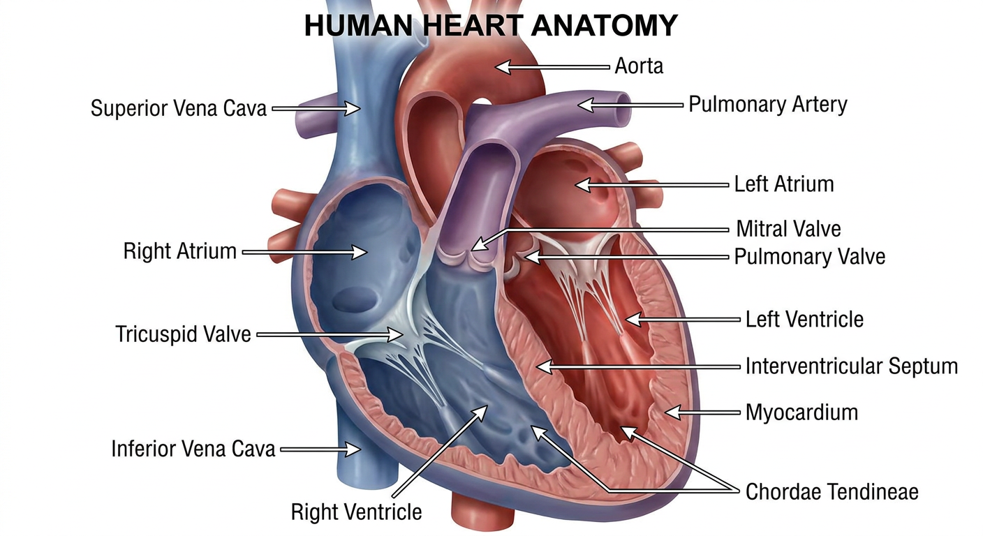
- Chambers - left/right atria and ventricles
- Valves - mitral, tricuspid, aortic, and pulmonary
- Major vessels - aorta, pulmonary artery, and venae cavae
- Internal structures - septum, myocardial walls, and chordae tendineae
Search for high-resolution cardiac imaging examples
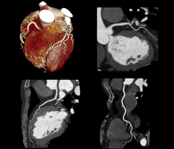
high resolution cardiac imaging examples echocardiography MRI CT angiography
cardiac MRI high resolution heart imaging gallery medical
Echocardiography (Echo)

Cardiac MRI


CT Angiography (CTA)
Chest X-ray with Cardiac Anatomy

Modality Comparison
| Modality | Radiation | Tissue Detail | Motion Assessment | Best For |
|---|---|---|---|---|
| Cardiac MRI | None | Excellent | Good | Myocardial function, viability, tumors |
| CT Angiography | Yes | Good (coronaries) | Limited | Coronary artery disease, bypass grafts |
| Echocardiography | None | Moderate | Excellent | Real-time valve/wall motion, bedside |
| PET/SPECT | Yes | Moderate | Limited | Perfusion, metabolic activity |
/generateimage 3d cutaway hand with detailed label
3D cutaway anatomical illustration of the human hand, cross-section revealing internal structures with detailed labels: bones (carpals, metacarpals, phalanges - proximal, middle, distal), tendons (flexor digitorum profundus, flexor digitorum superficialis, extensor tendons), muscles (thenar, hypothenar, interossei, lumbricals), nerves (median nerve, ulnar nerve, radial nerve branches), arteries (radial artery, ulnar artery, palmar arches, digital arteries), ligaments (collateral ligaments, volar plate), synovial sheaths, skin layers. Medical textbook illustration style, clean white background, precise anatomical labels with leader lines, high detail 3D rendering with transparent skin layer showing interior structures.
/generateimage 3d cutaway hand anatomy with detailed label
3D cutaway anatomical illustration of the human hand showing internal structures with detailed labels. Cross-section reveals: bones (carpals, metacarpals, proximal/middle/distal phalanges), flexor and extensor tendons, thenar and hypothenar muscles, interossei and lumbrical muscles, median nerve, ulnar nerve, radial and ulnar arteries, palmar arterial arches, digital arteries, synovial sheaths, collateral ligaments, and skin layers. Medical textbook illustration style, precise label lines pointing to each structure, 3D rendering with semi-transparent skin revealing interior anatomy, white background, high resolution.
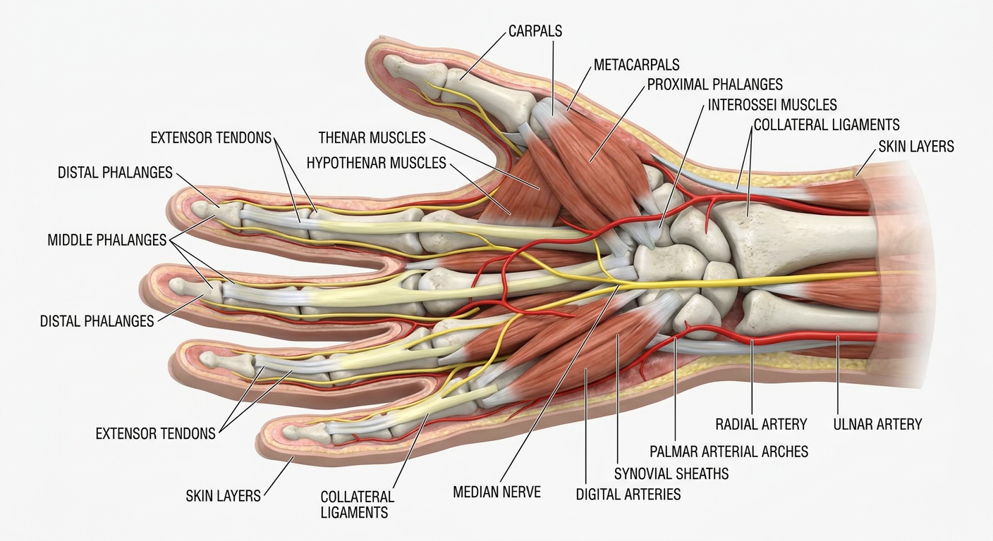
- Carpals, metacarpals, and phalanges (proximal, middle, distal)
- Flexor/extensor tendons, thenar & hypothenar muscles, interossei, lumbricals
- Median, ulnar & radial nerves; radial/ulnar arteries; palmar arches; digital arteries
- Synovial sheaths, collateral ligaments, skin layers
Create an anatomical poster of hand nerve pathways
Anatomical medical poster of hand nerve pathways. Shows both palmar and dorsal views of the hand with clearly illustrated nerve distributions. Median nerve highlighted in yellow covering the thenar eminence and lateral 3.5 fingers, ulnar nerve in blue covering hypothenar region and medial 1.5 fingers, radial nerve in green covering dorsal lateral hand. Includes: carpal tunnel cross-section inset, nerve branch labels with leader lines, sensory distribution color map, motor branch annotations for thenar muscles and interossei, recurrent branch of median nerve, deep branch of ulnar nerve. Title 'HAND NERVE PATHWAYS' at top, professional medical illustration style, clean layout, white background, poster format.
429 Too Many Requests. {'message': '{\n "error": {\n "code": 429,\n "message": "Resource exhausted. Please try again later. Please refer to https://cloud.google.com/vertex-ai/generative-ai/docs/error-code-429 for more details.",\n "status": "RESOURCE_EXHAUSTED"\n }\n}\n', 'status': 'Too Many Requests'}
We encountered an error while processing your request. Please try again or reach out to us for help.