
Answer with diagrams and all the necessary details
I'd be happy to help! However, your question is quite broad. Could you clarify what you'd like me to explain with diagrams?
Asking for Preferences
What topic would you like me to explain with diagrams and full details?
Describe in detail the MAMMARY GLAND under the following headings: a. Extent b. Relations c. Structure d. Blood Supply e. Lymphatic drainage f. Applied Anatomy
Reading File
Now let me search the medical library for mammary gland content across multiple headings simultaneously.
Finding Sources
Finding Sources
Finding Sources
Good. Now let me read the key sections from Gray's Anatomy and other sources in parallel.
Reading File
Reading File
Reading File
Now let me get the main breast anatomy section from Gray's Anatomy, and the histology section, in parallel.
Finding Sources
Reading File
Reading File
Now let me read the main Gray's Anatomy breast section for extent, relations, structure, and blood supply.
Reading File
Reading File
Now let me read the dedicated breast section more thoroughly - the section on structure, blood supply, and lymphatics from Gray's - and fetch some key diagrams.
Reading File
Loading Image
Loading Image
Excellent! I now have rich textbook content and two excellent diagrams. Let me fetch a few more diagrams - the axillary process diagram and the lymph nodes diagram.
Loading Image
Loading Image
Reading File
Loading Image
Loading Image
I now have all the content and diagrams needed. Let me compile the comprehensive answer.
The Mammary Gland - Detailed Description
Sources: Gray's Anatomy for Students (9780323934237), Creasy & Resnik's Maternal-Fetal Medicine (9780323828499), Histology: A Text and Atlas (9781975181512)
a. Extent
The mammary gland is a modified sweat gland situated in the superficial fascia of the pectoral region, anterior to the thoracic wall. It lies between skin and the deep pectoral fascia, from which it is separated by the retromammary space - a layer of loose connective tissue that allows limited mobility.
Vertical extent: Rib II (2nd rib) to Rib VI (6th rib)
Horizontal extent: Lateral border of the sternum to the midaxillary line
Nipple position: At the center of the areola, typically at the level of the 4th intercostal space, just below the midclavicular line
Axillary (Spence's) Tail: The superolateral region of the breast extends along the inferior margin of pectoralis major toward the axilla. In some individuals, it passes around the margin of the muscle, perforates the deep fascia, and enters the axilla - though it rarely reaches the apex of the axilla.
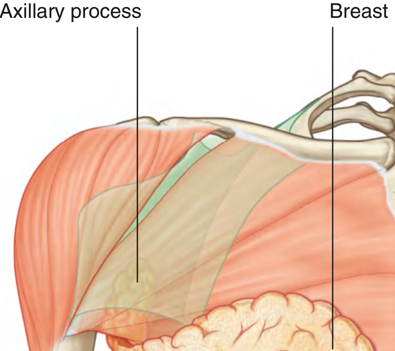
b. Relations
Anterior
- Skin with overlying superficial fascia
- Nipple and areola at the 4th intercostal space level
Posterior (Deep surface)
- Retromammary space (loose connective tissue) - allows the breast to slide on underlying structures
- Deep (pectoral) fascia covering pectoralis major (medially), serratus anterior (laterally), and external oblique/rectus abdominis (inferiorly)
Superior
- Clavicle
- Branches of the supraclavicular nerves
Inferior
- Upper anterior abdominal wall
Medial
- Sternum
Lateral
- Axilla (with the axillary tail projecting into it)
Muscles related to the breast:
| Position | Muscle |
|---|---|
| Central/medial deep | Pectoralis major |
| Lateral deep | Serratus anterior |
| Inferolateral deep | External oblique |
| Inferomedial | Rectus abdominis |
c. Structure
The mammary gland is a compound tubuloalveolar gland derived from modified sweat glands of the ectoderm.
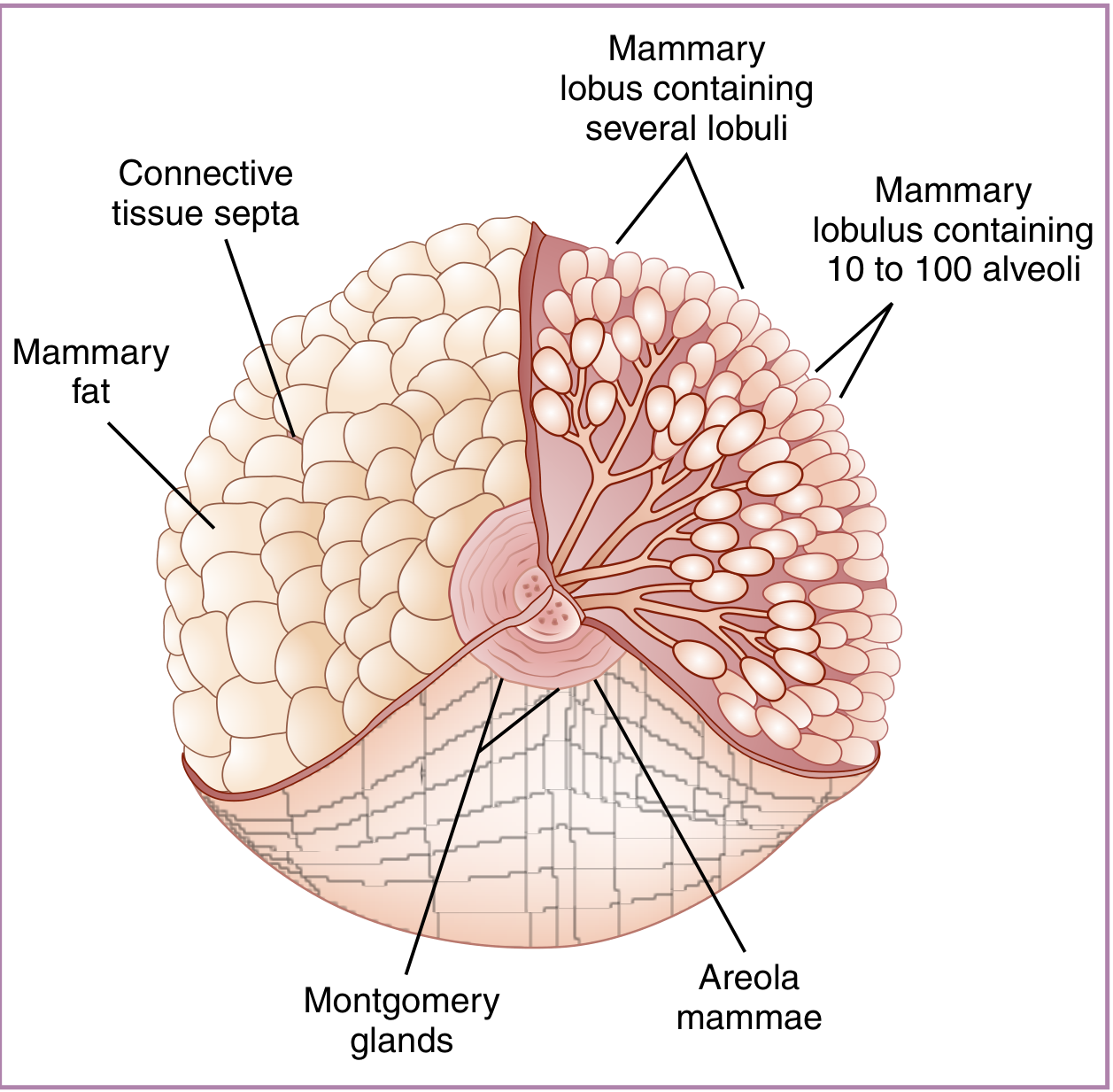
Parenchyma (Glandular tissue)
- 15-20 lobes arranged radially around the nipple, separated by connective tissue septa (Cooper's ligaments)
- Each lobe contains several lobules
- Each lobule contains 10-100 alveoli (secretory units)
- Each lobe drains via a lactiferous duct that opens independently at the nipple tip
- Just before opening at the nipple, each lactiferous duct dilates into a lactiferous sinus (ampulla), which acts as a temporary reservoir for milk
Stroma
- Dense irregular connective tissue (interlobular) and loose connective tissue (intralobular)
- Cooper's (Suspensory) Ligaments: Fibrous bands that extend from the dermis of the skin to the deep fascia, providing structural support. Distortion or shortening of these by carcinoma produces characteristic skin dimpling
- Adipose tissue: The predominant component in non-lactating women; fat surrounds the glandular elements and gives the breast its smooth, rounded contour
Nipple and Areola
- Nipple: Conical elevation at the center of the areola; contains smooth muscle fibers (erectile), richly innervated with sensory and pain fibers; contains 4-18 lactiferous duct openings (5-8 main ducts)
- Areola: Circular pigmented zone (avg. 15-16 mm diameter, enlarges in pregnancy); contains Montgomery glands (modified sebaceous/sweat glands) that hypertrophy during pregnancy and lactation to lubricate and protect the nipple
- Verrucous surface of nipple; has sebaceous and apocrine sweat glands but no hair
Histological Stages
| Stage | Features |
|---|---|
| Inactive (resting) | Sparse parenchyma; mainly ducts; intralobular loose CT + interlobular dense CT with adipocytes; alveoli absent (represented as cellular thickenings) |
| Pregnant/Lactating | Extensive alveolar proliferation; alveoli lined by cuboidal secretory and myoepithelial cells; lipid droplets in cells; reduced stroma |
| Post-weaning (Involution) | Alveoli regress; gland returns to near-resting state |
In males
- Rudimentary breast consisting of small ducts (often cords of cells) that do not extend beyond the areola
d. Blood Supply
The breast has a rich, multi-source blood supply reflecting its dual anatomical relationship with both the thoracic wall and upper limb.
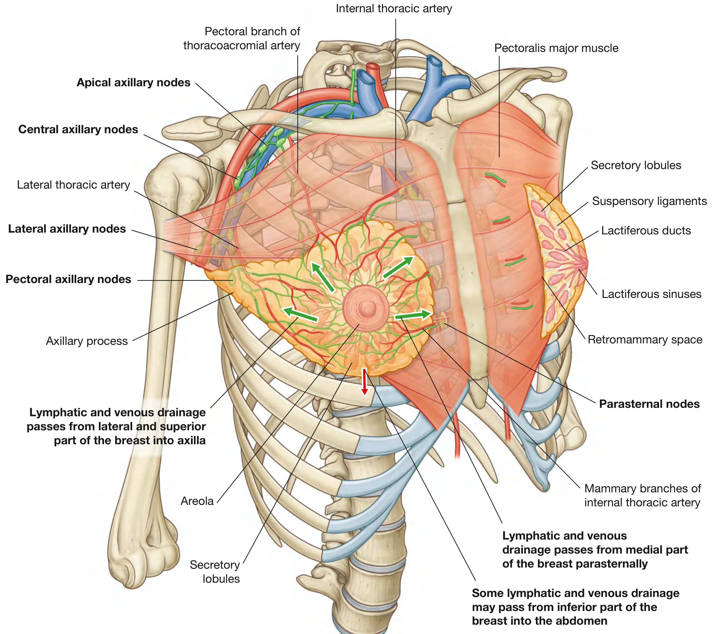
Arterial Supply
| Source | Contribution |
|---|---|
| Internal thoracic artery (internal mammary artery) - perforating branches (2nd-4th spaces) | Anteromedial breast (major supply ~60%) |
| Lateral thoracic artery (branch of axillary artery) | Lateral and superior breast |
| Thoracoacromial artery - pectoral branch | Superior breast |
| Superior thoracic artery (branch of axillary artery) | Superior breast |
| Subscapular artery (branch of axillary artery) | Lateral breast |
| Anterior intercostal arteries (2nd-4th branches) | Lateral perforating branches |
| Posterior intercostal arteries (2nd-5th) | Deep breast tissue |
The internal mammary artery and lateral thoracic artery are the primary sources of blood supply.
Venous Drainage
Veins follow a pattern parallel to the arteries and drain into:
- Internal thoracic (mammary) vein - anteromedially
- Axillary vein - laterally and superiorly
- Intercostal veins - posteriorly and inferiorly
Note: The intercostal veins connect with the vertebral venous plexus (Batson's plexus), providing a route for haematogenous metastasis of breast cancer to the vertebral column, skull, and pelvis - without going through the pulmonary circulation.
Innervation
- Anterior and lateral cutaneous branches of the 2nd-6th intercostal nerves
- Nipple: specifically innervated by the 4th intercostal nerve
- Efferent nerves are sympathetic adrenergic; no parasympathetic or cholinergic supply
- Sensory stimulation of nipple/areola triggers release of prolactin (adenohypophysis) and oxytocin (neurohypophysis)
e. Lymphatic Drainage
Lymphatic drainage is of paramount clinical importance due to its role in the spread of breast carcinoma. The lymphatics of the breast originate in lymph capillaries of the mammary connective tissue.
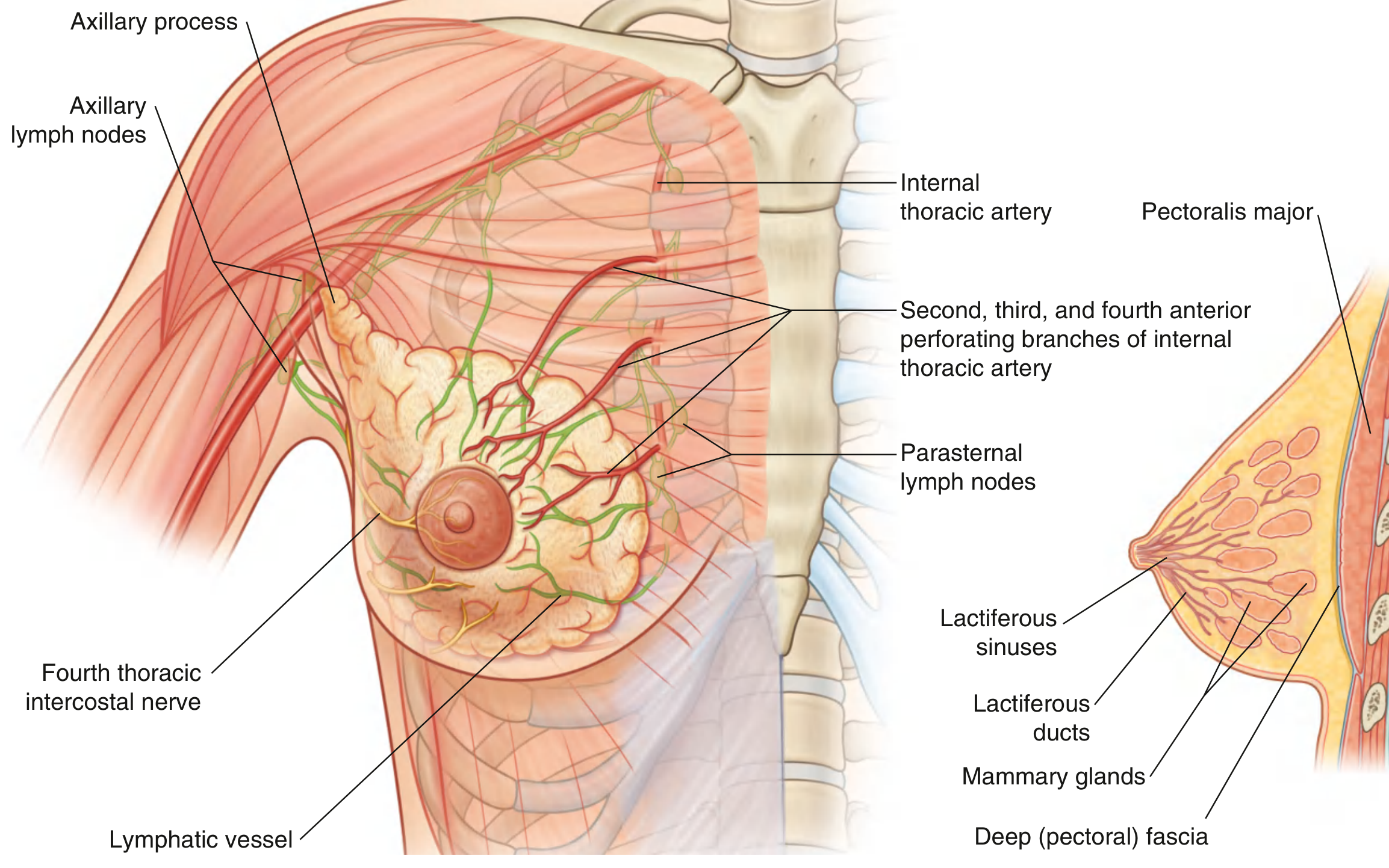
Primary Drainage Routes
1. Axillary Route (~75% of total drainage)
- Drains the lateral, superior, and central parts of the breast
- Drains into the axillary lymph nodes, which are divided into 5 groups:
| Group | Location | Receives from |
|---|---|---|
| Pectoral (anterior) | Along lateral thoracic vessels | Most of anterior chest wall, most of the breast |
| Subscapular (posterior) | Along subscapular vessels | Posterior thoracic wall, posterior shoulder |
| Humeral (lateral) | Along axillary vein | Upper limb (arm) |
| Central | Embedded in axillary fat | Pectoral, subscapular, humeral groups |
| Apical | Most superior, medial to pectoralis minor | All other axillary groups + superior breast; drains via subclavian trunk |
Efferent pathway: Apical nodes → Subclavian trunk → Joins subclavian vein at junction with internal jugular vein (right side) or joins thoracic duct (left side)
2. Parasternal (Internal Mammary) Route (~20-25%)
- Drains the medial and central parts of the breast
- Lymphatics accompany perforating branches of the internal thoracic artery
- Drain into parasternal (internal mammary) nodes along the internal thoracic vessels on the deep surface of the anterior thoracic wall
- Parasternal nodes drain into the bronchomediastinal trunks
3. Intercostal Route (minor)
- Some drainage from the posterior and inferior breast
- Via lymphatics that follow lateral branches of posterior intercostal arteries
- Drain into intercostal nodes near the heads and necks of ribs
- Intercostal nodes drain into the thoracic duct or bronchomediastinal trunks
4. Inferior (Subdiaphragmatic) Route
- Small amount of drainage from the inferior breast
- May communicate with subdiaphragmatic lymphatics and hepatic lymphatics (via the falciform ligament)
Summary Diagram (Lymphatic Routes)
BREAST LYMPHATIC DRAINAGE
|
┌───────────────┼───────────────┐
▼ ▼ ▼
AXILLARY (~75%) PARASTERNAL INTERCOSTAL
(lateral/sup.) (~20-25%) (minor, post.)
| (medial) |
Pectoral nodes Parasternal Intercostal
→ Subscapular nodes nodes
→ Humeral | |
→ Central Bronchomediastinal Thoracic duct /
→ Apical trunk Bronchomediastinal
| trunk
Subclavian trunk
|
Venous junction
(subclavian + IJ vein)
f. Applied Anatomy
1. Breast Carcinoma
Breast cancer is one of the most common malignancies in women. It arises from:
- Acini (terminal duct lobular units - TDLU) - lobular carcinoma
- Lactiferous ducts - ductal carcinoma (most common)
Spread routes:
- Lymphatic: To axillary nodes (most common), parasternal nodes, supraclavicular nodes, and contralateral breast
- Haematogenous: Via Batson's vertebral venous plexus (through intercostal veins) → vertebral column, skull, pelvis; or via axillary/internal thoracic veins → lungs, liver, bone
- Direct invasion: Into pectoral muscles, chest wall, skin
Staging requires:
- Size of primary tumor
- Exact site
- Number and sites of lymph node spread
- Distant metastasis (lungs, liver, bone - detected by CT, bone scan, PET-CT)
2. Skin Changes in Carcinoma
| Sign | Mechanism |
|---|---|
| Skin dimpling / pitting | Carcinoma invades and shortens Cooper's (suspensory) ligaments |
| Peau d'orange (orange-peel appearance) | Lymphatic obstruction causes skin oedema; hair follicles tethered to skin produce pitting |
| Nipple retraction / inversion | Carcinoma invades and shortens the lactiferous ducts |
| Nipple discharge | Intraductal carcinoma or Paget's disease |
3. Lymphoedema After Mastectomy
- Axillary lymph node clearance (ALND) or radiotherapy to the axilla disrupts lymphatic drainage of the upper limb
- Results in chronic lymphoedema of the arm (brawny, non-pitting oedema)
- Clinicians now favor sentinel lymph node biopsy (SLNB) to reduce this complication
4. Sentinel Lymph Node Biopsy (SLNB)
- The sentinel node is the first lymph node to receive lymphatic drainage from the primary tumor
- Identified by injecting blue dye or radioactive tracer around the tumor
- If sentinel node is clear of metastasis, full axillary clearance is avoided
- Reduces risk of lymphoedema significantly
5. Axillary Tail (Spence's Tail)
- Clinicians must examine the axillary tail when assessing breast lumps; pathology (carcinoma, fibroadenoma) can occur in this extension
- It may be mistaken for an axillary lymph node
6. Mammography
- The primary imaging modality for breast cancer screening
- Uses low-dose X-rays; the breast is compressed between plates
- Can detect microcalcifications (early sign of ductal carcinoma in situ - DCIS) and soft tissue masses
7. Paget's Disease of the Nipple
- Eczema-like change of the nipple/areola due to underlying intraductal carcinoma
- Paget's cells (large clear cells) invade the nipple epidermis
- Associated with subjacent ductal carcinoma in situ (DCIS) or invasive ductal carcinoma
8. Gynaecomastia
- Benign proliferation of male breast glandular tissue
- Can occur physiologically (neonatal, pubertal, elderly) or due to hormone imbalance, drugs (spironolactone, digitalis, cimetidine, exogenous estrogens), liver cirrhosis, or testicular tumors (hCG-secreting)
9. Supernumerary Breasts / Nipples (Polymastia / Polythelia)
- Due to failure of involution of the milk line (mammary ridge - a bilateral ectodermal thickening extending from axilla to groin during fetal development)
- Accessory nipples most commonly appear along the milk line, particularly below and medial to the normal breast
10. Abscess of the Breast
- Most common during lactation (puerperal mastitis)
- Superficial abscess: In the subcutaneous tissue; drains anteriorly
- Deep (retromammary) abscess: In the retromammary space; presents with deep fluctuance; may point inferiorly
Sources: Gray's Anatomy for Students, p. 168-176; Creasy & Resnik's Maternal-Fetal Medicine, p. 242-244; Histology: A Text and Atlas, p. 2361
Answer with the cadaveric image ANTERIOR COMPARTMENT OF ARM 1. Demonstrate the muscles of the front of ARM 2. Discuss the Origin and insertion of Biceps muscle 3. Demonstrate musculocutaneous nerve 4. Discuss the nerve Supply of the muscles of front of arm 5. Discuss the Origin, Insertion, Nerve supply and action of Brachialis muscle 6. Demonstrate Brachial artery, Median nerve and Ulnar nerve
Reading File
This is a visual anatomy topic. Let me search for all relevant headings and paragraphs in parallel.
Finding Sources
Finding Sources
Finding Sources
Reading File
Reading File
Finding Sources
Reading File
Reading File
Reading File
Excellent content found. Now let me fetch the key images and get the musculocutaneous nerve + brachial artery sections.
Loading Image
Loading Image
Reading File
Now let me fetch the nerve diagram and find/fetch the brachial artery + cadaveric images.
Loading Image
Finding Sources
Finding Sources
Reading File
Reading File
Loading Image
Now fetch the brachial artery surface anatomy diagram and get the THIEME atlas content on the anterior arm compartment for a cadaveric-style view.
Loading Image
Finding Sources
Reading File
I now have all the content and images needed. Let me compile the comprehensive, well-illustrated answer.
Anterior Compartment of the Arm
Sources: Gray's Anatomy for Students (9780323934237), Imaging Anatomy: Bones, Joints, Vessels and Nerves (9781626239845), THIEME Atlas of Anatomy (9781626237186)
1. Muscles of the Front (Anterior Compartment) of the Arm
The anterior compartment of the arm contains three muscles, all of which are flexors and all innervated primarily by the musculocutaneous nerve:
- Biceps Brachii (most superficial)
- Brachialis (deepest, directly on humerus)
- Coracobrachialis (proximal/medial)
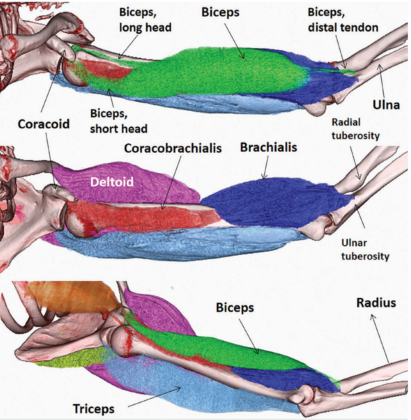
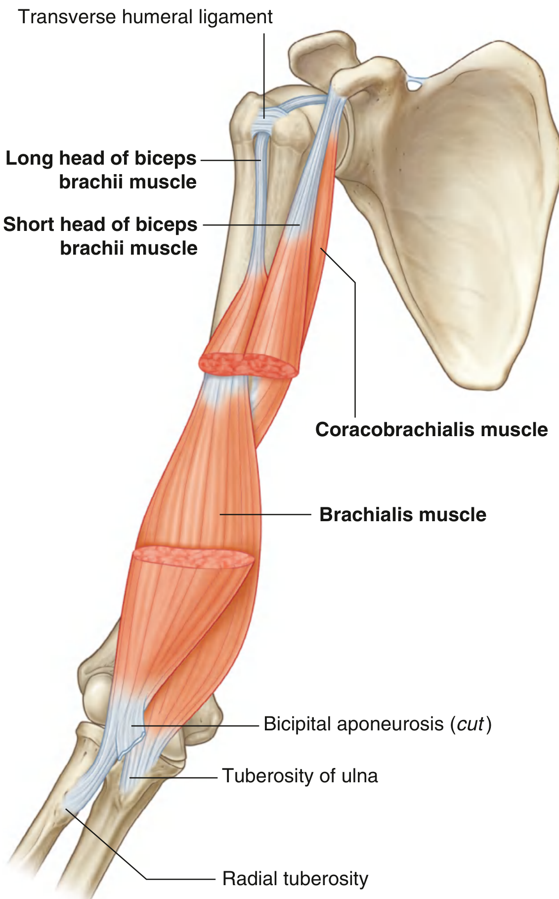
Summary Table: Muscles of the Anterior Compartment
| Muscle | Origin | Insertion | Nerve | Action |
|---|---|---|---|---|
| Coracobrachialis | Apex of coracoid process (with short head of biceps) | Linear roughening on medial aspect of midshaft humerus | Musculocutaneous n. (C5, C6, C7) | Flexion of arm at glenohumeral joint |
| Biceps Brachii | Long head: supraglenoid tubercle; Short head: coracoid process | Radial tuberosity + bicipital aponeurosis into deep fascia of forearm | Musculocutaneous n. (C5, C6) | Powerful elbow flexion + forearm supination; accessory shoulder flexor |
| Brachialis | Distal half of anterior humerus (medial & lateral surfaces) + intermuscular septa | Ulnar tuberosity (distal to coronoid) | Musculocutaneous n. (C5, C6); radial n. (C7) variable lateral portion | Powerful elbow flexion in all forearm positions |
2. Origin and Insertion of the Biceps Brachii
The biceps brachii is a biarticular, two-headed muscle that crosses both the shoulder and elbow joints.
Long Head
- Origin: Supraglenoid tubercle of the scapula (and blends with the superior glenoid labrum - forming the bicipitolabral complex)
- The tendon passes through the glenohumeral joint (intra-articular but extrasynovial), over the head of the humerus, through the rotator interval (between supraspinatus and subscapularis), then enters the intertubercular (bicipital) groove on the anterior humerus
- Held in the groove by the transverse humeral ligament (spanning between the greater and lesser tubercles)
Short Head
- Origin: Apex of the coracoid process of the scapula (shared with coracobrachialis as a conjoint tendon)
- Passes vertically through the axilla
Convergence and Insertion
- Both heads join in the distal arm, forming a single flattened tendon approximately 7 cm above the elbow
- The two tendons twist 90° as they descend (long head moves anteriorly, short head posteriorly), assisting in supination mechanics
- Insertion (primary): Radial tuberosity (posterior margin) of the proximal radius
- Insertion (secondary): Bicipital aponeurosis - a flat fibrous sheet that fans medially from the tendon to merge with the deep fascia of the forearm over the flexor muscles
Actions
| Joint | Action |
|---|---|
| Elbow | Flexion (powerful) |
| Forearm | Supination - most powerful when elbow is flexed at 90° |
| Shoulder | Flexion (accessory); long head resists superior translation of humeral head |
Key point: The biceps is the most powerful supinator of the forearm (more powerful than supinator muscle itself), but only when the elbow is flexed. This is because with elbow flexion the tendon's lever arm is nearly perpendicular to the axis of pronation/supination.
3. The Musculocutaneous Nerve
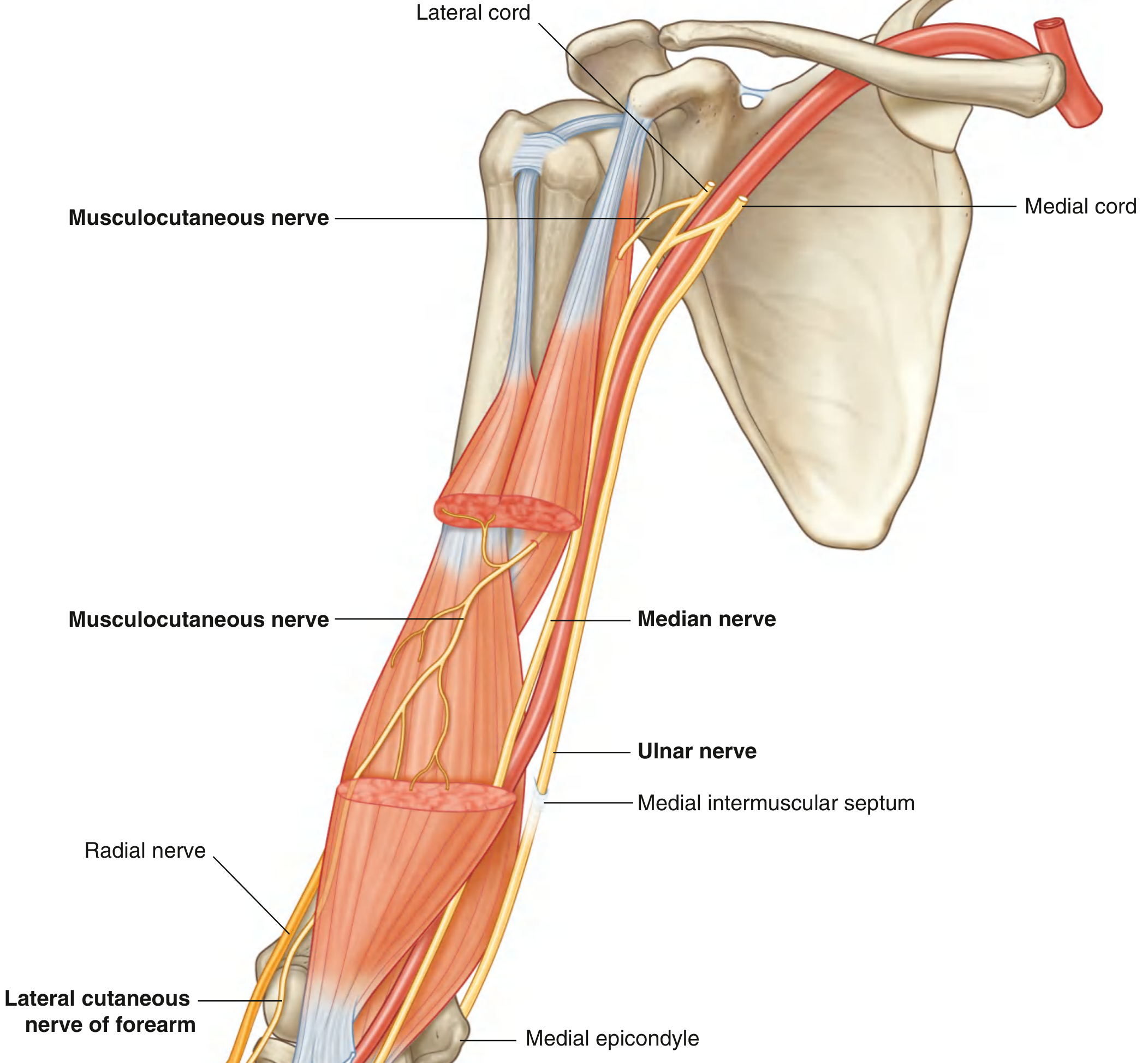
Origin
- Arises from the lateral cord of the brachial plexus (C5, C6, C7)
Course
- Exits the axilla by piercing through the coracobrachialis muscle (which it innervates)
- Passes diagonally down the arm in the plane between the biceps brachii (anteriorly) and brachialis (posteriorly)
- At the elbow, emerges laterally to the biceps tendon
- Pierces the deep fascia and continues into the forearm as the lateral cutaneous nerve of the forearm
Distribution
| Function | Territory |
|---|---|
| Motor | All 3 muscles of the anterior compartment: coracobrachialis, biceps brachii, brachialis |
| Sensory | Skin over the lateral surface of the forearm (as the lateral cutaneous nerve of the forearm) |
4. Nerve Supply of Muscles of the Front of the Arm
All three muscles are primarily innervated by the musculocutaneous nerve (C5, C6, C7):
| Muscle | Primary Nerve | Root Values | Additional Supply |
|---|---|---|---|
| Coracobrachialis | Musculocutaneous | C5, C6, C7 | None |
| Biceps Brachii | Musculocutaneous | C5, C6 | None |
| Brachialis | Musculocutaneous | C5, C6 | Radial nerve (C7) to lateral portion (~81% of cases); Median nerve to lower quarter (~15%) |
Important: Brachialis is the only muscle in the anterior compartment with dual nerve supply - its inferolateral fibers (deep head) commonly receive branches from the radial nerve as it passes the radial tunnel. This is significant: even with musculocutaneous nerve palsy, some elbow flexion is retained via the radial nerve supply to brachialis.
Reflex Testing
- Biceps reflex (C5, C6): Tapping the biceps tendon in the cubital fossa
- Brachioradialis reflex (C5, C6): Tests the brachioradialis (not an anterior arm muscle, but commonly tested alongside)
5. Brachialis Muscle - Full Account
Origin
- Distal half of the anterior surface of the humerus (from deltoid tuberosity downward)
- Medial and lateral intermuscular septa
- Two heads:
- Superficial head: originates near the deltoid tuberosity (larger, more proximal origin)
- Deep head: from distal third of the humeral shaft
Insertion
- Superficial head: Ulnar tuberosity, distal to the coronoid process
- Deep head: Coronoid process of the ulna (all but its tip); via three units - medial aponeurosis, lateral aponeurosis, and direct muscle fibers into the ulna
Nerve Supply
- Primary: Musculocutaneous nerve (C5, C6) - runs between brachialis and biceps brachii, lying on the surface of brachialis
- Variable: Radial nerve (C7) to inferolateral deep head fibers (~81% of individuals)
- Occasional: Median nerve to lower quarter (~15% of individuals)
Action
- Powerful flexor of the elbow joint - regardless of forearm position (pronated or supinated)
- The workhorse of elbow flexion - it is the largest contributor to elbow flexion force
- Also provides anterior stability to the coronoid process, contributing to resistance against posterior dislocation of the elbow
Key Characteristics
- Lies directly on the humerus - it is a pure elbow flexor with no rotatory action (unlike biceps which also supinates)
- Often called the "workhorse" of elbow flexion because it acts in all forearm positions
- The coronoid process is one of the anterior constraints providing buttress against posterior dislocation; the deep head of brachialis reinforces this
6. Brachial Artery, Median Nerve, and Ulnar Nerve
Brachial Artery
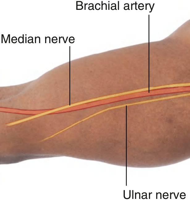
Course:
- Begins as a continuation of the axillary artery at the lower border of teres major
- Travels through the anterior compartment in the medial neurovascular bundle
- Proximal arm: Lies on the medial side, compressible against the humerus
- Distal arm: Moves laterally to lie midway between the medial and lateral epicondyles
- At the elbow (cubital fossa): Lies medial to the biceps tendon; bifurcates into radial and ulnar arteries just distal to the elbow joint
Branches in the arm:
- Profunda brachii artery (deep artery of arm) - largest branch, passes posteriorly with radial nerve through triangular interval, runs in the radial groove
- Superior ulnar collateral artery
- Inferior ulnar collateral artery
- Muscular branches and nutrient artery to humerus
Clinical: Brachial artery is the site for blood pressure measurement (compressed against humerus by sphygmomanometer cuff; Korotkoff sounds auscultated over cubital fossa); also used for arterial blood sampling and cardiac catheterization (Sones technique).
Median Nerve
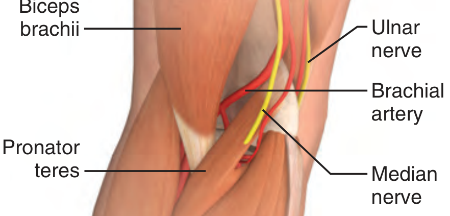
Formation: Formed anterior to the third part of the axillary artery by union of:
- Lateral root from the lateral cord (C6, C7)
- Medial root from the medial cord (C8, T1)
Course in the arm:
- Enters the arm at the lower border of teres major, initially lateral to brachial artery
- In the middle of the arm: crosses over the brachial artery (anterior) to lie on its medial side
- At the elbow: lies medial to the brachial artery anterior to the elbow joint
- Has no branches in the arm (occasional branch to pronator teres may arise proximal to elbow)
Ulnar Nerve
Course in the arm:
- Enters the arm medial to the axillary artery (posterior to the brachial artery)
- In the proximal arm: medial to brachial artery in the anterior compartment
- In the middle of the arm: pierces the medial intermuscular septum and enters the posterior compartment, where it lies anterior to the medial head of triceps
- Passes posterior to the medial epicondyle (in the ulnar groove/cubital tunnel) - this is where it is palpable and vulnerable to injury
- Enters the anterior compartment of the forearm between the two heads of flexor carpi ulnaris
- Has no major branches in the arm
Summary Diagram: Neurovascular Bundle of the Anterior Arm
AXILLA (entering arm at lower border of teres major)
│
├── BRACHIAL ARTERY (medial → moves laterally distally)
│ ├─ Profunda brachii → enters posterior compartment
│ ├─ Superior ulnar collateral
│ └─ Inferior ulnar collateral
│
├── MEDIAN NERVE (lateral → crosses to medial side at mid-arm)
│ └─ No branches in arm
│
└── ULNAR NERVE (medial → pierces medial intermuscular septum)
└─ Enters posterior compartment at mid-arm
→ wraps around medial epicondyle
→ enters forearm (cubital tunnel)
MUSCULOCUTANEOUS NERVE (separate - lateral cord):
Pierces coracobrachialis → runs between biceps and brachialis
→ emerges as LATERAL CUTANEOUS NERVE OF FOREARM
Applied Points
| Structure | Clinical Relevance |
|---|---|
| Brachial artery | BP measurement, blood gas sampling, site of injury in humeral fractures, brachial embolism |
| Median nerve | "Ape hand" deformity if injured; loss of thenar eminence and lateral 3.5 finger sensation |
| Ulnar nerve at medial epicondyle | "Cubital tunnel syndrome" - most common site of ulnar nerve compression; "claw hand" |
| Musculocutaneous nerve | Rare isolated injury; results in loss of biceps and brachialis action (weak elbow flexion), absent biceps reflex, loss of lateral forearm sensation |
| Bicipital aponeurosis | Protects brachial artery and median nerve in cubital fossa; becomes tense during resisted supination |
Sources: Gray's Anatomy for Students, pp. 852-880; Imaging Anatomy: Bones, Joints, Vessels & Nerves, pp. 109-113; THIEME Atlas of General Anatomy, pp. 2903-2950
This is a shared conversation. Sign in to Orris to start your own chat.