Here is your comprehensive clinical reference covering all the topics you asked about:
Fever - Complete Clinical Overview
1. Causes of Acute, Subacute, and Chronic Fever
Acute Fever (< 2 weeks)
- Infections: Most bacterial infections (pneumonia, UTI, cellulitis, meningitis), viral illnesses (influenza, dengue, COVID-19), malaria, typhoid (early phase), leptospirosis
- Non-infectious: Drug fever, transfusion reactions, pulmonary embolism, deep vein thrombosis, acute myocardial infarction, hematoma, post-operative fever
Subacute Fever (2-6 weeks)
- Infections: Infective endocarditis, brucellosis, tuberculosis (early), typhoid (prolonged), enteric fever, fungal infections, viral hepatitis
- Non-infectious: Malignancy (lymphoma), connective tissue diseases (SLE, vasculitis), drug fever
- Common in ICU: ~80% of febrile episodes are infectious - pneumonia, catheter-related infections
Chronic Fever (> 6 weeks / Fever of Unknown Origin)
- Infections: Tuberculosis, brucellosis, deep-seated abscesses, infective endocarditis, HIV, fungal infections (histoplasmosis - classified by duration into acute/subacute/chronic)
- Non-infectious: Malignancies (lymphoma, renal cell carcinoma, hepatocellular carcinoma), connective tissue diseases (SLE, Adult Still disease, vasculitis), granulomatous diseases (sarcoidosis, Crohn's)
- Drug fever can occur at any duration
Sources: Goldman-Cecil Medicine; Histoplasmosis chapter, Red Book 2021
2. Types of Fever - Patterns with Examples
| Type | Definition | Classic Examples |
|---|
| Continuous | Temperature stays elevated throughout 24 hours, does not touch normal; variation < 1°C | Typhoid fever (Salmonella typhi), lobar pneumonia, urinary tract infection |
| Remittent | Temperature remains elevated but touches near-normal values; variation > 1°C but does NOT reach normal | Infective endocarditis, many viral infections, brucellosis |
| Intermittent | Temperature spikes to fever then returns to NORMAL (afebrile) during the same day | Malaria (tertian = P. vivax/ovale every 48h; quartan = P. malariae every 72h), pyogenic abscess, septicemia, kala-azar (double daily spike = Pel-Ebstein not applicable here) |
| Relapsing / Periodic | Days of fever alternating with days of completely normal temperature | Borrelia relapsing fever, malaria (relapses from P. vivax/ovale hypnozoites), Pel-Ebstein fever in Hodgkin's lymphoma, brucellosis (undulant fever) |
3. Fevers Common in India - What to Suspect
In the Indian subcontinent, the following should be strongly suspected in a febrile patient:
Infectious (most common):
- Malaria (P. vivax most common; P. falciparum - severe/cerebral)
- Typhoid / Enteric fever (Salmonella typhi/paratyphi)
- Dengue fever (Aedes mosquito-borne)
- Chikungunya (fever + severe arthralgia)
- Leptospirosis (post-flood, occupational exposure)
- Scrub typhus (Orientia tsutsugamushi; eschar is pathognomonic)
- Rickettsial fever (spotted fevers, tick-borne)
- Tuberculosis (pulmonary and extrapulmonary; chronic low-grade fever)
- Kala-azar / Visceral leishmaniasis (Bihar, Jharkhand, Bengal, Assam endemic belt)
- Brucellosis (cattle farming areas, dairy workers)
- Viral hepatitis A/E (hepatitis with fever in endemic areas)
- Japanese encephalitis (Culex mosquito; Bihar, UP, Assam)
- Melioidosis (Northeast India, coastal areas)
- Enteric fever-related complications
Non-infectious to consider:
- Connective tissue diseases (SLE, adult Still's disease)
- Lymphomas
- Drug fever
4. Fever with Arthralgia - Differential Diagnosis
| Diagnosis | Key Features |
|---|
| Chikungunya | Bilateral symmetrical, very severe joint pain (incapacitating); maculopapular rash; Aedes aegypti |
| Dengue | Bone-crushing myalgia + arthralgia; retro-orbital pain; thrombocytopenia |
| Acute Rheumatic Fever | Migratory polyarthritis; Jones criteria (major: carditis, chorea, erythema marginatum, subcutaneous nodules, arthritis) |
| Rheumatoid Arthritis (reactive/early) | Symmetrical small joint involvement; morning stiffness |
| Serum sickness / Drug reaction | Fever + arthralgia + rash + urticaria |
| Infective Endocarditis | Fever + arthralgia + cardiac murmur |
| Primary HIV seroconversion (ARS) | Fever, pharyngitis, lymphadenopathy, rash, arthralgia; resembles glandular fever |
| Viral hepatitis (HBV) | Serum sickness-like prodrome: fever + arthralgia + rash before jaundice |
| SLE | Non-erosive arthralgia, malar rash, multi-system involvement |
| Adult Still's disease | Daily spiking fever, evanescent salmon-colored rash, arthralgia |
| Lyme disease | Tick exposure; erythema migrans rash; migratory arthralgia, carditis, neuroborreliosis |
| Brucellosis | Undulant fever; occupational/dietary exposure; sacroiliitis |
Sources: Fuster & Hurst's The Heart; Fitzpatrick's Dermatology; Goldman-Cecil Medicine
5. Fever with Rash - Differential Diagnosis
By Rash Type:
Maculopapular rash + fever:
- Measles (cephalocaudal spread, Koplik spots)
- Rubella (milder, lymphadenopathy)
- Dengue
- Chikungunya
- Infectious mononucleosis (EBV)
- Roseola infantum (HHV-6; rash appears as fever breaks)
- Primary HIV (ARS)
- West Nile virus
- Zika virus
- Parvovirus B19 (slapped cheek + lacy reticular rash)
- Scarlet fever (sandpaper rash, strawberry tongue, desquamation of palms/soles)
- Rocky Mountain spotted fever (centripetal spread, palms/soles involvement)
- Ehrlichiosis
- Drug reactions (exanthematous)
- Kawasaki disease
Petechiae/Purpura + fever (EMERGENCIES):
- Meningococcemia (Neisseria meningitidis) - non-blanching purpura, rapidly progressive; EMERGENCY
- Rocky Mountain spotted fever (may progress to purpura)
- Disseminated gonococcal infection
- Infective endocarditis (petechiae + Osler nodes + Janeway lesions)
- Dengue hemorrhagic fever
- Thrombotic thrombocytopenic purpura (TTP)
- Vasculitis (HSP/IgA vasculopathy)
Vesicular/Pustular rash + fever:
- Varicella (chickenpox)
- Herpes zoster (dermatomal)
- Smallpox (all lesions in same stage - historical)
- Herpes simplex
- Enteroviral hand-foot-mouth disease
Other specific rashes:
- Erythema migrans - Lyme disease
- Erythema marginatum - Acute rheumatic fever
- Butterfly (malar) rash - SLE
- Erythema nodosum - TB, sarcoidosis, streptococcal infection, Crohn's
- Eschar - Scrub typhus, rickettsial infections
- Rose spots - Typhoid fever (abdomen/chest; faint salmon-colored)
Source: Goldman-Cecil Medicine, Textbook of Family Medicine, Harrison's Principles of Internal Medicine 22E
6. Janeway Lesions
Definition: Janeway lesions are non-tender, erythematous or hemorrhagic macular/papular lesions on the palms and soles, caused by septic emboli in infective endocarditis.
Key features:
- Location: palms and soles (plantar aspect, thenar/hypothenar eminence)
- Non-tender (unlike Osler nodes which are PAINFUL)
- Caused by infected microemboli with microabscess formation
- Associated with acute infective endocarditis (especially Staphylococcus aureus)
Comparison with Osler Nodes:
| Feature | Janeway Lesions | Osler Nodes |
|---|
| Tenderness | Non-tender | Painful/tender |
| Location | Palms, soles | Fingertips, pulp of digits |
| Mechanism | Septic emboli (microabscesses) | Immune complex deposition |
| Association | Acute IE (Staph aureus) | Subacute IE (Strep viridans) |
Other signs of infective endocarditis: Fever (80-90%), heart murmur (75-85%), splinter hemorrhages (subungual), Roth spots (retinal), conjunctival hemorrhages, petechiae.
Sources: Textbook of Family Medicine; Dermatology 2-Volume Set 5e; Braunwald's Heart Disease
7. Fever with Hepatosplenomegaly - Conditions
Infectious:
- Malaria (P. vivax/falciparum - massive splenomegaly in chronic/hyperreactive malaria)
- Kala-azar / Visceral Leishmaniasis (massive progressive splenomegaly; the largest spleens seen in clinical practice)
- Typhoid fever (Salmonella; splenomegaly in 2nd-3rd week)
- Infectious mononucleosis (EBV; teenager with sore throat + lymphadenopathy + hepatosplenomegaly)
- Viral hepatitis (A, B, E)
- Infective endocarditis (bacterial emboli)
- Brucellosis
- Leptospirosis (Weil's disease)
- Schistosomiasis (portal hypertension, massive splenomegaly)
- Miliary tuberculosis
- Typhus/rickettsial disease
- Cytomegalovirus (CMV)
- HIV/AIDS
Non-infectious:
- Lymphomas (Hodgkin's and Non-Hodgkin's)
- Leukemias (CML - massive splenomegaly)
- Hemolytic anemias (thalassemia, sickle cell)
- Storage diseases (Gaucher's, Niemann-Pick)
- Autoimmune diseases (SLE, Still's disease)
- Sarcoidosis
- Portal hypertension (cirrhosis - enlargement of both; liver may actually shrink in cirrhosis)
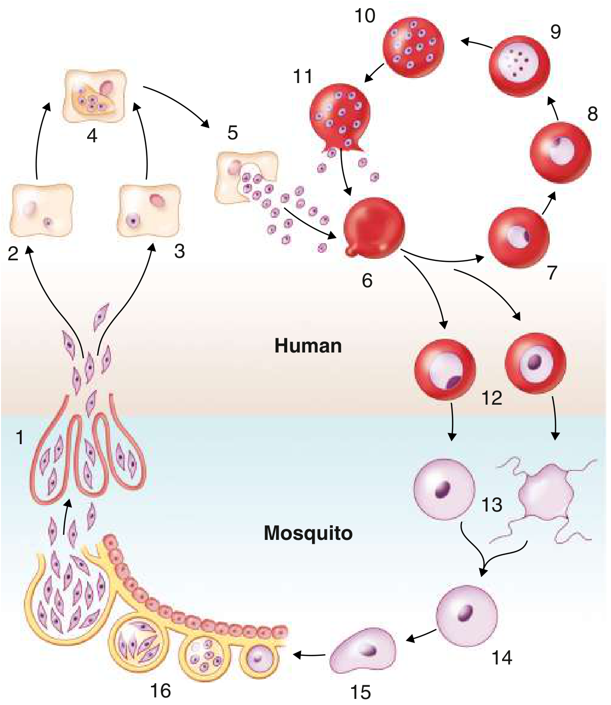
8. Life Cycle of Malaria
IN THE HUMAN HOST:
1. Exoerythrocytic (Liver) Phase:
- Female Anopheles mosquito bites → injects sporozoites into bloodstream (Step 1)
- Sporozoites travel to liver within 1 hour → enter hepatocytes
- Develop into tissue schizonts (Step 4) → each releases up to 30,000 merozoites (Step 5)
- Hypnozoites (Step 3): In P. vivax and P. ovale only - some sporozoites remain dormant in liver → can reactivate months/years later causing relapse
2. Erythrocytic Phase (symptomatic):
- Merozoites invade RBCs (Step 6) → ring form → trophozoite (Step 9) → schizont (Step 10) → rupture releases new merozoites (Step 11)
- Cycle duration: 48 hrs (P. falciparum, P. vivax, P. ovale) → tertian fever; 72 hrs (P. malariae) → quartan fever
- Schizont rupture causes fever spikes (pyrogens released)
3. Gametocyte Phase:
- Some merozoites develop into male and female gametocytes (Step 12) - taken up by mosquito
IN THE MOSQUITO HOST:
- Gametocytes → gametes → fertilization → zygote (Step 14) → ookinete (Step 15) → oocyst on gut wall (Step 16) → rupture → sporozoites migrate to salivary gland (Step 1) → ready to infect next human
Clinically important species differences:
| Feature | P. vivax | P. falciparum | P. malariae | P. ovale |
|---|
| Fever cycle | 48h (tertian) | 48h (malignant tertian) | 72h (quartan) | 48h |
| Hypnozoites (relapse) | YES | No | No | YES |
| Severe disease | Less common | Most dangerous | Rare | Rare |
| RBC preference | Reticulocytes | All RBCs | Old RBCs | Reticulocytes |
Source: Goldman-Cecil Medicine 22E; Medical Microbiology 9e
9. Abdominal Tuberculosis
Definition: TB involving any abdominal organ, accounting for ~15% of extrapulmonary TB cases.
Forms:
- Tuberculous lymphadenopathy - MOST COMMON form; mesenteric lymph nodes most frequently involved; can develop central caseation and calcification
- Peritoneal TB - Three subtypes:
- Wet ascitic type (most common): large free or loculated ascites; exudative; high SAAG
- Fixed fibrotic type: omental/mesenteric involvement, matted bowel loops on imaging
- Dry plastic type: fibrous peritoneal reaction, nodules, adhesions
- GI TB - Most commonly involves the ileocecal region (MC site), followed by the jejunum
- Morphological types: ulcerative, hypertrophic, stricturing
- Difficult to distinguish from Crohn's disease
- Visceral TB - Solid organs; genitourinary most frequent
Clinical Features:
- Abdominal pain (most common), fever, weight loss (present for months)
- Right lower quadrant mass in ~25%
- Ascites on cross-sectional imaging strongly suggests abdominal TB over Crohn's
Key distinguishing features from Crohn's:
- TB: transverse ulcers, patulous ileocecal valve, ascites, granulomas with caseation (>400 µm, large giant cells)
- Crohn's: left colonic involvement, longitudinal ulcers, cobblestoning, skip lesions, rectal involvement
Histology: Caseating granulomas (coalescent, >400 µm, large giant cells); AFB staining, PCR, or culture confirms
Diagnosis: High suspicion in risk groups (immigrants from endemic areas, HIV, malnourished). Anti-TNF therapy for Crohn's can worsen unrecognized TB - must rule out TB before starting biologics.
Source: Yamada's Textbook of Gastroenterology 7e; Grainger & Allison's Diagnostic Radiology
10. Fever with Altered Sensorium - Where We See It
This is a serious clinical presentation. Key conditions:
Infections of CNS:
- Bacterial meningitis (N. meningitidis, S. pneumoniae, L. monocytogenes) - fever + neck stiffness + altered sensorium (triad)
- Viral encephalitis (Herpes simplex - most common treatable cause; Japanese encephalitis in India; West Nile)
- Cerebral malaria (P. falciparum - parasitized RBCs occlude cerebral vessels; severe manifestation)
- Tubercular meningitis (subacute onset, cranial nerve palsies)
- Bacterial brain abscess (focal signs + fever)
- Cerebral toxoplasmosis (HIV patients)
- Rickettsial encephalitis (Scrub typhus with meningoencephalitis)
Systemic infections with CNS involvement:
- Leptospirosis (Weil's disease) - encephalitis, altered sensorium, psychosis, seizures, hemiplegia - quoted directly from Kaplan & Sadock's Comprehensive Textbook of Psychiatry
- Typhoid encephalopathy ("muttering delirium" - 2nd-3rd week)
- Septic encephalopathy (any severe sepsis - multiorgan dysfunction)
- Dengue encephalitis (rare manifestation of dengue)
- Rabies (hydrophobia, aerophobia, agitation)
- Neuro-syphilis (chronic, VDRL positive)
- Plasmodium falciparum - cerebral malaria (specific to P. falciparum)
- Polyarteritis Nodosa (PAN) - catastrophic intracranial hemorrhage with altered sensorium has been described (especially pediatric PAN)
Non-infectious:
- Liver failure (hepatic encephalopathy - asterixis, elevated ammonia)
- Uremic encephalopathy
- Anti-NMDA receptor encephalitis (autoimmune; fever + psychiatric + movement disorder)
- Heat stroke (exertional or classic)
- Drug/toxin ingestion
- Metabolic derangements (hyponatremia, hypoglycemia, DKA)
Sources: Bradley and Daroff's Neurology; Kaplan & Sadock's Comprehensive Textbook; Goldman-Cecil Medicine
11. Leukoencephalopathy
Progressive Multifocal Leukoencephalopathy (PML)
Definition: PML is a demyelinating brain disease caused by lytic replication of JC polyomavirus in oligodendrocytes (the cells that produce myelin) in subcortical white matter and, rarely, gray matter neurons.
Pathogen: JC polyomavirus (human polyomavirus 2)
Pathobiology:
- JC virus replicates in oligodendrocytes → cell lysis → loss of myelin sheaths
- Affected oligodendrocytes: enlarged nuclei with eosinophilic intranuclear inclusions
- Lesions start in subcortical white matter, less commonly cerebellum or basal ganglia
- As demyelination expands: lesions range from <1 mm to several cm; multiple zones coalesce
Epidemiology:
- General population: ~0.3 per 100,000 person-years (North America/Europe)
- HIV/AIDS (pre-ART era): ~2.4 per 1,000 patient-years; prevalence 1-7%
- Natalizumab (for MS): ~2.13 per 1,000 patient-years; rises to 11.1 per 1,000 in JC-seropositive patients treated >24 months
- Also seen with: lymphoproliferative malignancies, other immunosuppressive biologics, idiopathic CD4 lymphocytopenia
Predisposing conditions (any state of impaired T- and B-cell immunity):
- HIV/AIDS (most common historically)
- Hematologic malignancies (lymphoma, CLL, multiple myeloma)
- Organ transplant recipients (on immunosuppressants)
- Natalizumab therapy (multiple sclerosis)
- Other monoclonal antibodies (rituximab, efalizumab)
- Sarcoidosis
Clinical Manifestations:
- Progressive motor weakness (most common)
- Speech abnormalities, cognitive deficits
- Headache, ataxia, aphasia, cranial nerve deficits
- Visual disturbances (optic agnosia, cortical blindness, oculomotor abnormalities)
- Seizures (more common with gadolinium-enhancing cortical lesions)
- Sensory deficits of hands (parietal involvement)
Diagnosis: MRI (T2/FLAIR hyperintense non-enhancing white matter lesions), JC virus PCR in CSF
Treatment:
- No specific anti-JC viral therapy proven effective
- For HIV patients: start/optimize antiretroviral therapy → immune reconstitution
- Withdraw offending immunosuppressive agent (e.g., stop natalizumab, plasmapheresis to accelerate removal)
- Risk of IRIS (immune reconstitution inflammatory syndrome) when immunity is restored
Prevention: In HIV/AIDS, ART reducing HIV to undetectable levels has dramatically reduced PML incidence.
Source: Goldman-Cecil Medicine International Edition; Firestein & Kelley's Textbook of Rheumatology; Bradley and Daroff's Neurology in Clinical Practice
Quick Summary Mnemonics
Fever patterns - "CRIR":
- Continuous = Typhoid, lobar pneumonia
- Remittent = Infective endocarditis, viral fevers
- Intermittent = Malaria, pyogenic abscess
- Relapsing = Borrelia, Hodgkin's (Pel-Ebstein), brucellosis
Fever + Rash EMERGENCIES to rule out first:
- Meningococcemia (non-blanching petechiae/purpura → septic shock)
- Rocky Mountain Spotted Fever (petechiae on palms/soles spreading centrally)
- Toxic Shock Syndrome (Staph/Strep)
- Staphylococcal Scalded Skin Syndrome
Janeway vs Osler:
- Janeway = No pain, on palms/soles = Acute IE (Staph aureus)
- Osler = Ouchy/painful, on digits = Subacute IE (Strep viridans)