I now have all the images and content needed. Let me compile the complete, comprehensive surgical exam notes.
COMPLETE SURGICAL EXAM NOTES
With Operative Steps, Images & Viva Traps
PART 1: HERNIA (ALL TYPES) - IN DEPTH
Definition
A hernia is the protrusion of a viscus or part of a viscus through a weakness or defect in its containing wall.
Parts of a Hernia
| Part | Description |
|---|
| Neck | The aperture through which it protrudes (most important - determines risk of strangulation) |
| Sac | A diverticulum of peritoneum |
| Contents | Omentum (most common), small bowel, large bowel, bladder, ovary |
| Coverings | Layers of abdominal wall through which the sac passes |
Classification
- Reducible: Contents can be returned to the cavity
- Irreducible/Incarcerated: Contents cannot be returned
- Obstructed: Bowel is obstructed but blood supply intact
- Strangulated: Blood supply to contents is compromised - SURGICAL EMERGENCY
- Richter's hernia: Only part of bowel wall caught in hernia - can strangulate WITHOUT obstruction
- Maydl's hernia: Two loops of bowel in sac ("W" hernia) - middle loop in abdomen strangulates first
- Littre's hernia: Meckel's diverticulum in the sac
1. INGUINAL HERNIA
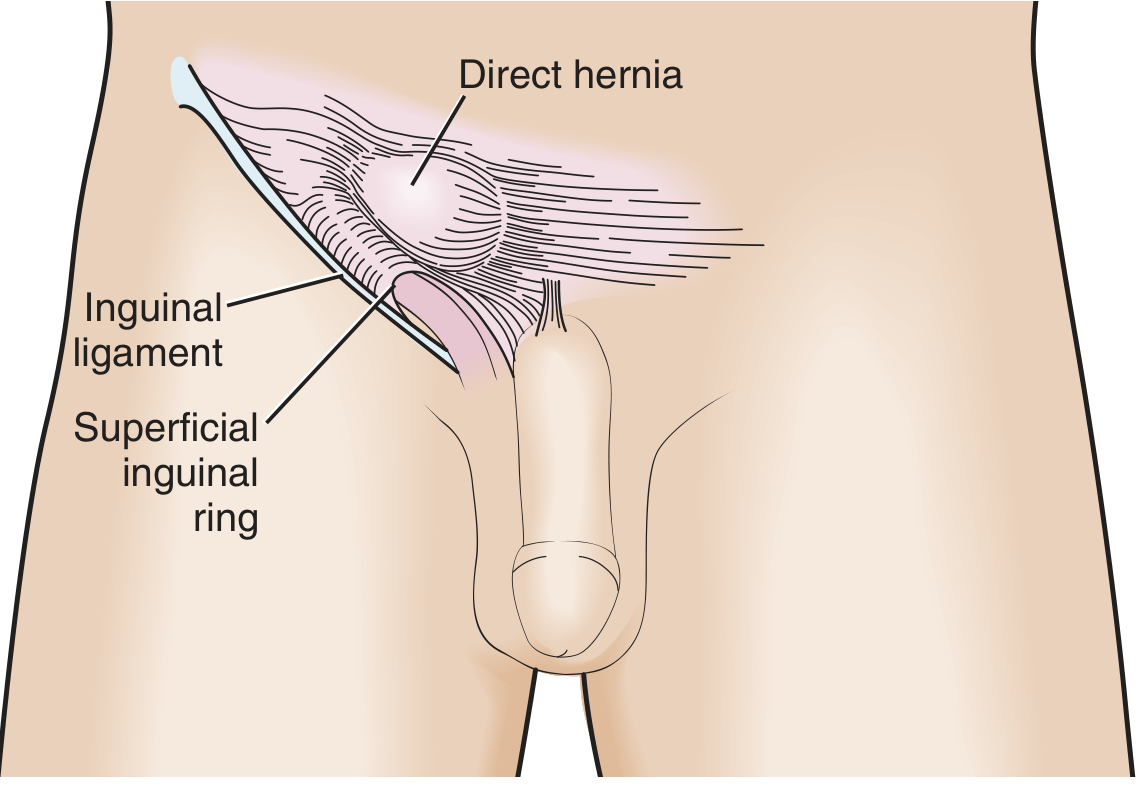
Direct inguinal hernia: protrudes medial to inferior epigastric vessels through Hesselbach's triangle
Anatomy of Inguinal Canal
- Length: 4 cm
- Direction: Medially and downward
- Deep ring: In transversalis fascia, 1.25 cm above midpoint of inguinal ligament
- Superficial ring: In external oblique aponeurosis, above pubic tubercle
- Boundaries:
- Anterior: External oblique (whole length) + internal oblique (lateral 1/3)
- Posterior: Transversalis fascia (whole length) + conjoint tendon (medial 1/3)
- Floor: Inguinal ligament + lacunar ligament medially
- Roof: Arching fibers of internal oblique and transversus abdominis
Hesselbach's Triangle
- Medially: Lateral border of rectus abdominis
- Laterally: Inferior epigastric vessels
- Inferiorly: Inguinal ligament
Indirect vs Direct Inguinal Hernia
| Feature | Indirect | Direct |
|---|
| Through | Deep inguinal ring | Hesselbach's triangle |
| Relation to inf. epigastric vessels | Lateral | Medial |
| Age | Any age | Older men |
| Patent processus vaginalis | Yes | No |
| Sac covered by | All 3 cord coverings | Transversalis fascia |
| Descent into scrotum | Yes | Rarely |
| Neck | Narrow (risk of strangulation) | Wide |
| Reduction with pressure at deep ring | Reduced | Not reduced |
Indirect Inguinal Hernia - Operative Steps (Open Herniotomy in Children)
- Position: Supine, slight Trendelenburg
- Incision: Skin crease incision over inguinal region, above the inguinal ligament
- Layers: Scarpa's fascia, Camper's fascia divided
- External oblique aponeurosis: Identified and incised in direction of fibers - opened along the canal
- Ilioinguinal nerve: Identified and preserved
- Cremaster muscle: Split to expose the cord
- Sac identification: White, glistening - anterior and medial to cord structures
- Sac dissection: Separated from vas deferens and testicular vessels
- Sac opened: Confirm contents, reduce
- Sac ligation: Twisted, transfixed, ligated at deep ring
- Repair: Posterior wall reinforcement if needed (Bassini/Shouldice/mesh)
- Closure: External oblique, Scarpa's, skin
Repair Types (Adult)
| Repair | Principle |
|---|
| Bassini | Conjoint tendon sutured to inguinal ligament |
| Shouldice | 4-layer continuous suture repair (gold standard for non-mesh) |
| Lichtenstein | Tension-free mesh onlay (most common open) |
| TEP | Totally extraperitoneal - mesh in pre-peritoneal space |
| TAPP | Trans-abdominal preperitoneal - enter peritoneum, then preperitoneal |
Laparoscopic: TEP and TAPP - 10×15 cm mesh covers deep ring, Hesselbach's triangle, and femoral canal.
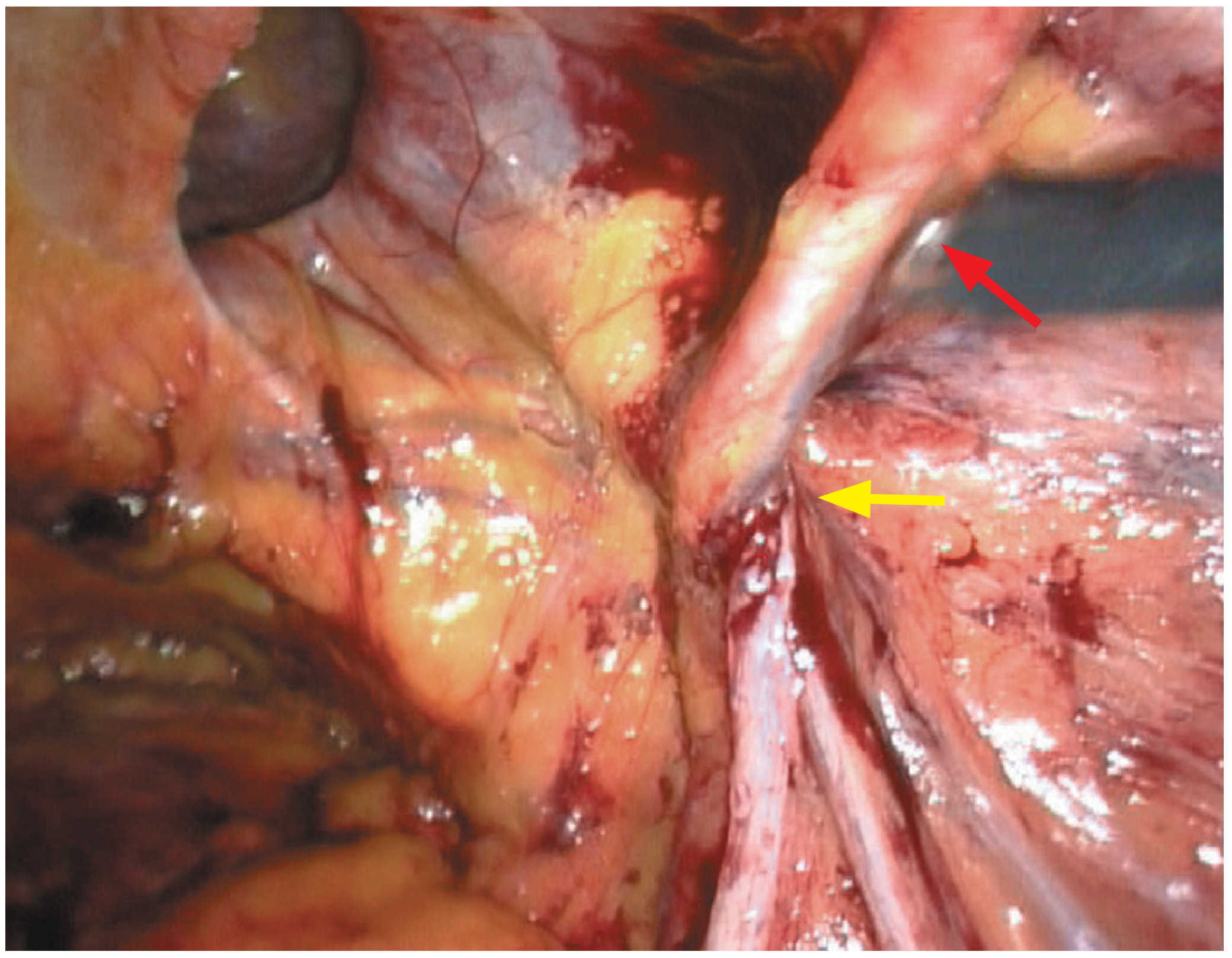
Laparoscopic view: right medial/direct inguinal hernia. Red arrow = inferior epigastric vessels; yellow arrow = spermatic cord at deep ring
Complications of Inguinal Hernia Repair
- Early: Haematoma, wound infection, urinary retention
- Late: Chronic groin pain (inguinodynia), recurrence, testicular atrophy, injury to vas deferens, neuralgia (ilioinguinal, iliohypogastric, genitofemoral nerves)
🚨 VIVA TRAPS - Inguinal Hernia
- Q: Which hernia is more common in females? A: Indirect inguinal (still, though femoral is proportionally more common in women)
- Q: What is the most common hernia in females? A: Inguinal (indirect), not femoral - femoral is just MORE common in women than in men
- Q: What nerve is most commonly injured in inguinal hernia repair? A: Ilioinguinal nerve (supplies scrotum/labia majora + medial thigh)
- Q: Why is a narrow neck dangerous? A: Risk of strangulation - blood supply cut off by tight neck
- Q: What is a sliding hernia (Hernie en glissade)? A: Part of the sac wall IS a viscus (e.g., caecum on right, sigmoid on left) - DO NOT excise the sac without checking
- Q: Why does strangulation risk more with indirect than direct? A: Narrow neck of indirect hernia
- Q: The key landmark to distinguish direct from indirect laparoscopically? A: Inferior epigastric vessels - direct is medial to them
2. FEMORAL HERNIA
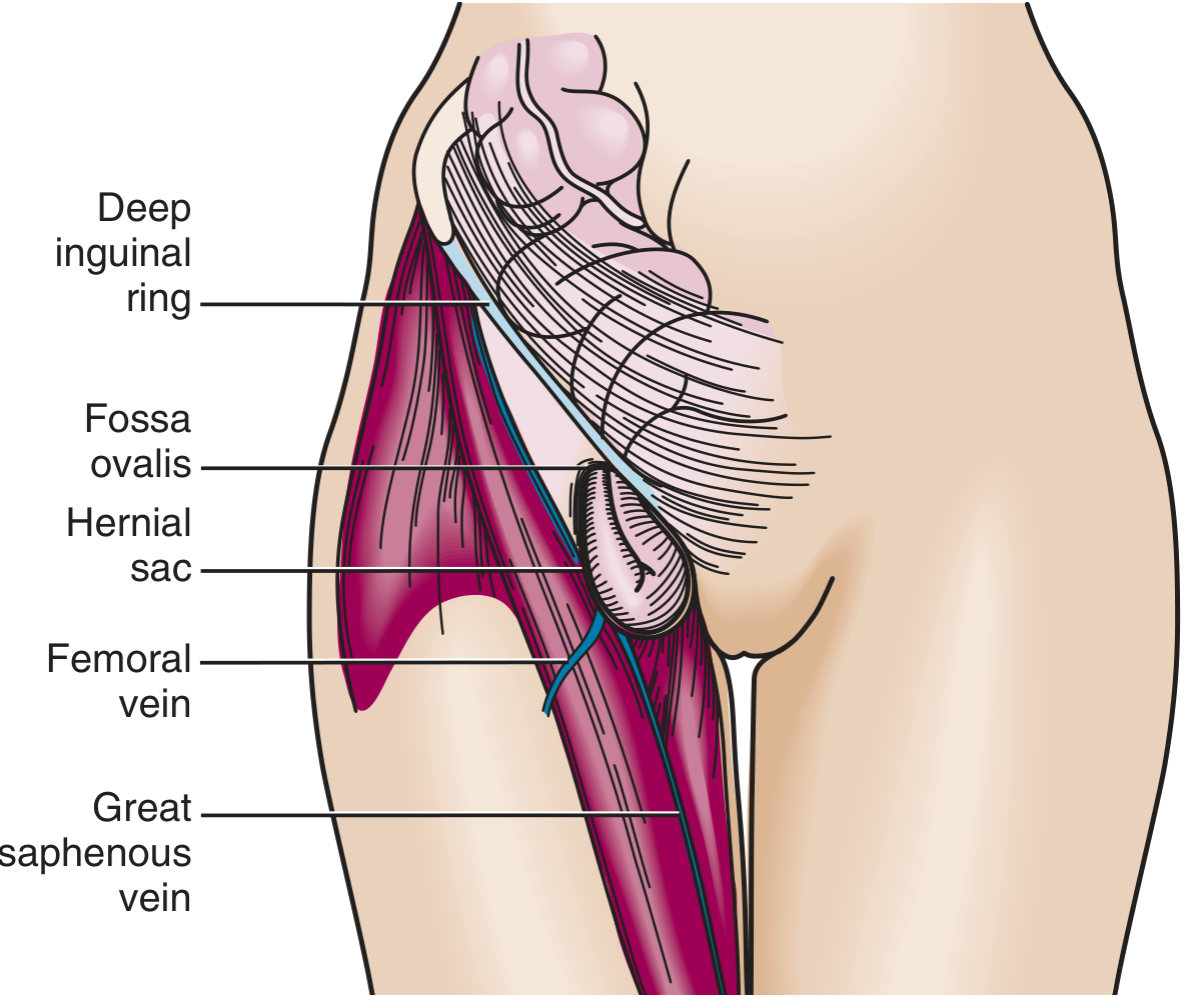
Femoral hernia: passes below and lateral to pubic tubercle, through femoral canal
Anatomy of Femoral Canal
- Boundaries:
- Anterior: Inguinal ligament
- Posterior: Pectineal (Cooper's) ligament and pectineus muscle
- Medial: Lacunar ligament
- Lateral: Femoral vein
- Emerges through fossa ovalis in deep fascia of thigh
- Contains lymph nodes of Cloquet/Rosenmüller
Key Clinical Features
- More common in women (wider pelvis)
- Lies below and lateral to pubic tubercle (inguinal hernia is above and medial)
- Highest risk of strangulation of all hernias (45% incarcerate)
- Strangulation may be silent due to wide omentum entering sac
- Richter's hernia is classically described at the femoral site
Operative Approaches
| Approach | Description |
|---|
| Low (Lockwood) | Below inguinal ligament - simplest, best for uncomplicated |
| High (McEvedy) | Through rectus sheath/vertical incision - best for strangulation |
| Inguinal (Lotheissen) | Through inguinal canal - best when unsure of diagnosis |
| Preperitoneal | Used in laparoscopic repair |
Repair Operative Steps (Lockwood - Low Approach)
- Transverse incision below inguinal ligament over sac
- Identify sac in femoral canal
- Reduce contents (if strangulated - open sac first, assess viability)
- Ligate and excise sac
- Close femoral canal: suture inguinal ligament to pectineal ligament (Cooper's) using non-absorbable suture
- Avoid lateral sutures - femoral vein injury risk
🚨 VIVA TRAPS - Femoral Hernia
- Q: Which hernia has the highest risk of strangulation? A: Femoral hernia
- Q: What are boundaries of femoral ring? Remember NAVEL from lateral to medial: N=Nerve (femoral), A=Artery (femoral), V=Vein (femoral), E=Empty space, L=Lymphatics (in canal)
- Q: Where is pubic tubercle in relation to femoral hernia? A: Femoral hernia is below and lateral to pubic tubercle; inguinal is above and medial
- Q: What is the structure that can be accidentally cut to enlarge the femoral ring (dangerous)? A: Lacunar ligament - but beware an aberrant obturator artery ("corona mortis") in ~20% that runs on its medial side
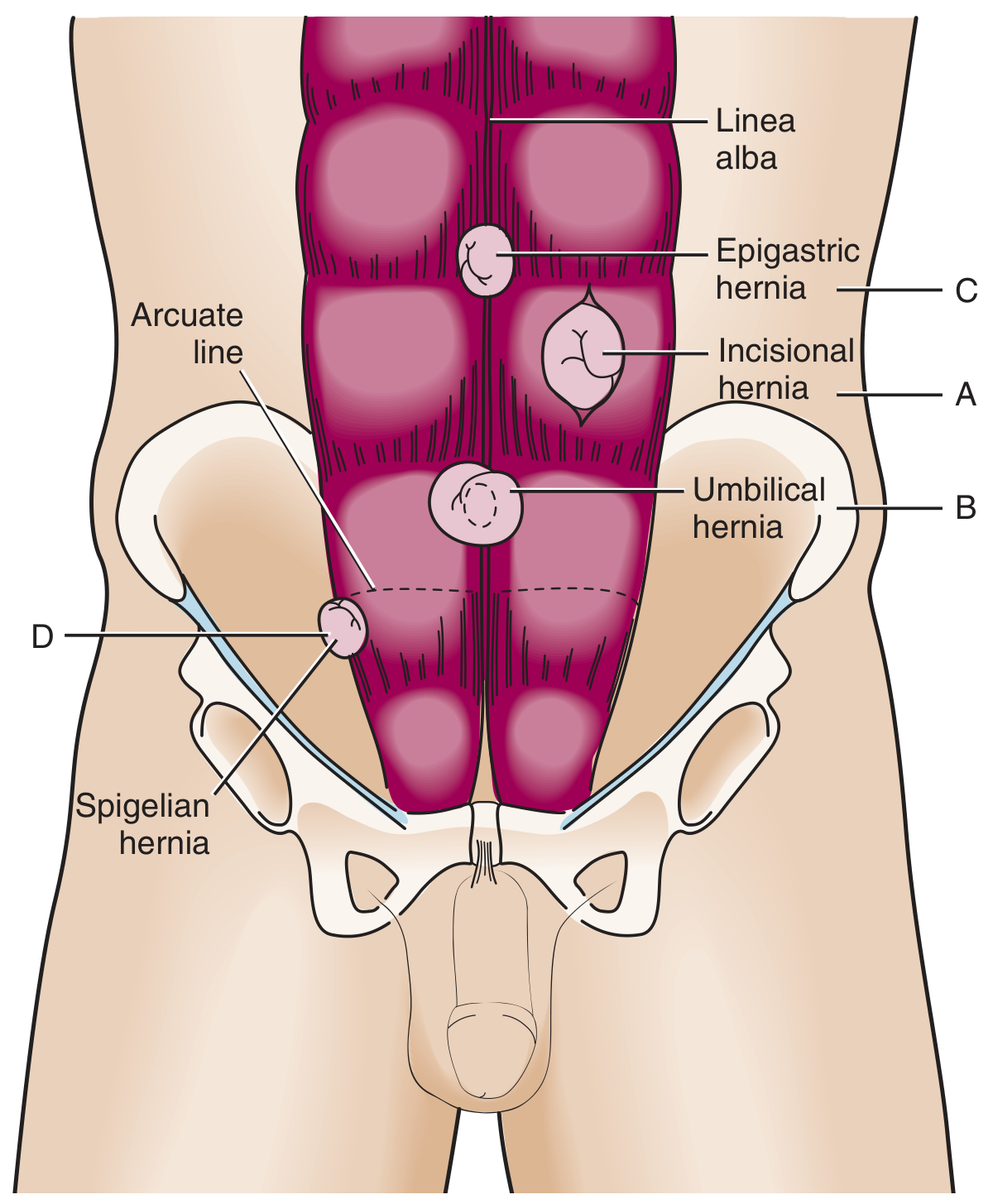
3. UMBILICAL HERNIA
Types
| Type | Features |
|---|
| Exomphalos (omphalocele) | Congenital, sac present, bowel outside abdomen at birth, associated anomalies 50% |
| Gastroschisis | Paraumbilical defect (usually right), NO sac, bowel thickened/matted |
| Infantile umbilical | Through umbilical scar, usually closes by age 5 |
| Adult/Acquired umbilical | Paraumbilical in adults, associated with obesity, ascites, multiparity |
Adult Paraumbilical Hernia - Operative Steps (Mayo's Repair)
- Elliptical incision around umbilicus
- Sac excised (umbilicus may be retained cosmetically)
- Mayo's "vest over pants" repair: Upper leaf of rectus sheath sutured over lower leaf as double-breasting, using non-absorbable sutures
- For large defects: mesh repair preferred
- Drain if large sac
🚨 VIVA TRAPS - Umbilical
- Q: What is the commonest complication of large umbilical hernia in cirrhosis? A: Spontaneous rupture (not strangulation)
- Q: Why not repair ascitic umbilical hernia electively? A: High risk - ascites causes wound leakage, recurrence; must control ascites first
- Q: Exomphalos vs gastroschisis key difference? A: Exomphalos has a SAC; gastroschisis has NO sac
4. INCISIONAL HERNIA
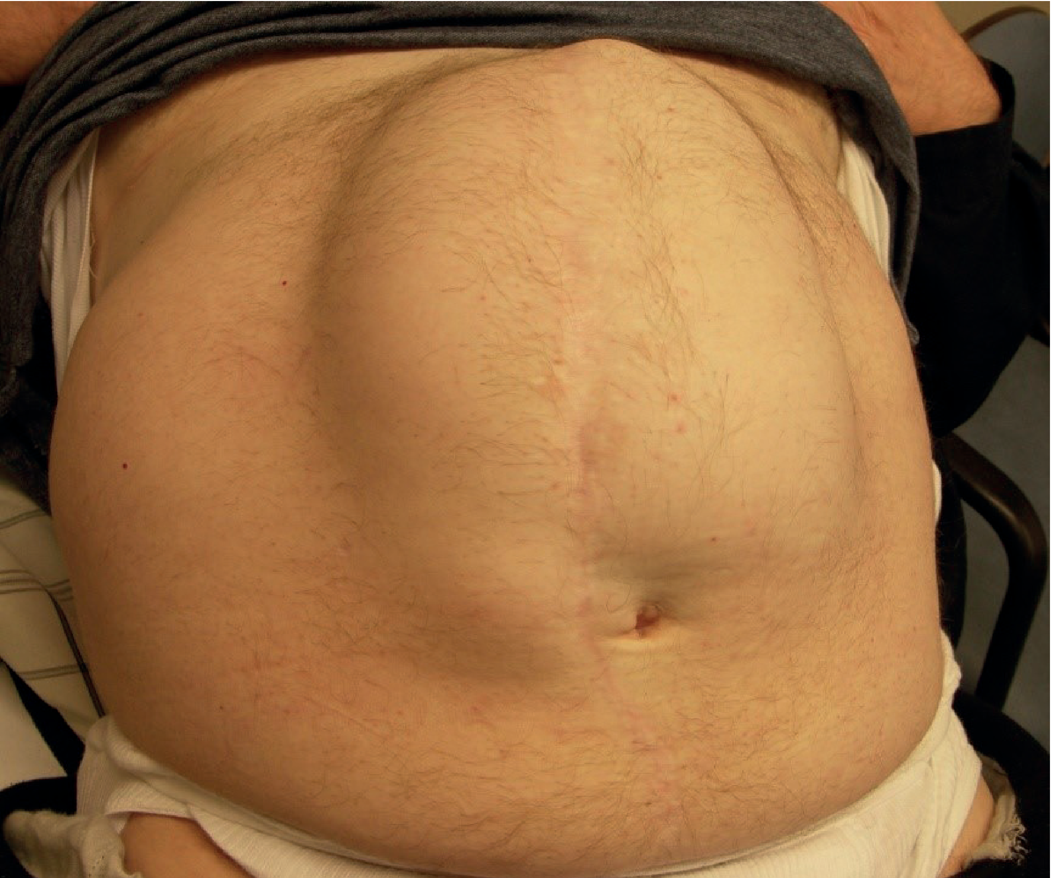
Large incisional hernia - note full-length bulge of entire laparotomy scar
Definition & Epidemiology
- Hernia through musculofascial layers at a previous operative scar
- Incidence: 10-50% of laparotomy wounds; 1-5% of laparoscopic ports
- Obstruction common; strangulation less common (wide neck, shallow)
Risk Factors
- Patient: Obesity, malnutrition, steroids, collagen disorders, chronic cough, cancer
- Wound: Infection, ischaemia, poor tissue quality
- Surgeon: Wrong suture, poor technique (mass closure preferable)
Repair
- Prehabilitation: Weight loss (7% body weight target), smoking cessation
- Open repair with mesh: IPOM (intraperitoneal onlay mesh), retromuscular (Rives-Stoppa), component separation
- Laparoscopic repair: Mesh intraperitoneally with overlap ≥4 cm all sides
- Recurrence after repair: 20-50%
🚨 VIVA TRAPS - Incisional
- Q: Most common time for incisional hernia to appear? A: 6 months to 2 years postoperatively
- Q: What is the "burst abdomen" (dehiscence) herald sign? A: Serosanguinous discharge on day 5-7 post op
- Q: Recommended suture length to wound length ratio? A: 4:1 (small-stitch, small-bite technique)
5. OTHER HERNIAS - QUICK REFERENCE
Spigelian Hernia
- Occurs at lateral edge of rectus muscle through the semilunar (Spigelian) line, at or below arcuate line
- Interparietal - hidden under intact external oblique; painful but invisible on surface
- Diagnosis: CT/USS
Obturator Hernia
- Through obturator foramen
- Mostly in thin, old, multiparous women
- Howship-Romberg sign: Pain down medial thigh (obturator nerve compression)
- Diagnosed at laparotomy/CT for bowel obstruction
Epigastric Hernia
- Through linea alba between xiphoid and umbilicus
- Small defect, contains extraperitoneal fat
- Painful despite small size - fat incarcerated
Internal Hernia
- Paraduodenal (Treitz), through epiploic foramen (Winslow's hernia), transmesenteric
- Presents as small bowel obstruction; diagnosed at CT
Richter's Hernia
- Part of the antimesenteric border of bowel wall caught in hernia
- Can strangulate WITHOUT bowel obstruction (no complete occlusion of lumen)
- High mortality - diagnosis often delayed
🚨 VIVA TRAP - Richter's
- Q: Why can Richter's hernia present without obstruction? A: Only part of the bowel wall is incarcerated - lumen remains patent
- Q: Where is Richter's most commonly found? A: Femoral canal
PART 2: VARICOSE VEINS
Definition
Dilated, tortuous, incompetent superficial veins due to valve failure, most commonly in the long (great) saphenous system.
Anatomy
- Great saphenous vein (GSV): Runs from medial malleolus to saphenofemoral junction (SFJ) in femoral triangle (1 cm below and lateral to pubic tubercle)
- Small saphenous vein (SSV): Runs from lateral malleolus to saphenopopliteal junction (SPJ) in popliteal fossa
- Normal veins drain upward; valves prevent retrograde flow
Pathophysiology
- Primary: Intrinsic valve failure or vein wall weakness
- Secondary: DVT damage, pregnancy (venous hypertension), AV fistula, pelvic tumors
CEAP Classification
| Class | Features |
|---|
| C0 | No visible/palpable signs |
| C1 | Telangiectasia/reticular veins |
| C2 | Varicose veins |
| C3 | Oedema |
| C4a | Pigmentation/eczema; C4b = lipodermatosclerosis |
| C5 | Healed venous ulcer |
| C6 | Active venous ulcer |
Symptoms
Aching, heaviness, throbbing, burning, itching - worse with prolonged standing, relieved by elevation.
Clinical Examination
Trendelenburg test (tourniquet test):
- Elevate leg, compress SFJ
- Veins fill when tourniquet released = SFJ incompetence
- Veins fill with tourniquet applied = lower perforator incompetence
Perthe's test: Tourniquet applied, patient walks - if varicosities reduce = deep vein patent; if increase = deep vein obstruction
Investigations
Duplex USS (gold standard):
- Identifies site of reflux
- Measures diameter
- Checks deep vein patency
- "Mickey Mouse" sign at SFJ: GSV and CFV medial to CFA in transverse view
Treatment Options
| Method | Mechanism |
|---|
| Compression hosiery | Conservative, class 2/3 |
| Sclerotherapy | Foam (UGFS) or liquid injection |
| Endovenous laser ablation (EVLA) | Thermal damage to vein wall; tumescent anaesthesia required |
| Radiofrequency ablation (RFA) | Similar to EVLA (ClosureFast) |
| Mechanochemical ablation (MOCA) | Spinning wire + sclerosant - NO tumescent needed |
| Endovenous glue (VenaSeal) | Cyanoacrylate adhesive |
| Open surgery | SFJ ligation + GSV stripping + avulsion phlebectomy |
Operative Steps: SFJ Ligation + GSV Stripping
- Oblique groin incision at groin crease, lateral to pubic tubercle
- Identify GSV, trace to SFJ
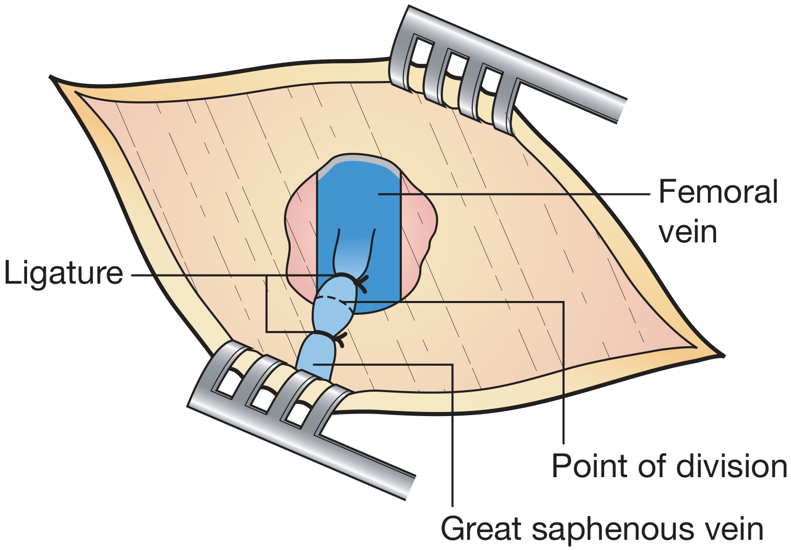
- Identify and ligate all 6 tributaries at SFJ flush (SIEV, SCIV, SEPV, DEPV, AAGSV, posteromedial)
- Flush ligation at SFJ - confirm femoral vein not accidentally divided
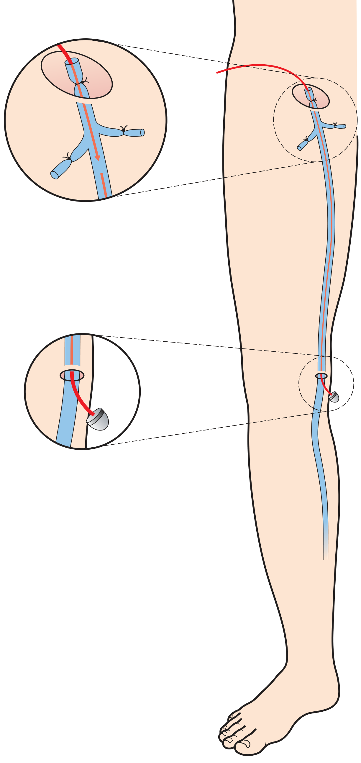
- GSV stripped retrogradely to knee level using stripper
- Avulsion phlebectomy (Muller's hook) of tributaries through mini-stab incisions
- Compression bandaging
6 Tributaries at SFJ (mnemomic: "Silent Sound Echo Does Penetrate Above")
- Superficial Inferior Epigastric Vein
- Superficial Circumflex Iliac Vein
- Superficial External Pudendal Vein
- Deep External Pudendal Vein
- Anterior Accessory GSV
- Posteromedial thigh vein
Complications of Varicose Vein Surgery
- Early: Haematoma, wound infection, DVT, saphenous nerve injury (medial leg below knee)
- Late: Recurrence (neovascularization commonest cause), sural nerve injury (SSV stripping)
🚨 VIVA TRAPS - Varicose Veins
- Q: Why strip the GSV only to the knee? A: Saphenous nerve runs close below knee - injury causes medial leg numbness
- Q: What does "flush ligation" mean? A: Ligating the GSV at its entry to the femoral vein WITHOUT leaving a stump (stump causes neovascularization and recurrence)
- Q: Commonest cause of recurrence? A: Neovascularization at groin (not leaving stump)
- Q: NICE guidelines - first line treatment for varicosities? A: Endothermal ablation (EVLA/RFA), then foam sclerotherapy, then surgery
- Q: Can you do varicose vein surgery if deep veins are blocked? A: NO - deep veins must be patent, as superficial veins are the only drainage route
PART 3: STOMA
Definition
An artificial opening of a hollow viscus onto the body surface.
Types of Stoma
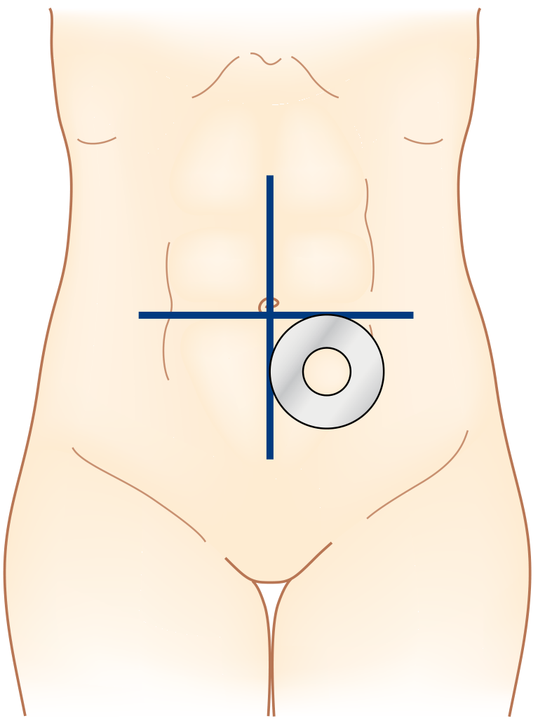
Stoma siting: ideal site is the infraumbilical fat mound, avoiding bony prominences, skin folds and scars
| Type | Site | Output | Shape | Use |
|---|
| Colostomy | Large bowel | Formed stool | Flush/everted | Hartmann's, APR, obstruction |
| Ileostomy | Ileum | Liquid (1-2L/day) | Spouted/everted (2-3 cm) | After proctocolectomy, protection of anastomosis |
| Loop ileostomy | Ileum loop | Liquid | Temporary | Protect distal anastomosis |
| Loop colostomy | Transverse colon | Semi-formed | Temporary | Obstruction, Hartmann's reversal |
| Urostomy (ileal conduit) | Ileum + ureters | Urine | Spouted | After cystectomy |
| Jejunostomy | Jejunum | High-output watery | Flush | Feeding/after oesophagectomy |
| Caecostomy | Caecum | Liquid | Flush | Rarely used |
Stoma Spouting
- Ileostomy: Must be spouted (2-3 cm) - proteolytic enzymes in liquid output damage skin
- Colostomy: Flush with skin is acceptable - formed stool less irritant
Siting of Stoma
- Must be sited preoperatively by stoma nurse
- In the infraumbilical fat mound (best site)
- Away from: bony prominences, umbilicus, old scars, skin folds, belt line
- Patient must be able to see and manage it
Stoma Complications
| Complication | Details |
|---|
| Retraction | Stoma sinks below skin level; ischemia, poor siting |
| Prolapse | Loop stomas > end stomas; transverse > other |
| Parastomal hernia | Most common long-term complication of colostomy |
| Stenosis | Ischaemia, Crohn's |
| Ischaemia/Necrosis | Early - inadequate blood supply |
| High output | Ileostomy >2L/day - dehydration, electrolyte imbalance |
| Skin excoriation | Poor appliance fit, especially ileostomy |
| Fistula | Crohn's, ischemia |
High Output Ileostomy (>2L/day)
- Causes: Short bowel, Crohn's, obstruction distal to stoma, sepsis
- Management: Low-fibre diet, oral rehydration, codeine phosphate, loperamide, PPI (reduces gastric secretions), octreotide (last resort)
🚨 VIVA TRAPS - Stoma
- Q: Why spout an ileostomy but not a colostomy? A: Ileal output contains active proteolytic enzymes that damage skin - spout directs output into the bag
- Q: Commonest long-term complication of colostomy? A: Parastomal hernia
- Q: Why avoid siting a stoma in the midline? A: Appliance will not seal and bag will leak
- Q: What is a loop ileostomy defunctioning stoma? A: Most common covering stoma for low anterior resection
- Q: Hartmann's operation = ? A: Sigmoid colectomy + end colostomy + rectal stump closure (no anastomosis)
PART 4: X-RAY INTERPRETATION (SURGICAL)
Plain Abdominal X-Ray (AXR) - Systematic Approach
- Patient details: Name, date, AP/PA, supine/erect
- Gas pattern: Normal (stomach + small bowel only)
- Bowel dilatation: Small bowel >3 cm (central, valvulae conniventes cross full width); large bowel >6 cm (caecum >9 cm), peripheral, haustrations
- Free gas: Erect CXR preferred - gas under diaphragm = perforation
- Calcification: Gallstones (10% radio-opaque), renal calculi (90%), aortic aneurysm, pancreatic calcification
- Soft tissues/masses: Psoas shadow loss = retroperitoneal pathology
- Bones: Fractures, metastases, osteoporosis
Key X-Ray Patterns in Surgery
| Pattern | X-Ray Finding | Diagnosis |
|---|
| Erect CXR - gas under right hemidiaphragm | Free peritoneal gas | Perforation (peptic ulcer, bowel) |
| Dilated small bowel (central, valvulae conniventes) | Ladder pattern | Small bowel obstruction |
| Dilated large bowel + coffee bean sign | Sigmoid volvulus | |
| Dilated caecum + coffee bean in RUQ | Caecal volvulus | |
| Grossly dilated transverse colon >6 cm | Toxic megacolon | |
| No gas in rectum + central dilated loops | Closed loop obstruction | |
| Thumb printing in LIF | Mucosal oedema | Ischaemic colitis |
| Rigler's sign (gas on both sides of bowel wall) | Massive pneumoperitoneum | |
| Stepladder pattern (multiple air-fluid levels) | Mechanical SBO | |
| Ground glass appearance | Free fluid/ascites | |
| Obliteration of psoas shadow | Retroperitoneal pathology | |
Chest X-Ray Post-Op
- Pneumothorax: Absence of lung markings
- Pleural effusion: Homogeneous opacity, meniscus sign
- Consolidation: Air bronchogram
- Mediastinal widening: Aortic dissection, rupture
- NG tube position: Should cross midline (not go to right = right main bronchus)
🚨 VIVA TRAPS - X-Ray
- Q: Gas under diaphragm - which side is normal? A: Right side (liver confirms right diaphragm; left has gastric bubble which can mimic)
- Q: Rigler's sign? A: Gas visible on BOTH sides of the bowel wall (both inner and outer surface) = massive pneumoperitoneum
- Q: Why is erect CXR better than AXR for pneumoperitoneum? A: Even 1 mL of gas visible under diaphragm on CXR; needs 500 mL on AXR
- Q: Coffee bean sign? A: Sigmoid volvulus - sigmoid colon bent on itself, both limbs dilated, seen in RUQ pointing to RUQ
PART 5: SURGICAL SPECIMENS
How to Describe a Specimen
- Type of specimen (bowel, stomach, thyroid, breast, etc.)
- Size and weight
- External surface: capsule, serosa, peritoneum, colour
- Attached structures: mesentery, lymph nodes, margins
- Cut surface: tumour, haemorrhage, necrosis, cysts
- Tumour features: site, size, shape, colour, borders, depth of invasion
- Resection margins: distance from margins
- Lymph nodes: number, suspicious nodes
Common Specimens You May Be Asked About
| Specimen | What to Look For |
|---|
| Right hemicolectomy | Caecum, ascending colon, terminal ileum; tumour in caecum/ascending colon |
| Sigmoid colectomy | Diverticular disease or adenocarcinoma |
| APR specimen | Rectum + anus + sphincters + levators; TME quality |
| Whipple specimen | Pancreatic head, duodenum, CBD, part of stomach; tumour at ampulla |
| Gastrectomy | Type of gastrectomy (subtotal/total), tumour, D1/D2 nodes |
| Cholecystectomy | Gallbladder - stones, thickened wall, cancer |
| Thyroid | Thyroid lobe/whole gland; nodule character, capsule |
| Mastectomy | Breast tissue, tumour quadrant, axillary nodes |
| Appendix | Faecolith, perforation, carcinoid (at tip), tumour |
TME (Total Mesorectal Excision) Quality Assessment
- Grade 1 (Complete): Intact mesorectal fascia, smooth surface, no defects
- Grade 2 (Near complete): Minor irregularities, no bare areas
- Grade 3 (Incomplete): Bare areas, inadequate surgical plane
PART 6: OPERATIVE PROCEDURES
1. SUPERFICIAL PAROTIDECTOMY
Indications
- Benign parotid tumours (pleomorphic adenoma, Warthin's tumour)
- Biopsy of parotid mass
- Removes the superficial lobe only (80% of gland)
Key Anatomy
- Facial nerve (CN VII) exits stylomastoid foramen → enters parotid → pes anserinus → 5 branches (TETMB: Temporal, Zygomatic, Buccal, Marginal mandibular, Cervical)
- Marginal mandibular branch = most commonly injured (unprotected, runs close to lower border)
- Greater auricular nerve (C2,3) - provides sensation to earlobe/parotid area
Operative Steps
- Position: Supine, head turned away from operative side, shoulder roll
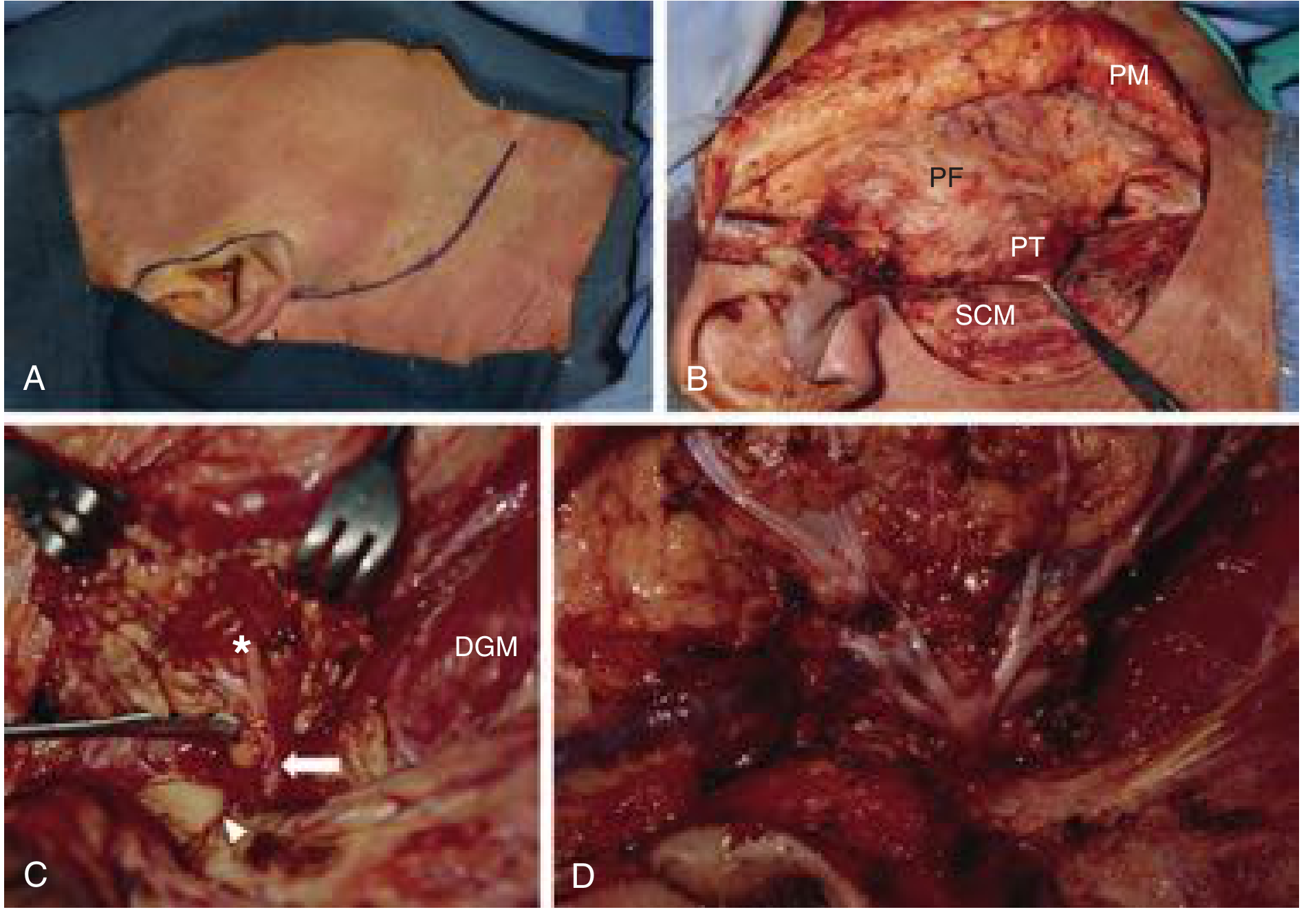
- Incision: Modified Blair incision - pre-auricular crease, around lobule, behind ear onto mastoid, then into neck skin crease (or facelift incision for cosmesis)
- Skin flaps: Elevated in plane superficial to parotid fascia anteriorly; subplatysmal in neck
- Greater auricular nerve: Identified over SCM - may need to be divided (or preserved if possible)
- Tail of parotid: Freed from SCM
- Identify facial nerve: Use tragal pointer - nerve is 1-1.5 cm deep and inferior to tragal pointer; OR follow posterior digastric to mastoid attachment. Tympanomastoid fissure = alternative landmark
- Dissection along nerve: All branches traced forward meticulously - "teasing" rather than cutting
- Superficial lobe removal: Parotid tissue lateral to nerve removed - pes anserinus displayed
- Haemostasis: Clip all small vessels
- Drain: Closed suction drain
- Closure: Platysma, subcutaneous, skin
Complications
- Facial nerve palsy: Temporary (neuropraxia) = most common; permanent = rare
- Frey's syndrome (auriculotemporal syndrome): Gustatory sweating - sweating and flushing over parotid area when eating (misdirected regeneration of auriculotemporal nerve to sweat glands); treatment = Botox
- Salivary fistula
- Greater auricular nerve numbness (lobule anaesthesia)
- Haematoma
- Scar
- Recurrence (pleomorphic adenoma - satellite nodules)
🚨 VIVA TRAPS - Parotidectomy
- Q: What is Frey's syndrome? A: Gustatory sweating - auriculotemporal nerve (parasympathetic secretomotor) misdirected to sweat glands after surgery
- Q: Why not enucleate a pleomorphic adenoma? A: Pseudocapsule with satellite nodules - rupture causes seeding and 50% recurrence
- Q: Most reliable landmark for facial nerve? A: Tragal pointer (nerve is 1-1.5 cm deep and inferior)
- Q: Branch most commonly injured? A: Marginal mandibular (runs superficial at lower border of mandible)
- Q: What is pes anserinus? A: Goose foot appearance of facial nerve branching in parotid gland
2. SUBTOTAL THYROIDECTOMY
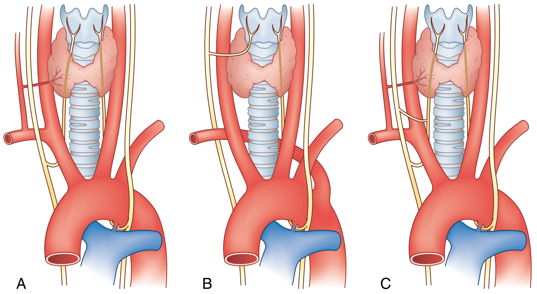
Anomalous variations of the RLN course - non-recurrent laryngeal nerve (right side) in ~1% cases
Indications for Subtotal vs Total Thyroidectomy
- Subtotal: Bilateral toxic goitre (Graves'), leaving ~4-7 g of tissue on each side
- Total: Thyroid cancer (papillary, follicular, medullary), large compressive goitre
- Hemithyroidectomy: Single nodule, one lobe, diagnostic excision
Key Anatomy
- Recurrent laryngeal nerve (RLN): Runs in tracheoesophageal groove; enters larynx posterior to cricothyroid joint; most commonly crosses POSTERIOR to inferior thyroid artery; RIGHT RLN hooks around right subclavian; LEFT hooks under ligamentum arteriosum
- External branch of superior laryngeal nerve (EBSLN): Runs with superior thyroid vessels; injury → loss of high pitch/projection
- Parathyroid glands: 4 glands; superior (at junction of upper 2/3 and lower 1/3 of thyroid, posterior) and inferior (variable, associated with inferior thyroid artery/thymic remnants)
- Tubercle of Zukerkandl: Lies lateral and anterior to RLN - useful landmark
Operative Steps (Subtotal Thyroidectomy)
- Position: Supine, neck extended (shoulder roll), table 30° head up
- Incision: Kocher's collar incision - 2-3 cm above sternal notch in skin crease (6-8 cm)
- Subplatysmal flaps: Raised superiorly to thyroid cartilage, inferiorly to sternal notch
- Midline division: Strap muscles (sternohyoid, sternothyroid) - divided in midline, retracted laterally OR divided horizontally if goitre large
- Superior pole: Superior thyroid vessels ligated close to the gland (to protect EBSLN)
- Lateral mobilisation: Middle thyroid vein ligated and divided
- RLN identification: Traced in tracheoesophageal groove - protected throughout
- Inferior thyroid artery: Divided medial to RLN entry point
- Parathyroids: Identified, preserved with their vascular pedicle
- Berry ligament: Divided carefully (RLN close here)
- Gland removal: Leaving a posterolateral cuff of 4-7 g on each side (subtotal) or complete removal (total)
- Haemostasis: Check for bleeding - hiss test with Valsalva
- Drain: Not always needed
- Strap muscles: Reapproximated
- Platysma and skin: Closed
Complications
| Complication | Notes |
|---|
| Haemorrhage (post-op) | Cervical haematoma - airway emergency, open wound at bedside |
| RLN injury (temporary) | Hoarseness, most recover in 6-8 weeks |
| RLN injury (permanent) | Unilateral: hoarseness; Bilateral: stridor, tracheostomy needed |
| Hypoparathyroidism | Hypocalcaemia - tingling, Chvostek's, Trousseau's; treat with calcium + vitamin D |
| Hypothyroidism | All cases after total thyroidectomy |
| Thyroid storm | In uncontrolled hyperthyroid - tachycardia, fever, confusion - treat with beta-blockers, propylthiouracil, Lugol's, steroids, cooling |
| Wound infection | |
| Keloid scar | |
🚨 VIVA TRAPS - Thyroidectomy
- Q: What is the most common cause of hoarseness after thyroidectomy? A: RLN neuropraxia (usually temporary, recovers in 6-8 weeks)
- Q: Why is the superior thyroid artery ligated close to the gland? A: To protect the EBSLN which runs with the artery
- Q: What is the non-recurrent laryngeal nerve? A: Right RLN in ~1% - aberrant right subclavian (arteria lusoria) - nerve does NOT recurse
- Q: Chvostek's sign? A: Tap over facial nerve anterior to tragus → ipsilateral facial muscle twitching (hypocalcaemia)
- Q: Trousseau's sign? A: BP cuff inflated above systolic → carpal spasm (hypocalcaemia)
- Q: Why not incise strap muscles horizontally routinely? A: Preserves nerve supply (ansa cervicalis from below)
3. THYROGLOSSAL CYST EXCISION (SISTRUNK OPERATION)
Definition
Cyst arising from persistence of the thyroglossal duct - the tract along which the thyroid descended from foramen cecum at base of tongue to its final position.
Key Facts
- Most common congenital neck mass in children
- Location: Midline, at or just below hyoid bone (most common); moves UP on tongue protrusion AND swallowing (distinguishes from other midline lumps)
- May present anywhere along thyroglossal tract from foramen cecum to pyramidal lobe
- Can become infected → may form a fistula to skin (thyroglossal fistula)
- Malignancy rare (1-2%): papillary carcinoma most common
- Before surgery: Confirm normal thyroid is present by USS (cyst may be the only thyroid tissue)
Sistrunk Operation - Operative Steps
- Position: Supine, neck extended
- Incision: Transverse incision over the cyst
- Cyst dissection: Cyst is carefully excised (avoid rupture to prevent recurrence)
- Tract traced superiorly: Above the hyoid
- Central portion of hyoid bone excised (middle 1/3) - this is the KEY step (tract passes THROUGH hyoid in many cases)
- Tract cored out: Through tongue base musculature up to foramen cecum using a core of muscle (a finger pressed into the vallecula from inside helps)
- Recurrence prevention: Complete excision including muscle core around the tract
- Closure: In layers
Why Remove the Hyoid?
- The thyroglossal duct passes through or immediately posterior to the hyoid bone
- Simple cyst excision without hyoid resection has >50% recurrence
- Sistrunk operation reduces recurrence to <5%
🚨 VIVA TRAPS - Thyroglossal Cyst
- Q: What makes thyroglossal cyst move upward? A: Tongue protrusion (attached to foramen cecum at base of tongue via tract)
- Q: Why must you always do USS before surgery? A: The cyst may be the only functional thyroid tissue - removing it causes hypothyroidism
- Q: What is the key step to prevent recurrence? A: Removal of central portion of hyoid bone (Sistrunk principle)
- Q: Most common carcinoma in thyroglossal cyst? A: Papillary thyroid carcinoma
- Q: What structure should be palpated for during surgery to ensure complete excision of the tract? A: Surgeon's finger in foramen cecum/vallecula guides excision up to base of tongue
4. MODIFIED RADICAL MASTECTOMY (MRM)
Definition
Removal of the entire breast + level I-II axillary lymph node dissection; preserves the pectoralis major muscle (unlike Halsted's radical mastectomy).
Halsted vs Patey vs Auchincloss
| Operation | Pec Major | Pec Minor | Axillary Nodes |
|---|
| Halsted (Radical) | Removed | Removed | All (I, II, III) |
| Patey | Preserved | Removed | I, II, III |
| Auchincloss (MRM) | Preserved | Preserved | I, II |
Indications
- Breast cancer with clinically/biopsy-proven positive lymph node involvement
- Large tumour not suitable for BCS
- Multifocal disease
- Patient preference
- Inflammatory breast cancer
Operative Steps (MRM)
- Position: Supine, arm abducted 90°, ipsilateral chest raised on sandbag
- Incision: Elliptical incision encompassing nipple-areolar complex and any previous biopsy scar; adequate skin to lie flat on chest (if ptotic = wider ellipse)
- Skin flaps raised: In plane of Cooper's ligaments (between glandular tissue and subcutaneous fat); upper to clavicle, lower to inframammary fold, medial to sternum, lateral to latissimus dorsi
- Breast dissection: Off pectoralis major fascia (fascia included in specimen)
- Axillary dissection:
- Axillary fascia opened
- Axillary contents cleared: Level I (lateral to pec minor), Level II (posterior to pec minor)
- Long thoracic nerve of Bell preserved (runs on serratus anterior - injury = winged scapula)
- Thoracodorsal nerve preserved (to latissimus dorsi - injury = weak shoulder extension)
- Intercostobrachial nerve (T2) - often divided → medial upper arm numbness
- Specimen: Breast + axillary contents removed en bloc
- Haemostasis, 2 drains (one breast, one axilla)
- Closure: Subcutaneous, skin
Complications
| Complication | Notes |
|---|
| Seroma | Most common (30-40%); aspirated |
| Haematoma | Re-exploration if large |
| Wound infection | |
| Lymphoedema | Chronic arm swelling after axillary clearance |
| Winged scapula | Long thoracic nerve injury |
| Weak shoulder extension | Thoracodorsal nerve injury |
| Numbness medial arm | Intercostobrachial nerve division |
| Flap necrosis | |
🚨 VIVA TRAPS - MRM
- Q: Which nerve injury causes winged scapula? A: Long thoracic nerve of Bell (C5,6,7) to serratus anterior
- Q: Which nerve must be preserved for latissimus dorsi flap reconstruction later? A: Thoracodorsal nerve
- Q: What nerve is routinely divided in axillary clearance? A: Intercostobrachial nerve (T2) - medial arm numbness expected
- Q: What is Level III axillary dissection? A: Nodes medial to pec minor (risks lymphoedema significantly)
- Q: What is "sentinel lymph node biopsy"? A: Identification of first draining node using blue dye ± radioisotope - if negative, full dissection avoided
5. TRACHEOESOPHAGEAL FISTULA REPAIR
Classification (Gross)
| Type | Description | Incidence |
|---|
| A | Pure EA without fistula | 8% |
| B | EA with proximal TEF | 3% |
| C | EA with DISTAL TEF | 85% (commonest) |
| D | EA with both proximal and distal TEF | <1% |
| E (H-type) | TEF without EA | 4% |
Associations: VACTERL
V-ertebral, A-norectal, C-ardiac (most common associated anomaly - 33%), T-racheal, E-sophageal, R-adial/Renal, L-imb
Diagnosis
- Failure to pass size 10F catheter beyond 10 cm
- Excessive drooling, respiratory distress, cyanosis during feeds
- CXR: Curled NG tube in blind pouch; gastric bubble (if distal fistula present)
Operative Steps (Repair of Type C - most common)
- Position: Left lateral decubitus
- Approach: Right posterolateral thoracotomy through 4th intercostal space (extrapleural approach preferred)
- Identify fistula: Trace to trachea
- Divide fistula: Close tracheal end with interrupted sutures
- Mobilise oesophageal ends
- Primary end-to-end anastomosis: If gap <3 cm (single layer interrupted absorbable sutures)
- Long-gap EA: >3 cm = delayed repair, Foker technique (external traction sutures), or interposition (colon, stomach)
- Drain: Extrapleural drain
- Post-op: Baby kept intubated 24-48h, parenteral nutrition, contrast swallow at 5-7 days before feeding
Complications
- Anastomotic leak (10-20%)
- Stricture (most common long-term)
- GORD (very common - majority of patients)
- Tracheomalacia
- Recurrent fistula
🚨 VIVA TRAPS - TEF
- Q: Commonest type of TEF? A: Type C - EA with distal TEF (85%)
- Q: Why is there a gastric bubble on CXR in Type C but not Type A? A: Distal fistula allows air from trachea into stomach in Type C
- Q: What is polyhydramnios in utero + this diagnosis? A: Classic antenatal finding - fetus cannot swallow amniotic fluid
- Q: What is the Foker technique? A: External traction sutures to elongate the two oesophageal ends before anastomosis (used in long-gap EA)
6. RADICAL GASTRECTOMY WITH SPLENECTOMY
Indications
- Gastric carcinoma of upper third/proximal stomach/fundus/greater curvature requiring en bloc splenectomy for D2 lymph node clearance (station 10/11)
- Note: Modern evidence shows routine splenectomy does NOT improve survival in proximal gastric cancer without direct involvement - controversial
D Lymph Node Dissection
| Level | Nodes included | Survival benefit |
|---|
| D1 | Perigastric nodes (stations 1-6) | Adequate for early gastric cancer |
| D2 | D1 + celiac, hepatic, left gastric, splenic artery nodes | Standard for resectable gastric cancer |
| D3 | D2 + para-aortic | No proven benefit |
Operative Steps (Total/Radical Gastrectomy with Splenectomy)
- Position: Supine, reverse Trendelenburg
- Incision: Upper midline/bilateral subcostal (roof-top)
- Exploration: Assess resectability - peritoneum, liver, nodes
- Greater omentum: Divided from transverse colon
- Lesser omentum: Opened, hepatogastric ligament divided
- Left gastric artery: Ligated at celiac axis origin (D2 dissection)
- Splenic vessels: Divided; spleen removed with tail of pancreas in extended procedures
- Right gastric and right gastroepiploic arteries: Ligated
- Duodenum: Divided 2 cm beyond pylorus (stapled or manual closure)
- Oesophageal transection: Divided above cardia
- Roux-en-Y reconstruction: 60 cm Roux limb anastomosed to oesophagus (oesophagojejunostomy); or Billroth I (subtotal)
- Feeding jejunostomy: Inserted
- Drains: Near anastomosis
🚨 VIVA TRAPS - Gastrectomy
- Q: Billroth I vs II vs Roux-en-Y? A: Billroth I = gastroduodenostomy (subtotal only); Billroth II = gastrojejunostomy (loop); Roux-en-Y = preferred for total gastrectomy - reduces bile reflux
- Q: What is dumping syndrome? A: Early dumping (30 min) = rapid emptying → fluid shift from blood to bowel; Late dumping (2-3 h) = reactive hypoglycaemia
- Q: Why splenectomy with proximal gastrectomy? A: Station 10 (hilar) and 11 (splenic artery) nodes - but current evidence does not support routine splenectomy if not involved
- Q: Anastomotic leak risk factors? A: Malnutrition, tension, ischaemia, infection, diabetes
7. TOTAL PROCTOCOLECTOMY
Definition
Removal of entire colon, rectum, and anus.
Indications
- Ulcerative colitis (with dysplasia or failure of medical treatment)
- Familial adenomatous polyposis (FAP)
- Synchronous colorectal cancers
Types
| Procedure | Continence | Stoma |
|---|
| Total proctocolectomy + end ileostomy (Brooke) | None needed | Permanent ileostomy |
| Restorative proctocolectomy + ileal pouch (IPAA) | Maintained | Temporary loop ileostomy (6-8 weeks) |
| Kock pouch (continent ileostomy) | Continent | Flush stoma + intubated to empty |
IPAA (Ileal Pouch-Anal Anastomosis) Steps
- Total abdominal colectomy (colon removed)
- Rectal excision with TME to dentate line
- J-pouch construction from distal 30-40 cm of ileum (most common shape)
- Stapled or hand-sewn anastomosis to anal canal at dentate line
- Covering loop ileostomy
- Ileostomy reversal at 6-8 weeks (contrast pouch study first)
🚨 VIVA TRAPS - Proctocolectomy
- Q: Why is UC cured by total proctocolectomy but Crohn's is not? A: UC is limited to the colon and rectum; Crohn's can affect any part of GI tract (skip lesions)
- Q: Commonest complication of IPAA? A: Pouchitis (inflammation of ileal pouch) - treated with metronidazole
- Q: What is the IPAA contraindicated in? A: Crohn's disease (anastomotic failure, pelvic sepsis, fistula), poor sphincter function
- Q: J vs S vs W pouch? A: J-pouch most widely used (simplest, good results); S-pouch allows longer reach but may need intubation; W-pouch largest capacity
8. ESOPHAGOPLASTY (COLONIC INTERPOSITION / OESOPHAGEAL REPLACEMENT)
Indications
- Long-gap oesophageal atresia
- Oesophageal stricture/burns (caustic injury)
- After oesophagectomy when stomach not available
Types of Conduit
| Conduit | Notes |
|---|
| Gastric tube/whole stomach | Most common after oesophagectomy |
| Right colon | Good length; ileocolic artery pedicle |
| Left colon | Most popular for benign disease; L colic + MCA pedicle |
| Free jejunum | For short cervical defects (microvascular) |
Colonic Interposition Steps
- Assess colon viability - no diverticular disease, colonoscopy
- Isoperistaltic left or right colon segment chosen (15-20 cm)
- Vascular pedicle based on middle colic or left colic artery
- Colon divided, transposed to chest (substernal or posterior mediastinal route)
- Proximal: Cervical oesophagocolic anastomosis
- Distal: Cologastric or colo-jejunal anastomosis
- Colon continuity restored (colocolostomy)
🚨 VIVA TRAPS - Esophagoplasty
- Q: What is the most common conduit after oesophagectomy? A: Stomach (gastric tube/pull-up)
- Q: What is the blood supply of the gastric conduit? A: Right gastroepiploic artery (the most important vessel to preserve)
- Q: Route for gastric conduit? A: Posterior mediastinal (orthotopic) = shortest; substernal = alternative
9. OPEN CHOLECYSTECTOMY
Indications
- Conversion from laparoscopic (bleeding, unclear anatomy, CBD injury)
- Emergency: Peritonitis, empyema, severe cholecystitis
Key Anatomy - Calot's Triangle
- Medially: Common hepatic duct
- Superiorly: Inferior surface of liver
- Inferiorly: Cystic duct
- Contains: Cystic artery (from right hepatic in 80%; variable), node of Lund
Operative Steps
- Position: Supine, slight right tilt
- Incision: Kocher's right subcostal OR upper midline
- Exposure: Xiphoid retracted, liver retracted up by assistant
- Identification of Calot's triangle: Hepatoduodenal ligament cleared
- Critical view of safety (CVS): Two structures (cystic duct and cystic artery) seen entering the gallbladder, base of Calot's triangle cleared of fat
- Cystic artery clipped and divided
- Cystic duct clipped and divided (cholangiogram if CBD unclear)
- Gallbladder dissected off liver bed (fundus first in open, or fundus-down)
- Haemostasis of liver bed
- Drain: Optional
🚨 VIVA TRAPS - Cholecystectomy
- Q: What is the Critical View of Safety? A: Two structures seen entering gallbladder only (cystic duct and cystic artery) after clearing the hepatocystic triangle - prevents CBD injury
- Q: Mirizzi syndrome? A: Stone impacted in Hartmann's pouch/cystic duct compresses CHD → obstructive jaundice - convert to open
- Q: Mucocele vs empyema of gallbladder? A: Mucocele = distension with mucus (clear); Empyema = pus (infected)
10. LAPAROSCOPIC CHOLECYSTECTOMY
Port Positions (4-port standard)
- 10mm at umbilicus (camera)
- 10mm epigastric (surgeon's right hand)
- 5mm RUQ anterior axillary line (retraction)
- 5mm RUQ mid-clavicular line (retraction)
Operative Steps
- Pneumoperitoneum: Veress needle or Hasson technique at umbilicus (CO₂, 12-15 mmHg)
- Ports: As above
- Trendelenburg + left tilt: Moves bowel away from field
- Gallbladder retraction: Fundus retracted cephalad over liver; Hartmann's pouch retracted laterally
- Peritoneum scored: Anterior and posterior leaves of hepatocystic triangle
- Critical View of Safety: Dissect until fat cleared, two structures only visible entering gallbladder
- Cystic artery: Clips applied + divided
- Cystic duct: Clips applied (3 clips: 2 on specimen side, 1 on common duct side); divided
- Gallbladder removed from liver bed using diathermy
- Suction/irrigation: Check for bleeding and bile leak
- Gallbladder retrieval: Via umbilical port (retrieval bag if stones spilled)
- Ports closed: Fascia at 10mm ports
Complications
| Complication | Management |
|---|
| CBD injury (0.3-0.5%) | Immediate repair if recognized; choledochojejunostomy |
| Bleeding | Packing, conversion |
| Bile leak | Drain, ERCP + stent |
| Port site hernia | Fascial closure of 10mm ports |
| Retained stone in CBD | ERCP post-op |
🚨 VIVA TRAPS - Lap Cholecystectomy
- Q: What is the Critical View of Safety? A: Clear the hepatocystic triangle until only TWO structures seen entering gallbladder - established by Strasberg
- Q: Most serious complication? A: CBD injury (0.3-0.5%)
- Q: Most common time of CBD injury? A: "Tent effect" when Hartmann's pulled laterally - CBD tented and mistaken for cystic duct
- Q: What is a bile lake? A: Bile collection in subhepatic space - diagnose with HIDA scan; treat with ERCP ± drain
11. CHOLEDOCHODUODENOSTOMY
Definition
Anastomosis between the common bile duct (CBD) and the first part of the duodenum.
Indications
- Common bile duct stones not amenable to ERCP
- Benign biliary stricture
- Choledochal cyst (rarely)
- Bile duct obstruction from chronic pancreatitis
- CBD diameter must be ≥15 mm (dilated) for best results
Contraindications
- Cholangitis (active)
- CBD not dilated (<15 mm) - sump syndrome risk
Operative Steps
- Kocherisation of duodenum (mobilise to expose CBD posteriorly)
- CBD identified on hepatoduodenal ligament
- Longitudinal incision on CBD (at least 15 mm)
- Transverse incision in first part of duodenum
- Side-to-side anastomosis: 2-layer interrupted absorbable sutures
- T-tube if desired
- Drain placed near anastomosis
Complication: Sump Syndrome
- Food, biliary sludge accumulates in CBD distal "sump" beyond anastomosis
- Presents with cholangitis, jaundice
- Prevention: Large anastomosis (>15mm), place anastomosis as distal as possible
🚨 VIVA TRAPS - CDD
- Q: Why must CBD be ≥15 mm for choledochoduodenostomy? A: Smaller anastomosis risks stricture + sump syndrome
- Q: What is sump syndrome? A: Food debris collects in distal blind-ending CBD between anastomosis and ampulla → cholangitis
- Q: What is the alternative to choledochoduodenostomy? A: Roux-en-Y choledochojejunostomy (avoids sump syndrome; used for malignant obstruction)
12. WHIPPLE'S PROCEDURE (PANCREATICODUODENECTOMY)
Indications
- Pancreatic head carcinoma
- Ampullary carcinoma
- Distal CBD carcinoma
- Duodenal carcinoma
- Chronic pancreatitis (severe, pain)
What is Removed
- Head and neck of pancreas
- Duodenum (all 4 parts)
- Distal CBD
- Gallbladder
- Distal stomach (or pylorus-preserving variant - PPPD)
Three Anastomoses in Whipple's (Reconstruction)
- Pancreaticojejunostomy (or pancreaticogastrostomy): Highest leak risk
- Hepaticojejunostomy: Bile duct to jejunum
- Gastrojejunostomy (or duodenojejunostomy in PPPD)
Operative Steps
- Position: Supine, reverse Trendelenburg
- Incision: Upper midline or bilateral subcostal
- Exploration: Assess resectability (no distant mets, no SMA encasement)
- Kocher manoeuvre: Duodenum mobilised, SMA and SMV assessed
- Cholecystectomy and CBD division
- Gastric transection: 40% gastrectomy (or pylorus preservation in PPPD)
- Jejunal transection: 15 cm beyond ligament of Treitz
- Pancreatic neck division: Tunnel behind pancreas over SMV/portal vein
- Uncinate process dissection: Off SMA (most critical step)
- Specimen removed
- Reconstruction: Child's technique (panc-J → hepatic-J → gastro-J all on same limb) OR Whipple's modification (separate limbs)
- Drains: Near pancreatic anastomosis
Complications
| Complication | Incidence | Notes |
|---|
| Delayed gastric emptying (DGE) | 20-30% | Most common; NGT, prokinetics |
| Pancreatic fistula | 10-15% | Most dangerous; amylase in drain >3x serum |
| Post-pancreatectomy haemorrhage | 5-10% | Late = eroded vessel by fistula |
| Bile leak | 5% | |
| Wound infection | Common | |
| Diabetes | Long-term | |
🚨 VIVA TRAPS - Whipple
- Q: Most common complication? A: Delayed gastric emptying (DGE)
- Q: Most dangerous complication? A: Pancreatic fistula/POPF (post-operative pancreatic fistula)
- Q: What is the "critical step"? A: Uncinate process dissection off SMA - risk of uncontrollable bleeding
- Q: What is PPPD? A: Pylorus-preserving pancreaticoduodenectomy - stomach and pylorus kept; avoids dumping, similar oncologic outcome
- Q: What is the "soft pancreas"? A: Soft pancreatic texture → higher fistula risk (vs hard fibrotic pancreas in chronic pancreatitis)
- Q: Borderline resectable pancreatic cancer definition? A: Tumour abutting (not encasing) SMA or SMV - may need neoadjuvant chemo
13. LOW ANTERIOR RESECTION (LAR)
Definition
Resection of upper and mid rectum with anastomosis in the pelvis, preserving the sphincter complex. Includes Total Mesorectal Excision (TME).
Indications
- Rectal cancer in upper or mid rectum
- Tumour ≥1-2 cm from dentate line (enough sphincter to preserve function)
TME (Total Mesorectal Excision)
- Sharp dissection in the avascular plane outside the mesorectal fascia
- Preserves the intact mesorectal envelope (containing lymph nodes, blood vessels)
- Reduces local recurrence from 25% → 5-8%
- Preserves autonomic nerves (hypogastric plexus, pelvic splanchnic nerves)
Operative Steps (Open LAR)
- Position: Lloyd-Davis (lithotomy-Trendelenburg)
- Incision: Midline laparotomy
- Exploration: Liver, peritoneum for mets
- Left colon mobilisation: White line of Toldt incised; splenic flexure taken down (provides length)
- IMA ligation: At origin (high tie) or distal to left colic (low tie - preserves vascularity)
- TME dissection: Sharp dissection in avascular "holy plane" behind mesorectum
- Lateral ligaments: Divided (containing middle rectal vessels + autonomic nerves)
- Anterior dissection: In front of Denonvilliers' fascia (male); behind vaginal wall (female)
- Rectum divided: At least 2 cm clear margin; transanal stapler (CEEA)
- Anastomosis: Colorectal or coloanal - double stapled technique
- Loop ileostomy: Defunctioning to protect anastomosis in low anastomosis
- Drain: Pelvic drain
🚨 VIVA TRAPS - LAR
- Q: What is the Holy Plane? A: Avascular plane between mesorectal fascia and presacral fascia - described by Heald
- Q: What nerves are at risk during LAR? A: Hypogastric nerves + pelvic splanchnic nerves → injury causes bladder and sexual dysfunction
- Q: Anastomotic leak risk factors? A: Low anastomosis, male, obesity, smoking, steroids, no defunctioning stoma
- Q: Why is defunctioning ileostomy used? A: Anastomotic leak after low anastomosis → pelvic sepsis → stoma reduces consequences of leak
- Q: What margin is required for rectal cancer? A: Distal margin ≥1 cm; circumferential radial margin (CRM) ≥1 mm
14. ABDOMINOPERINEAL RESECTION (APR)
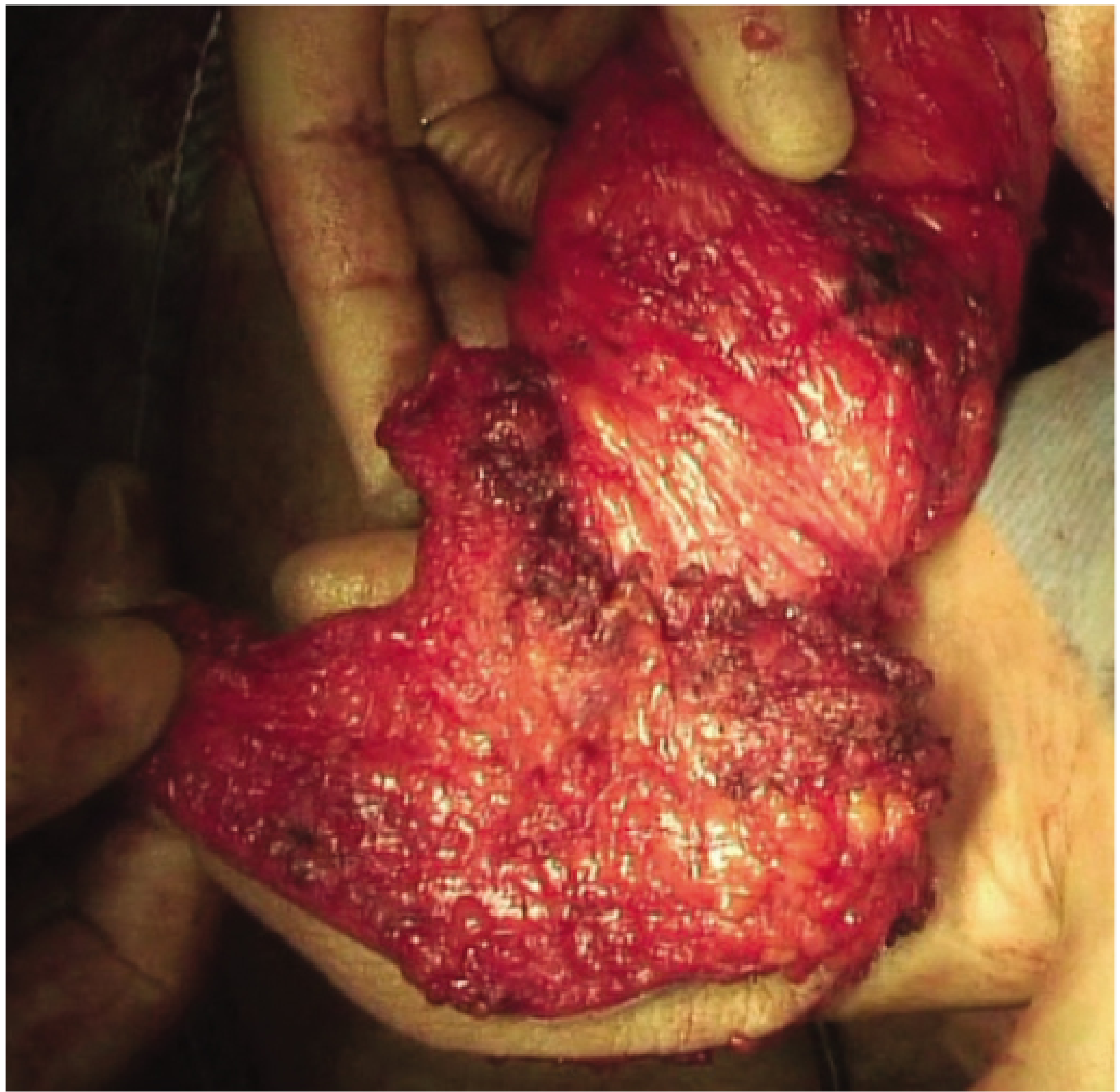
Perfect ELAPE specimen: intact mesorectal fascia above, sphincter + levator wrap around distal specimen
Definition
Removal of sigmoid colon, rectum, anus, sphincters, and levators. Results in permanent end colostomy.
Indications
- Low rectal cancer within 1-2 cm of dentate line (too low for LAR)
- Anal canal carcinoma (squamous cell) after failed chemoradiotherapy
- Recurrent anal/rectal cancer
Extralevator APR (ELAPE) - Modern Technique
- Dissection continues outside the levators (not within) to avoid "waisting" of the specimen
- Reduces CRM positivity
- Perineal phase in prone jackknife position for better visualization
Operative Steps
Abdominal Phase:
- Position: Lloyd-Davis initially, then prone for perineal phase
- Mobilise left colon, divide IMA
- TME as for LAR but without dividing rectum
- Mobilise distally - divide levators close to their origins (ELAPE)
- Divide sigmoid, form end colostomy in left iliac fossa
Perineal Phase (prone jackknife):
- Elliptical incision around anus
- Dissect through ischiorectal fat
- Levators divided close to their pelvic wall origins
- Specimen delivered through perineum
- Perineal wound closed (or reconstructed with flap if large defect)
- Colostomy matured
🚨 VIVA TRAPS - APR
- Q: What permanent stoma results from APR? A: End sigmoid colostomy in left iliac fossa
- Q: What is "waisting" in APR specimen? A: Specimen becomes narrow at level of levators - means surgeon dissected WITHIN levators → positive CRM
- Q: Why prone jackknife for perineal phase? A: Better visualization of perineal anatomy; lower injury to neurovascular bundles
- Q: After APR for squamous cell anal carcinoma - what is first line treatment? A: Nigro protocol = chemoradiotherapy (5-FU + mitomycin C + radiation); APR reserved for residual/recurrent disease
15. STANDARD APPENDICECTOMY (OPEN)
Anatomy
- Appendix arises from posteromedial caecum at confluence of 3 taeniae coli
- Blood supply: Appendicular artery (branch of ileocolic artery) in mesoappendix
- Position: Retrocaecal (most common, 65%), pelvic (30%), subcaecal, paracolic, pre/post-ileal
Alvarado Score (Diagnosis)
| Feature | Score |
|---|
| Migration of pain to RIF | 1 |
| Anorexia | 1 |
| Nausea/vomiting | 1 |
| RIF tenderness | 2 |
| Rebound tenderness | 1 |
| Elevated temperature | 1 |
| Leucocytosis | 2 |
| Left shift (neutrophils) | 1 |
| Total | 10 |
Score ≥7 → appendicectomy; 5-6 → observe; <5 → unlikely appendicitis
Operative Steps (Open - Gridiron Incision)
- Position: Supine
- Incision: Gridiron (McBurney's point - 1/3 from ASIS to umbilicus) OR Lanz (transverse, better cosmesis)
- Layers: External oblique (split), internal oblique (split), transversus abdominis (split)
- Peritoneum: Opened, any pus sent for culture
- Caecum delivered: Followed taeniae to base of appendix
- Mesoappendix divided: Appendicular artery ligated (clips or ties)
- Appendix base: Crushed, ligated with absorbable tie
- Appendix excised
- Stump: Buried in purse-string suture (traditional) or left inverted (current practice varies)
- Wound: Peritoneum closed, muscle layers closed, skin (close if non-perforated; leave open if perforated)
🚨 VIVA TRAPS - Appendicectomy
- Q: What is McBurney's point? A: 1/3 of the way from ASIS to umbilicus - site of maximum tenderness + incision
- Q: What is Rovsing's sign? A: Pressure in LIF causes pain in RIF (due to movement of gas) → suggests peritoneal irritation
- Q: What is a "white appendix"? A: Normal appendix found at surgery - must search for Meckel's (50 cm from ileocaecal valve), Crohn's, mesenteric adenitis, ovarian pathology, Yersinia
- Q: Commonest tumour of appendix? A: Carcinoid tumour (at tip) - if <2 cm, appendicectomy curative; if >2 cm, right hemicolectomy
- Q: Gridiron vs Lanz incision? A: Gridiron = muscle split along fibers (stronger, classical); Lanz = transverse (better cosmesis, same access)
16. LAPAROSCOPIC APPENDICECTOMY
Port Positions (Standard)
- 10mm umbilical (camera)
- 12mm suprapubic (working/stapler port)
- 5mm LIF (assistant/traction)
Steps
- Pneumoperitoneum: Veress or Hasson
- Exploration: Check pelvis, small bowel, ileocaecal region
- Caecum grasped: Appendix identified
- Mesoappendix: Divided using LigaSure/clip/diathermy
- Base: 2 endoloops or stapler (Endo-GIA) applied at base and divided
- Appendix extracted: Via umbilical port in retrieval bag
- Peritoneal toilet: Suction irrigation if perforation/pus
- Ports closed
Advantages Over Open
- Less wound infection
- Faster recovery
- Better cosmesis
- Better diagnostic ability (especially women - check ovaries)
- Less pain
🚨 VIVA TRAPS - Lap vs Open
- Q: Which is better in perforated appendicitis? A: Laparoscopic is appropriate - better peritoneal lavage, fewer SSI
- Q: What is "stump appendicitis"? A: Remaining appendiceal stump inflamed after incomplete appendicectomy - rare, presents as recurrent RIF pain
17. ORCHIDOPEXY FOR UNDESCENDED TESTIS (UDT)
Key Facts
- 90% of boys with UDT have a patent processus vaginalis → risk of hernia
- Histological changes begin at 1 year of age
- Surgery timing: 6-18 months of age
-
70% of cryptorchid testes are palpable
- 30% non-palpable → diagnostic laparoscopy first
Consequences of UDT if Not Treated
- Infertility (1 undescended: risk x2; bilateral: x6)
- Testicular malignancy (seminoma, 5-10x risk)
- Torsion (abnormal mesentery)
- Hernia
- Psychological
Operative Steps (Standard Orchidopexy)
- Position: Supine
- Incision: Short inguinal crease incision over deep ring
- External oblique: Opened in direction of fibers
- Cord structures: Identified, cremasteric muscle divided
- Sac (patent processus vaginalis): Identified, separated from vas and vessels, ligated at deep ring
- Three manoeuvres to gain length:
- Ligate processus vaginalis
- Divide cremaster muscle
- Divide lateral fibrous bands inside deep ring
- Dartos pouch: Created between dartos and scrotal skin
- Testis placed in pouch: Fixed without tension
- Incisions closed
For Non-Palpable Testis (Laparoscopic)
- If testis found laparoscopically: One-stage or two-stage Fowler-Stephens orchidopexy
- Fowler-Stephens: Testicular vessels divided (testis survives on vasal artery + cremasteric artery) - 2-stage preferred
🚨 VIVA TRAPS - UDT
- Q: When should orchidopexy be done? A: 6-18 months of age (histological changes start at 1 year)
- Q: What are the 3 steps to gain length? A: 1) Ligate processus vaginalis, 2) Divide cremaster, 3) Divide lateral fibrous bands at deep ring
- Q: Retractile vs true undescended testis? A: Retractile testis can be milked INTO the scrotum; true UDT cannot
- Q: Risk of malignancy? A: 5-10x increased even after orchidopexy - orchidopexy does NOT abolish risk but enables self-examination and early detection
- Q: Impalpable testis management? A: Laparoscopy first - if blind-ending vessels found = testis absent; if vessels enter deep ring = explore inguinal canal
18. LAPAROSCOPIC GASTROJEJUNOSTOMY WITH VAGOTOMY
Indications
- Unresectable gastric outlet obstruction (duodenal/pyloric tumour)
- Peptic ulcer disease with pyloric stenosis (now rare)
- Bypass for unresectable pancreatic cancer
Vagotomy Types
| Type | Division | Drainage required |
|---|
| Truncal | Both vagal trunks at oesophagus | Yes (pyloroplasty/GJ) |
| Selective | Hepatic + coeliac branches spared; only gastric branches divided | Yes |
| Highly selective (HSV/proximal gastric) | Only fibres to acid-secreting fundus/body; crow's foot preserved | No |
Laparoscopic Steps
- Ports: 5 ports (umbilical camera, epigastric, LUQ, RUQ, subcostal)
- Oesophageal mobilisation: Both vagal trunks identified (anterior and posterior)
- Truncal vagotomy: Both trunks divided + 2 cm resected
- Gastrojejunostomy: Posterior retrocolic isoperistaltic preferred
- Jejunum 40-60 cm from Treitz, brought retrocolic
- Anastomosis to posterior wall of stomach (greater curvature)
- Linear stapler (60mm) used for side-to-side anastomosis
- Common enterotomy closed with running suture
- Test: Check anastomosis is patent, no twist in jejunal loop
🚨 VIVA TRAPS - GJ + Vagotomy
- Q: Why add vagotomy to gastrojejunostomy for peptic ulcer? A: Without vagotomy, acid continues → marginal ulcer at anastomosis
- Q: Why is highly selective vagotomy preferred over truncal + drainage? A: No dumping, no diarrhea, preserves pyloric function
- Q: What is "Crow's foot"? A: Last 5-7 cm of gastric branches of anterior vagus that supply pyloric antrum - preserved in HSV to maintain gastric emptying
- Q: Complication of truncal vagotomy? A: Diarrhea, dumping, gastric stasis
19. HELLER'S MYOTOMY
Indication
Achalasia (failure of lower oesophageal sphincter relaxation) - types I, II, III (extended myotomy for type III)
Pathophysiology of Achalasia
- Loss of inhibitory neurons (VIP, NO) in Auerbach's plexus
- LOS fails to relax; absent peristalsis
- HRM: Classic = aperistalsis + incomplete LOS relaxation; type III = spastic contractions
Operative Steps (Laparoscopic Heller Myotomy)
- Position: Supine, French position (surgeon between legs), reverse Trendelenburg
- Ports: 5 (similar to fundoplication)
- Phrenoesophageal ligament: Divided, distal 5-6 cm of oesophagus mobilised
- Myotomy: Anterior oesophageal myotomy starting 6 cm above OGJ extending 2-3 cm onto gastric cardia
- Both outer longitudinal and inner circular muscles divided; mucosa must be clearly seen but NOT perforated
- Test: If perforation occurred → close with mucosal suture
- Partial fundoplication: Dor (anterior) or Toupet (posterior) fundoplication added to reduce GORD risk
- 360° Nissen is CONTRAINDICATED (aperistaltic oesophagus cannot overcome resistance)
- GORD occurs in up to 40% without fundoplication
🚨 VIVA TRAPS - Heller's Myotomy
- Q: Why is Nissen contraindicated in Heller's? A: Aperistaltic oesophagus cannot overcome 360° wrap resistance → severe dysphagia
- Q: Length of myotomy? A: 6 cm on oesophageal side, 2-3 cm on gastric cardia
- Q: Which partial fundoplication preferred? A: Dor (anterior) - also covers the myotomy/mucosa
- Q: POEM vs Heller's? A: POEM = peroral endoscopic myotomy - similar efficacy but higher GORD rate (no fundoplication)
- Q: What is achalasia type III? A: Spastic achalasia - requires longer proximal myotomy extending to thoracic oesophagus; POEM preferred
20. IVOR LEWIS ESOPHAGECTOMY
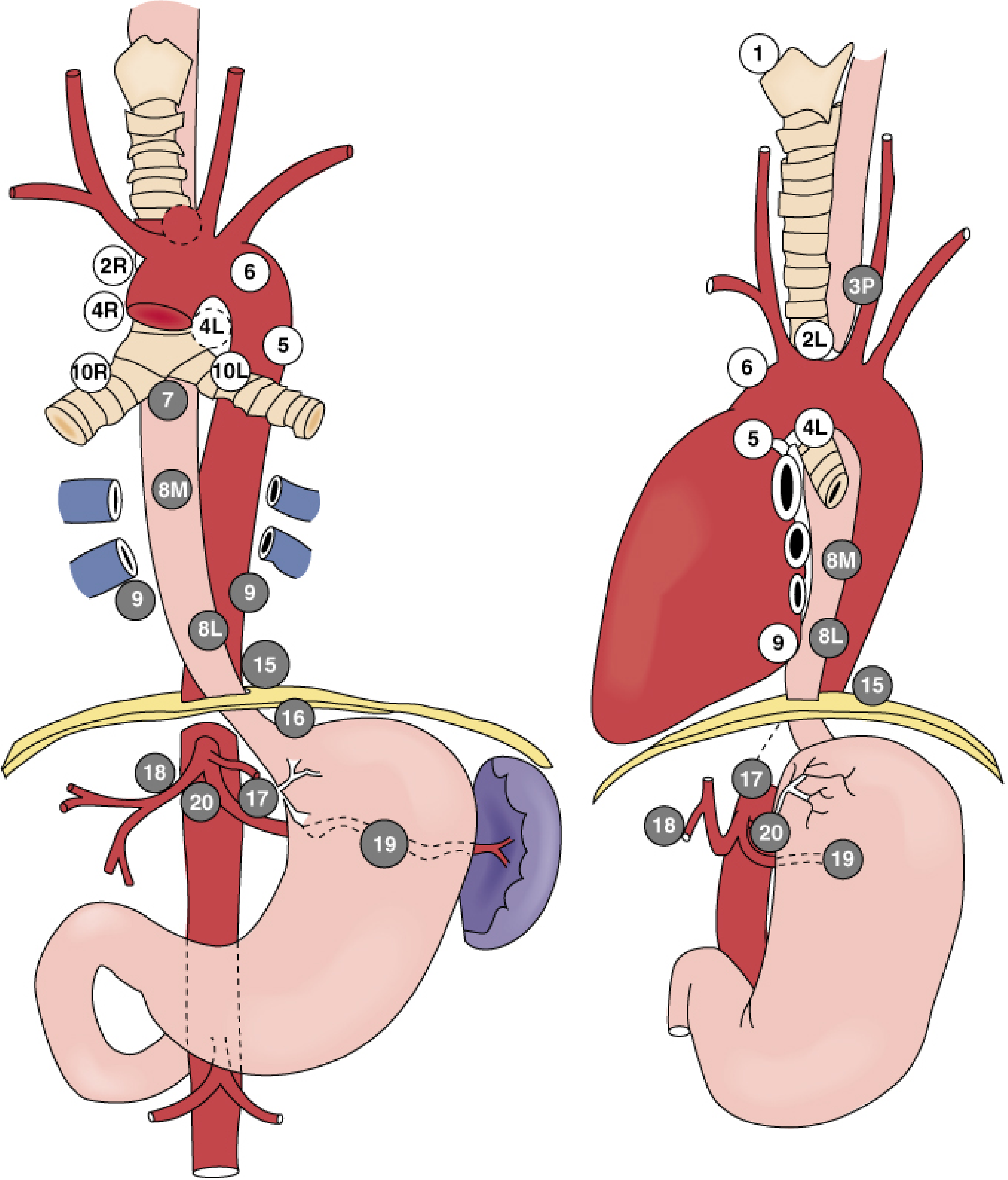
Regional lymph node stations for oesophageal cancer (AJCC staging)
Indication
Oesophageal carcinoma of mid and distal oesophagus (adenocarcinoma or squamous cell).
Ivor Lewis = Two-Stage Operation
- Stage 1 (Abdominal): Supine position - gastric mobilisation, D2 node dissection
- Stage 2 (Thoracic): Left lateral decubitus - right posterolateral thoracotomy (5th ICS)
Conduit
- Gastric tube: 3-5 cm wide, based on right gastroepiploic artery
- Left gastric artery and short gastric vessels divided
- Pyloroplasty or Botox to pylorus (conduit drainage)
Operative Steps
Abdominal Phase:
- Upper midline laparotomy (or 5 laparoscopic ports)
- Explore - no peritoneal/liver mets
- Divide gastrohepatic ligament, expose right crus
- D2 lymphadenectomy (celiac, left gastric, hepatic nodes)
- Left gastric artery divided
- Gastrocolic ligament divided preserving right gastroepiploic
- Short gastrics divided; fundus freed
- Pyloroplasty/Botox pylorus
- Gastric conduit fashioned (3-5 cm wide), stapler from incisura to fundus
- Feeding jejunostomy inserted
Thoracic Phase:
- Patient repositioned - left lateral decubitus
- Right posterolateral thoracotomy 5th ICS (or 4-port VATS right thorax)
- Right lung collapsed (double-lumen ETT or capnothorax)
- Pulmonary ligament divided to inferior pulmonary vein
- Mediastinal pleura opened, oesophagus mobilised with nodes
- Oesophagus divided at apex of thorax
- Gastric conduit pulled up through hiatus
- Oesophagogastric anastomosis: right chest (end-to-side or end-to-end; circular stapler or hand-sewn)
- Chest drain, NG tube confirmed position
- Closure
Complications
| Complication | Management |
|---|
| Anastomotic leak | Most serious - mediastinitis; drain, nil by mouth, antibiotics |
| Recurrent laryngeal nerve injury | Right RLN at risk in thoracic phase |
| Chylothorax | Leakage from thoracic duct → lymph in pleural space; nil by fat diet then ligation |
| Aspiration pneumonia | Conduit empties poorly |
| Anastomotic stricture | Dilate endoscopically |
| Conduit ischaemia/necrosis | Most catastrophic |
| Cardiac arrhythmia | AF very common post-thoracotomy |
🚨 VIVA TRAPS - Ivor Lewis
- Q: Blood supply of gastric conduit? A: Right gastroepiploic artery (right gastric artery is a minor contribution)
- Q: Why pyloroplasty/pyloric drainage? A: Vagotomy effect from dividing vagal trunks → impaired gastric emptying → conduit distension
- Q: What is anastomotic leak after Ivor Lewis? A: Mediastinitis - may present with neck swelling/crepitus, fever, chest pain, drain bile/salivary fluid
- Q: What is chylothorax? A: Thoracic duct injury → lymph (milky fluid) in pleural space; confirm with pleural fluid triglycerides >1.24 mmol/L
- Q: McKeown vs Ivor Lewis? A: McKeown = 3-stage (laparotomy + thoracotomy + neck dissection); cervical anastomosis; leak = less dangerous but more strictures. Ivor Lewis = intrathoracic anastomosis; leak = mediastinitis
21. RADICAL NEPHRECTOMY
Indications
- Renal cell carcinoma (T1b-T4 depending on size/laterality)
- Locally advanced renal tumour
- Partial nephrectomy preferred for T1a (<4 cm) to preserve nephrons
What is Removed (Radical Nephrectomy)
- Kidney + perinephric fat + Gerota's fascia
- Ipsilateral adrenal gland (if involved)
- Regional lymph nodes (lymphadenectomy up to renal hilar nodes)
- Thrombus from renal vein/IVC if present
Operative Steps (Open Radical Nephrectomy)
- Position: Lateral decubitus (kidney position) with kidney bridge raised, table flexed
- Incision: Flank (loin) through 11th or 12th rib OR anterior subcostal/midline
- Gerota's fascia: Opened - dissection outside the fascia to ensure complete removal
- Renal artery: Ligated and divided FIRST (reduces blood loss, prevents tumour embolization)
- Renal vein: Ligated and divided next
- Adrenal gland: Clipped and divided if involved
- Ureter: Divided in mid/lower third
- Lymph node dissection: From crus to bifurcation
- Specimen out
- Drains, closure
For Renal Vein Thrombus / IVC Extension
- Right thrombus: Need vascular clamps on IVC, contralateral renal vein
- Level III/IV thrombus: Cardiopulmonary bypass required
🚨 VIVA TRAPS - Radical Nephrectomy
- Q: Why ligate renal artery first? A: Reduces bleeding; prevents tumour cell dissemination (venous pressure raised before artery = tumour emboli)
- Q: Left vs right renal vein differences? A: Left renal vein is longer (crosses aorta); receives left gonadal vein, left adrenal vein, left lumbar vein. Right is shorter, drains directly to IVC
- Q: What is a Robson stage IV renal tumour? A: Tumour extending into IVC or adjacent organs - surgery complex, may need IVC resection
- Q: Paraneoplastic syndromes of RCC? A: Polycythaemia (erythropoietin), hypertension (renin), hypercalcaemia (PTHrP), Stauffer syndrome (non-metastatic hepatic dysfunction)
- Q: What are absolute indications for nephron-sparing (partial nephrectomy)? A: Solitary kidney, bilateral RCC, hereditary RCC (VHL syndrome)
QUICK VIVA MASTERCLASS: COMMON TRAPS
Top 10 Most Asked Viva Traps Across All Topics
| Question | Answer |
|---|
| Which hernia has highest strangulation risk? | Femoral |
| Commonest cause of varicose vein recurrence? | Neovascularization |
| Why spout an ileostomy? | Proteolytic enzymes in liquid output damage skin |
| Commonest complication of ileostomy? | Skin excoriation (long term: high output) |
| CBD injury during cholecystectomy prevention? | Critical View of Safety (Strasberg) |
| Most dangerous complication of Whipple's? | Pancreatic fistula (POPF) |
| Most common complication of Whipple's? | Delayed gastric emptying |
| Why is Nissen contraindicated in Heller's? | Aperistaltic oesophagus cannot overcome 360° wrap |
| Commonest type of TEF? | Type C - EA with distal fistula (85%) |
| Facial nerve landmark in parotidectomy? | Tragal pointer (nerve 1-1.5 cm deep and inferior) |
Sources: Bailey & Love's Surgery 28th Ed, Sabiston Surgery, Fischer's Mastery of Surgery, Current Surgical Therapy 14e, Cummings Otolaryngology, Mulholland & Greenfield Surgery 7e