Hii
Hi! How can I help you today?
Explain TMT test
Reading File
Finding Sources
Finding Sources
Searching the Web
Trail Making Test TMT neuropsychological assessment explained
Reading File
Reading File
Reading File
Now I have enough information to give a comprehensive answer.
Trail Making Test (TMT)
The Trail Making Test (TMT) is a neuropsychological assessment tool used to evaluate attention, visual scanning, psychomotor speed, and executive function. It is part of the Halstead–Reitan Neuropsychological Battery and is considered one of the most sensitive measures of general brain dysfunction.
Overview
Originally published as the Partington Pathways Test in the 1930s, the TMT has two parts:
Part A — Psychomotor Speed & Visual Scanning
- The patient is presented with circled numbers (1–25) scattered on a page.
- Task: Connect them in sequential order (1 → 2 → 3 → ... → 25) as fast as possible.
- Measures:
- Visual scanning
- Visuospatial sequencing
- Motor speed
- Sustained attention
Part B — Executive Function & Cognitive Flexibility
- The patient is presented with both numbers (1–13) and letters (A–L) scattered on a page.
- Task: Alternate between numbers and letters in order (1 → A → 2 → B → 3 → C → ...) as fast as possible.
- Measures:
- Cognitive flexibility (set-shifting)
- Divided attention
- Working memory
- Mental processing speed
What It Assesses
| Domain | Part A | Part B |
|---|---|---|
| Attention | ✓ | ✓ |
| Visual scanning | ✓ | ✓ |
| Motor speed | ✓ | ✓ |
| Cognitive sequencing | ✓ | ✓ |
| Executive function / set-shifting | — | ✓ |
| Cognitive flexibility | — | ✓ |
B − A difference score: Subtracting Part A time from Part B time isolates the executive/cognitive flexibility component, controlling for motor and scanning speed.
Clinical Significance
- Highly sensitive to virtually any form of neurocognitive injury or dysfunction.
- Impairment is seen in:
- Alzheimer's disease and other dementias
- Frontotemporal dementia
- Parkinson's disease (subcortical dysfunction)
- Traumatic brain injury (Post-Traumatic Dysexecutive Syndrome)
- Depression (psychomotor slowing)
- Frontal lobe lesions and frontal–subcortical circuit disruption
- Part B specifically tests frontal–subcortical circuit integrity.
Scoring
- Scored by time to completion (seconds) — lower time = better performance.
- Normative data is age- and education-adjusted.
- Scores at < 1st percentile indicate severe impairment.
- The D-KEFS (Delis–Kaplan Executive Function System) includes a modified version of the TMT with additional subtests (e.g., Motor Speed subtest) to further differentiate pure motor slowing from cognitive slowing.
Summary
The TMT is quick, easy to administer, and extremely sensitive to brain dysfunction. Part A primarily taps attentional and psychomotor speed, while Part B adds executive set-shifting. Together they provide a powerful window into frontal lobe and overall brain function.
Sources: Kaplan & Sadock's Comprehensive Textbook of Psychiatry; Kaplan and Sadock's Synopsis of Psychiatry; Adams and Victor's Principles of Neurology, 12th Ed.
Explain ecg
Finding Sources
Finding Sources
Reading File
Reading File
Reading File
Reading File
Loading Image
Searching Images
normal ECG waveform P wave QRS T wave intervals
Reading File
Now I have comprehensive information for a full ECG explanation. Let me compile it all.
Electrocardiogram (ECG)
What is an ECG?
An ECG (electrocardiogram) is a recording of the electrical activity of the heart, captured by placing electrodes on the skin surface. When a cardiac impulse travels through the heart, electrical current spreads to surrounding tissues and reaches the body surface — electrodes detect these small potentials, and the recording is the ECG.
The Cardiac Conduction System (Basis of ECG)
The ECG reflects the sequential electrical activation of the heart:
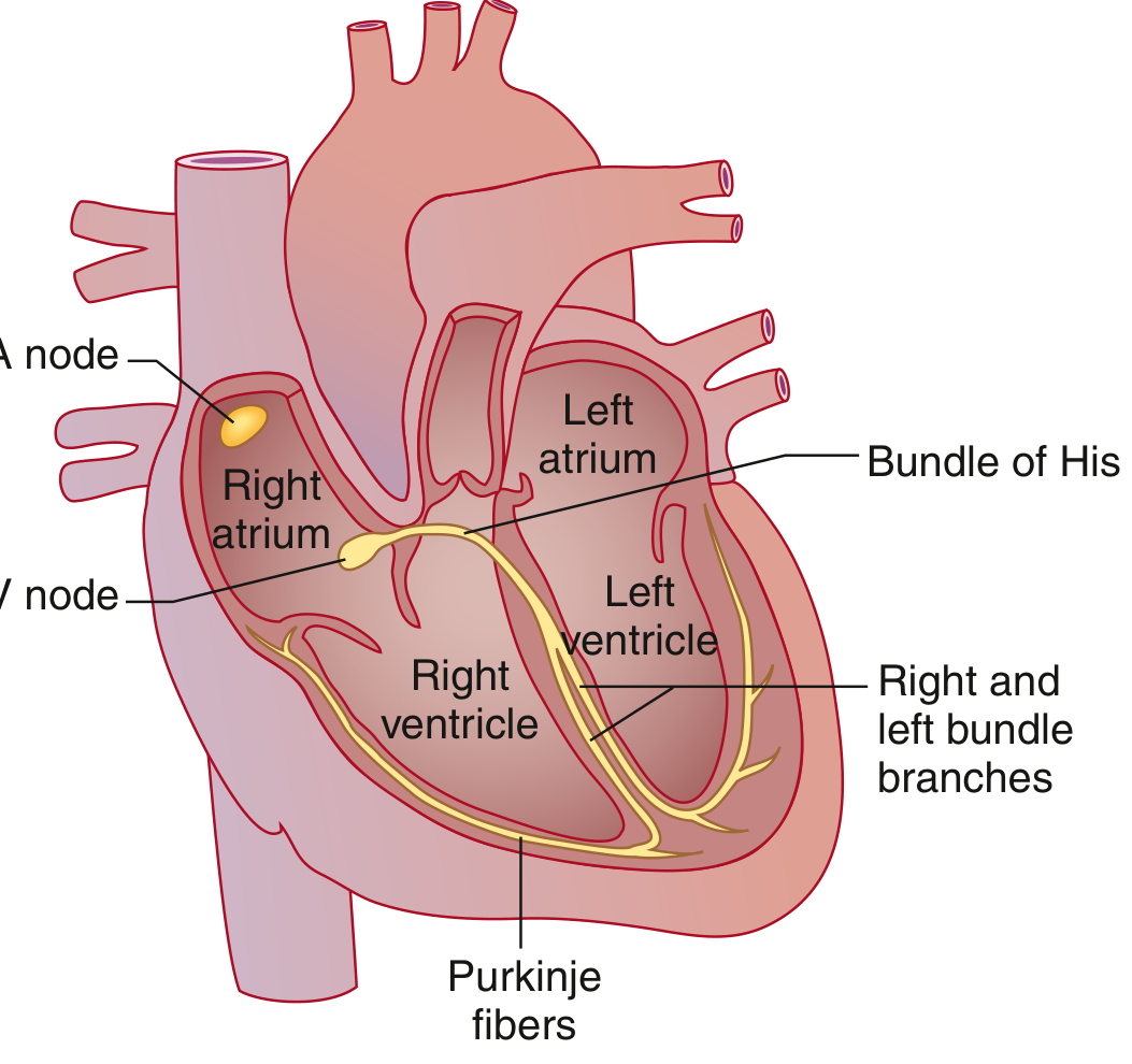
- SA Node (sinoatrial node) — dominant pacemaker in the right atrium; spontaneously depolarizes and initiates each heartbeat.
- Atria — the wave spreads across both atria (via Bachmann bundle to the left atrium), triggering atrial contraction.
- AV Node — the only normal electrical connection between atria and ventricles; introduces a physiologic delay.
- Bundle of His → Right & Left Bundle Branches → Purkinje Fibers — rapidly conduct impulse through ventricular myocardium.
- Ventricles depolarize → contract → then repolarize.
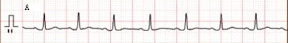
ECG Waveforms
A normal ECG consists of: P wave → QRS complex → T wave
P Wave
- Represents atrial depolarization (muscular activation of both atria)
- Triggers atrial contraction
- Small, rounded deflection
- Atrial repolarization is hidden beneath the QRS (too small to see)
PR Interval
- From onset of P wave → onset of QRS
- Represents the AV nodal delay (time for impulse to travel from atria to ventricles)
- Normal: 0.09–0.20 sec (90–200 ms)
- Prolonged PR = 1st degree AV block; Short PR = preexcitation (e.g., WPW syndrome)
QRS Complex
- Represents ventricular depolarization
- Three components:
- Q wave — initial small negative deflection
- R wave — large positive deflection
- S wave — terminal negative deflection
- Capital letters (Q, R, S) = large (≥5 mm); lowercase (q, r, s) = small (<5 mm)
- Much larger than P wave because ventricular mass >> atrial mass
- Normal duration: 75–110 ms; prolonged = bundle branch block
ST Segment
- From end of QRS (J point) to start of T wave
- Represents early ventricular repolarization
- Should be at the isoelectric baseline
- Elevation = myocardial infarction or pericarditis; Depression = ischemia
T Wave
- Represents ventricular repolarization (electrical recovery)
- Normally upright in most leads
- Occurs 0.25–0.35 sec after depolarization
QT Interval
- From onset of QRS → end of T wave
- Represents total ventricular electrical activity (depolarization + repolarization)
- Must be corrected for heart rate using Bazett's formula:
QTc = QT / √RR
- Normal QTc: ≤450 ms (males), ≤460 ms (females)
- Prolonged QTc → risk of Torsades de Pointes (dangerous arrhythmia)
Normal ECG Intervals at a Glance
| Parameter | Normal Value |
|---|---|
| Heart Rate | 50–100 bpm |
| P wave duration | <120 ms |
| PR interval | 90–200 ms |
| QRS duration | 75–110 ms |
| QTc (males) | 390–450 ms |
| QTc (females) | 390–460 ms |
| QRS axis | −30° to +90° |
The 12-Lead ECG
A standard ECG uses 12 leads (vantage points) derived from 10 electrodes:
- 6 limb electrodes → 6 frontal plane leads:
- Bipolar limb leads: I, II, III
- Augmented unipolar leads: aVR, aVL, aVF
- 6 precordial (chest) electrodes: V1–V6
This gives a 3D picture of the heart's electrical activity.
Heart Rate Calculation from ECG
- HR = 60,000 ÷ RR interval (in ms)
- Quick method (regular rhythm): count large boxes between R waves
- 2 boxes = 150 bpm | 3 = 100 | 4 = 75 | 5 = 60 | 6 = 50
Clinical Uses of ECG
| Condition | ECG Finding |
|---|---|
| Myocardial infarction | ST elevation, Q waves |
| Ischemia | ST depression, T-wave inversion |
| Atrial fibrillation | Absent P waves, irregular RR |
| AV block | Prolonged/absent PR |
| Bundle branch block | Wide QRS (>120 ms) |
| Hypokalemia | Flattened T, prominent U wave |
| Hyperkalemia | Peaked T wave, wide QRS, sine wave |
| WPW syndrome | Short PR, delta wave |
| Long QT syndrome | Prolonged QTc |
| Hypothermia | J (Osborn) wave |
Sources: Goldman-Cecil Medicine; Guyton and Hall Textbook of Medical Physiology; Miller's Anesthesia, 10th Ed.
This is a shared conversation. Sign in to Orris to start your own chat.