
J
Teach me peritonitis in detail and also gastric and duodenal perforations in detail for ms general surgery theory exam with all the recent evidence and trials
peritonitis management surgery
peptic ulcer perforation surgical management
secondary peritonitis abdominal sepsis management
laparoscopic open repair perforated peptic ulcer outcomes
PMID: 39361132
PMID: 38838070
PMID: 36252181
PMID: 40221691
Boey score peptic ulcer perforation prognosis
PMID: 41507810
PMID: 42031639
perforated peptic ulcer free gas under diaphragm chest X-ray
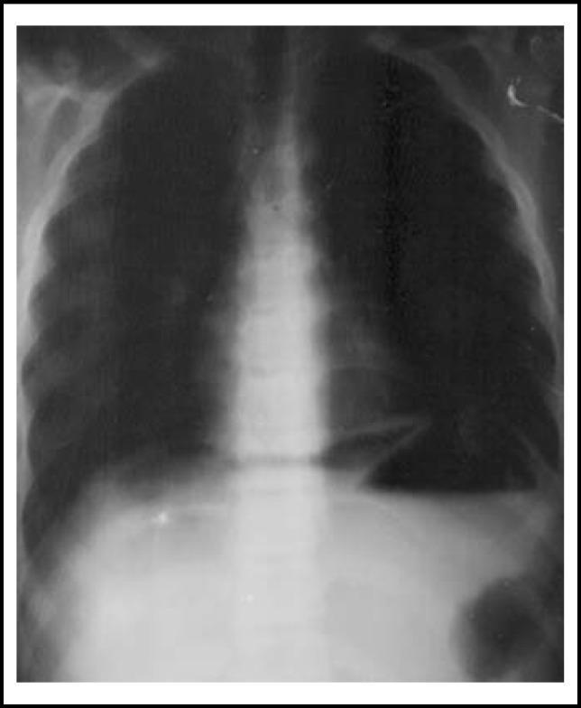
This diagnostic image is an upright plain chest/abdominal X-ray demonstrating a classic sign of pneumoperitoneum. The radiograph clearly shows crescent-shaped radiolucent areas of free air located immediately beneath both the right and left domes of the diaphragm, often referred to as 'free air under the diaphragm'. The diaphragm appears as a thin, curvilinear radio-opaque line separating the pleural cavity from the abdominal cavity. The right hemidiaphragm is elevated by the gas collection, which is positioned between the liver and the diaphragm. The mediastinal shadow, rib cage, and spine are visible, providing anatomical context. This finding is highly indicative of a hollow viscus perforation, such as a perforated peptic ulcer. The image serves as a critical educational example for surgical triage and radiological identification of gastrointestinal emergencies in pediatric or adult populations.
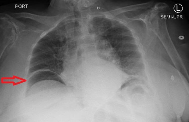
A semi-upright anteroposterior (AP) chest X-ray demonstrating a large pneumoperitoneum. The primary diagnostic finding is a crescent-shaped radiolucency (free air) located subdiaphragmatically under the right dome of the diaphragm, highlighted by a red arrow. This free air clearly silhouettes the superior margin of the diaphragm and separates it from the underlying liver shadow. The left hemidiaphragm is also visible, with underlying gastric bubble and potentially additional free gas. The lung fields are relatively clear without focal consolidation or pleural effusion. The cardiac silhouette is within normal limits for this projection, though the AP view limits precise assessment of cardiothoracic ratio. Bony structures including the ribs and clavicles appear intact. This classic radiographic sign, known as the cupola sign or subdiaphragmatic free air, is a surgical emergency highly suggestive of a perforated viscus (e.g., perforated peptic ulcer or diverticulitis).

This diagnostic image is an erect anterior-posterior (AP) X-ray of the chest and upper abdomen, demonstrating a classic radiographic sign of pneumoperitoneum. The primary finding is a prominent, curvilinear radiolucent (dark) area representing free intraperitoneal gas located beneath the left hemidiaphragm (indicated by a white arrow). This gas collection is situated superior to the stomach, causing inferior displacement of the gastric fundus and creating a clear demarcation between the diaphragm and the abdominal organs. The right hemidiaphragm does not show a similarly clear crescent, though some subphrenic lucency is present. Supporting anatomical structures visible include the thoracic rib cage, the cervical and thoracic spine, and mottled bowel gas patterns in the mid-abdomen. This imaging is highly clinically significant for identifying hollow viscus perforation, such as from a perforated peptic ulcer or trauma. It is an essential educational resource for medical students and clinicians for recognizing the 'gas under the diaphragm' sign in acute abdominal emergencies.
Peritonitis, Gastric & Duodenal Perforations - MS General Surgery Theory
PART I: PERITONITIS
Definition
- Bailey and Love's Short Practice of Surgery 28th Ed., p.1109
Classification
- Localised (walled off by omentum/bowel)
- Diffuse/Generalised (life-threatening)
| Type | Examples |
|---|---|
| Primary (Spontaneous) | SBP in cirrhosis, pneumococcal peritonitis in children |
| Secondary | GI perforation, ischaemic bowel, anastomotic leak |
| Tertiary | Persistent/recurrent peritonitis after treatment of secondary type; nosocomial flora |
- Bacterial (GI/non-GI origin)
- Chemical (bile, barium, gastric acid)
- Allergic (starch peritonitis)
- Traumatic (operative handling)
- Ischaemic (strangulated bowel, vascular occlusion)
- Miscellaneous (Familial Mediterranean Fever)
- GI perforation (ulcer, appendix, diverticulum)
- Transmural translocation without perforation (pancreatitis, ischaemic bowel, primary bacterial peritonitis)
- Exogenous contamination (drains, peritoneal dialysis, trauma)
- Female genital tract (PID - Chlamydia, gonococci; Fitz-Hugh-Curtis syndrome)
- Haematogenous spread (rare)
Localised Peritonitis
- Parietal peritoneum involved → somatic pain localised to the area
- Involuntary guarding and rebound tenderness = peritonism
- Referred phrenic pain (shoulder tip) if sub-diaphragmatic involvement (C5 dermatome)
- Pelvic peritonitis: signs may be minimal abdominally; detected on PR/PV exam
- Signs attenuated in obese patients or those on immunosuppressants
Diffuse (Generalised) Peritonitis
- Pressure-related perforation of viscus (obstructed colon)
- Large volume blood entry (ruptured AAA)
- Sustained leak (perforated duodenal ulcer, anastomotic leak)
Clinical Features of Peritonitis (Summary Box)
- Abdominal pain, worse on movement, coughing, deep respiration
- Constitutional upset: anorexia, malaise, fever, lassitude
- GI upset: nausea ± vomiting
- Pyrexia (may be absent early), raised pulse rate
- Tenderness ± guarding / rigidity / rebound
- Board-like rigidity of entire abdomen
- Hippocratic facies - gravely ill, lies still
- Absent or reduced bowel sounds
- Generalised ileus → abdominal distension
- Scaphoid abdomen in thin patients (rectus contraction)
- Septic shock (SIRS/MODS) in later stages
- PR/PV tenderness in pelvic peritonitis
Investigations
- Erect CXR - free gas under diaphragm (present in >50% of perforated PU; 75% on abdominal film)
- CT scan - investigation of choice; most accurate; identifies source
- Lateral decubitus X-ray - if patient too unwell for erect position
- FBC, LFT, U&E, glucose, coagulation
- Serum amylase - elevated in perforated PU but not as high as pancreatitis; CT distinguishes both
- Blood cultures
- Ultrasound - limited specificity; useful for tubo-ovarian pathology
- Laparoscopy - if above inconclusive
Management of Peritonitis
Resuscitation ("ABCDE" + source control)
- IV access - large bore, two lines
- Fluid resuscitation - crystalloids (follow Surviving Sepsis guidelines)
- Foley catheter + nasogastric tube
- Analgesia - do not withhold (adequate analgesia actually makes signs more obvious)
- Oxygen supplementation
- Monitoring: urine output >0.5 mL/kg/hr
- Broad-spectrum IV antibiotics targeting gram-negatives and anaerobes
- Typical regimens: Piperacillin-tazobactam, or Cefuroxime + Metronidazole, or Meropenem (in resistant/tertiary peritonitis)
- De-escalate based on culture results
- ICU care for MODS/septic shock
- Vasopressors if refractory hypotension
- Source control - remove or exclude the cause
- Peritoneal lavage ± drainage
- Timing: urgent (within hours) once resuscitation initiated
- Damage control surgery in unstable patients: control contamination, close abdomen temporarily (open abdomen technique with VAC dressing), definitive repair in 24-48 hours
Spontaneous Bacterial Peritonitis (SBP)
- Occurs in cirrhotic patients with ascites
- Organisms: Gram-negatives (E. coli, Klebsiella), Streptococcus pneumoniae
- Diagnosis: ascitic fluid PMN > 250 cells/mm³
- Treatment: Cefotaxime 2g IV 8-hourly for 5 days; albumin infusion reduces AKI
- Prophylaxis: Norfloxacin 400 mg/day in high-risk patients
PART II: GASTRIC AND DUODENAL PERFORATIONS (Perforated Peptic Ulcer)
Epidemiology
-
Steady increase in age of patients
-
Increase in females - PPU now most common in elderly females
-
NSAIDs responsible for most perforations (especially elderly)
-
Duodenal > Gastric, but rising proportion of GU perforations in elderly NSAID users
-
Globally: up to 5% of all PUD patients present with perforation
-
30-day mortality remains significant: ~6-30% depending on setting
-
Bailey and Love 28th Ed., p.1186; Sleisenger & Fordtran p.X
Aetiology
| Factor | Notes |
|---|---|
| H. pylori | Most common cause of PUD; less directly linked to perforation |
| NSAIDs/Aspirin | Major cause of perforation, especially in elderly |
| Steroids | Risk when combined with NSAIDs |
| Smoking | Predisposes to ulceration, increases relapse rate |
| Zollinger-Ellison Syndrome | Gastrinoma with extreme acid hypersecretion |
| Stress ulcers | ICU patients; major injury/surgery |
Pathology
- Most common on anterior wall of first part of duodenum (perforate into peritoneal cavity)
- Posterior wall DU penetrate into pancreas → can erode gastroduodenal artery → haemorrhage
| Type | Site | Notes |
|---|---|---|
| I | Lesser curvature (body, incisura) | Low acid; most common; not associated with DU |
| II | Body + duodenal ulcer | High acid |
| III | Prepyloric | High acid; behaves like DU |
| IV | High on lesser curve near GEJ | Rare; high risk malignancy |
| V | Anywhere; NSAID-induced | Diffuse distribution |
Clinical Features
- Sudden onset, severe generalised abdominal pain - patient may specify exact moment of onset ("knife-like/tearing pain")
- Initially chemical peritonitis from gastric acid → bacterial peritonitis supervenes over hours
- Board-like rigidity of entire abdomen
- Abdomen does not move with respiration
- Patient lies still (peritoneal irritation worsens with movement)
- Hippocratic facies - gravely ill, anxious, sunken features
- Initially shocked + tachycardic; pyrexia not usually present until hours later
- Referred shoulder tip pain (diaphragmatic irritation)
- Less dramatic (steroid/NSAID use blunts signs)
- Board-like rigidity may be absent
- "Contained" leak: pain in epigastrium + right iliac fossa (fluid tracking down right paracolic gutter)
- Self-sealing perforations can occur (inflammatory response + adhesions)
- NSAID switch from opioids → rising frequency in younger patients (recent epidemiological shift)
- Both present similarly; GU more likely to be contaminated with food/bacteria
- Free peritoneal gas more reliable in GU (larger stomach volume)
Investigations
- FBC (leucocytosis), CRP elevated
- U&E, LFT, glucose, coagulation, cross-match
- Serum amylase - may be elevated (up to 3× normal) in PPU, but not as high as pancreatitis; CT distinguishes
- Erect CXR - free gas under diaphragm in >50% of cases (Bailey & Love); 75% detectable on abdominal film (Fordtran)
- CT abdomen + pelvis - most accurate; demonstrates:
- Extraluminal free air
- Fluid/pus collections
- Edema around antrum/duodenum
- May identify perforation site
- Preferred investigation
- Lateral decubitus X-ray - alternative if patient too unwell to stand
- Endoscopy is CONTRAINDICATED when PPU suspected - insufflation can convert sealed perforation to free perforation
Prognostic Scoring
| Risk Factor | Score |
|---|---|
| Shock on admission (SBP < 100 mmHg) | 1 |
| Confounding medical illness (ASA class III-IV) | 1 |
| Delayed presentation (>24 hours) | 1 |
- Score 0: mortality ~0-1%
- Score 1: ~10%
- Score 2: ~45%
- Score 3: ~100%
- Includes age, ASA class, shock, co-morbidities, perforation size, time to surgery
Treatment
Initial Resuscitation (All patients)
- IV fluids - aggressive crystalloid resuscitation
- Analgesia (do not withhold - titrate the dose)
- Broad-spectrum IV antibiotics
- NGT decompression
- Foley catheter (monitor UO)
- Blood tests + cross-match
- Urgent surgical consultation
Conservative (Non-operative) Management
- Patient haemodynamically stable
- No signs of generalised peritonitis
- Radiologically demonstrated sealed perforation (CT/water-soluble contrast study)
- Age < 70 (relative)
- Presentation < 24 hours
- Strict NBM
- IV fluids
- NGT suction
- IV broad-spectrum antibiotics
- PPI infusion
- Serial clinical review - any deterioration = immediate surgery
Surgical Management
- Open (Upper midline incision) - traditional standard
- Laparoscopic - now preferred where expertise available
Laparoscopic vs. Open Repair - KEY EXAM EVIDENCE
- Systematic review + meta-analysis + trial sequential analysis of 9 RCTs, 670 patients
- Laparoscopic repair vs. open repair
- Results:
- Mortality: RR 0.37 (p=0.03) - significantly lower with laparoscopic
- Total complications: RR 0.57 (p=0.0009) - significantly lower
- Ileus: RR 0.43 (p=0.04) - lower
- Wound complications: RR 0.36 (p<0.0001) - lower
- Length of stay: MD -2.37 days (p=0.0003) - shorter
- No significant difference in: leak rate, abdominal collection, sepsis, respiratory complications, reoperation, operating time
- Conclusion: "Laparoscopic approach should be the management of choice subject to laparoscopic expertise."
- SR & MA, 29 studies, 5,311 patients (LOPR vs. OOPR)
- LOPR associated with: lower 30-day mortality (OR 0.57), lower overall morbidity (OR 0.31), lower SSI (OR 0.27), shorter LOS (MD -2.84 days)
- Postoperative leakage was comparable (OR 1.06, p=0.90)
- Note: LOPR performed mainly by consultants vs. trainees for open → selection bias acknowledged
Operative Technique
- Omental patch repair (Graham patch) - most commonly performed
- Close ulcer with 2-3 interrupted absorbable sutures (transversely, as in pyloroplasty)
- Do NOT tie so tight that sutures cut through; ensure adequate tissue in each bite
- Place pedicled omental patch over closure; tie sutures gently (preserve omental blood supply)
- If primary closure difficult: omental patch alone (plug technique)
- Definitive surgery (now rarely done):
- Patch + Highly Selective Vagotomy (HSV) - in stable patient with chronic symptoms or failed medical therapy
- Patch + Truncal Vagotomy + Drainage (V+D)
- Patch + Truncal Vagotomy + Antrectomy (V+A) - lowest recurrence but highest morbidity
- Biopsy MANDATORY (exclude malignancy) even if perforation repaired
- Type I (most common): Distal gastrectomy in stable low-risk patient; patch + biopsy in high risk
- Type II/III: Patch + Vagotomy + Drainage or distal gastrectomy + vagotomy
- Type IV (near GEJ): Excision with Roux-en-Y reconstruction
- Type V (NSAID): Patch + biopsy; stop NSAIDs
- Massive perforation (GU or DU): Roux-en-Y distal gastrectomy
- Haemodynamic instability + exudative peritonitis (>24h) → simple patch closure only
- Stable patient, chronic symptoms, failed medical therapy → patch + HSV
- V+D acceptable definitive procedure but disability possible
- Stable, low risk → distal gastrectomy (operation of choice; removes ulcer for histology)
- Unstable/high risk → patch + biopsy, or local excision + closure, or biopsy + closure + TV+D
Post-operative Care
- NGT on suction until bowel function returns
- PPI IV → oral (promotes healing)
- H. pylori testing and mandatory eradication therapy (triple/quadruple regimen)
- Stop NSAIDs permanently; if unavoidable, add long-term PPI
- Lifelong PPIs are a reasonable option after perforation
- Water-soluble contrast swallow on Day 5-7 to check for leak before feeding
- SR & MA of 6 RCTs, 356 patients
- Fast-track recovery after PPU surgery significantly:
- Shortened LOS (MD -3.50 days, p≤0.00001)
- Reduced superficial + deep SSIs (p=0.002)
- Fewer pulmonary complications (p=0.004)
- No increase in overall complications
- Conclusion: Fast-track protocols should be adopted post-PPU surgery
Laparoscopic Barbed Sutures (Emerging Evidence)
- Meta-analysis of 5 studies, 436 patients
- Barbed sutures vs. conventional sutures in laparoscopic repair
- Barbed sutures: significantly reduced operative time (SMD -0.89, p<0.001) and suturing time (SMD -1.31, p<0.001)
- No difference in postoperative complications (leakage, obstruction, abscess)
- Conclusion: Barbed sutures simplify laparoscopic repair without compromising safety
Complications of PPU/Peritonitis
- Persistent leak / suture line dehiscence
- Subphrenic/subhepatic abscess
- Enterocutaneous fistula
- Wound infection
- Pulmonary complications (atelectasis, pneumonia, ARDS)
- Septic shock / MODS
- Recurrent ulceration
- Gastric outlet obstruction (from scarring)
- Adhesions / adhesive intestinal obstruction
- Dumping syndrome (post-gastrectomy)
- Nutritional deficiencies (post-gastrectomy)
Outcomes / Mortality Predictors
- Presentation > 24 hours (4× higher complication odds)
- Shock on admission (SBP < 100 mmHg)
- Advanced age
- Significant co-morbidities (ASA III-IV)
- Large perforation size
- Faeculent peritonitis
Quick Summary for MS Exam "Must Know" Points
| Topic | Key Point |
|---|---|
| Most common site of DU perforation | Anterior wall, 1st part duodenum |
| Most common GU perforation type | Type I (lesser curvature) |
| Sign of free air on CXR | Present in >50% (Bailey) / 75% (Fordtran) |
| Investigation of choice | CT abdomen |
| Contraindicated investigation | Endoscopy (risk of converting sealed to free perforation) |
| Operation of choice (DU) | Laparoscopic omental patch repair (Graham patch) |
| Operation of choice (GU, stable) | Distal gastrectomy with biopsy |
| Mandatory post-op measure | H. pylori eradication |
| Prognostic score | Boey score (0-3); PULP score equally accurate (2026 meta-analysis) |
| Laparoscopic advantage | Lower mortality (RR 0.37), lower complications, shorter LOS - Sokhal 2025 |
| Fast-track recovery | Reduces LOS by 3.5 days, fewer SSI and pulmonary complications - Zeyara 2024 |
| Barbed sutures | Reduce operative time without increasing complications - Sun 2026 |
| GU biopsy | Mandatory in ALL gastric ulcers - exclude malignancy |
| Conservative Rx criteria | Stable, no peritonitis, sealed perforation on imaging |
Recent Evidence Table (Exam Ready)
| Study | Year | PMID | Finding |
|---|---|---|---|
| Sokhal BS et al. | 2025 | 39361132 | Lap repair: lower mortality (RR 0.37), lower complications, shorter LOS vs. open |
| Chan KS et al. | 2023 | 36252181 | LOPR: lower mortality (OR 0.57), morbidity (OR 0.31), LOS vs. OOPR |
| Zeyara A et al. | 2024 | 38838070 | Fast-track recovery: LOS -3.5 days, fewer SSI and pulmonary complications |
| Christanto et al. | 2026 | 42031639 | PULP vs. Boey: comparable accuracy; PULP marginally better but not significant |
| Sun Q et al. | 2026 | 41507810 | Barbed sutures: less operative + suturing time; no increase in complications |
| Endeshaw D et al. | 2025 | 40221691 | Pooled mortality 6.68%; delayed presentation (>24h) → 4× higher complications |
| Pope R et al. | 2024 | 37927010 | Conservative management feasible for contained/marginal perforations |
Explain more detail between grahams repair,cellan jones and modified grahams repair
Graham patch Cellan-Jones omental repair perforated peptic ulcer technique
Graham's patch vs Cellan Jones repair vs modified Graham's repair perforated peptic ulcer technique difference surgical steps