
Abdominal layers deatil
abdominal wall layers anatomy cross section diagram
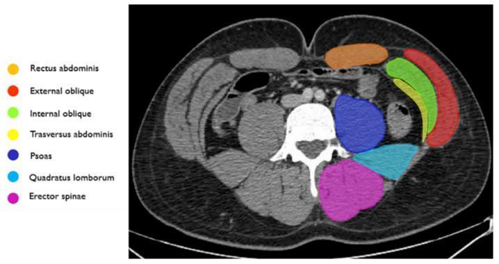
This diagnostic image is an axial CT scan of the abdomen, specifically annotated to demonstrate the anatomy of the abdominal wall musculature. The cross-section provides a clear view of the bilateral symmetry and layered arrangement of the core muscles. Anteriorly, the rectus abdominis is highlighted in orange. Moving laterally, the three distinct layers of the lateral abdominal wall are identified: the external oblique (red) as the most superficial layer, the internal oblique (green) in the middle, and the transversus abdominis (yellow) as the deep layer. In the posterior compartment, the psoas muscle (dark blue) is shown adjacent to the vertebral body. Posterior and lateral to the psoas is the quadratus lumborum (light blue), while the erector spinae group (magenta) is situated most posteriorly along the spine. This anatomical diagram serves as a key reference for identifying muscle boundaries in clinical imaging, essential for diagnosing abdominal wall hernias, hematomas, or planning surgical and interventional procedures like embolization for active bleeding.
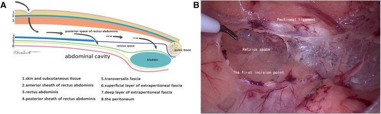
This composite educational material consists of an anatomical diagram (A) and a corresponding intraoperative clinical photograph (B) illustrating the surgical approach to the preperitoneal space during hernia repair. Diagram A provides a sagittal cross-section of the abdominal wall layers, including the skin, rectus abdominis, posterior rectus sheath, transversalis fascia, and peritoneum, highlighting the entry pathway into the Retzius space above the bladder and pubic bone. Intraoperative image B shows the laparoscopic view of the 'first incision point' within the transversalis fascia, located approximately 1 cm above the pectineal ligament (Cooper's ligament). The Retzius space is visible as a cavitary area with glistening, translucent connective tissue and fibrous networks. The pectineal ligament is identified as a distinct whitish band at the superior margin. This visual material is designed for surgical trainees to understand the 'transversalis fascia reference plane' and membrane anatomy during Totally Extraperitoneal (TEP) laparoscopic hernia repair, emphasizing safe entry into the Retzius space to avoid vascular or bladder injury.
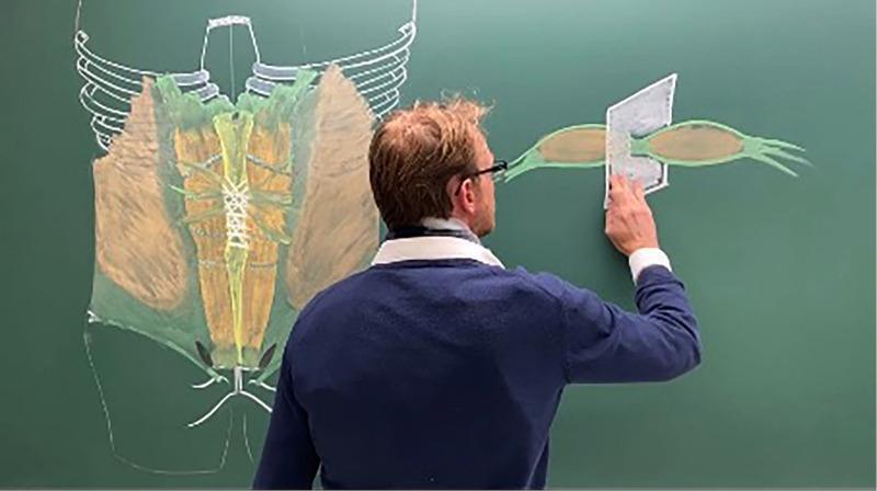
This educational image depicts a medical professional presenting anatomical diagrams of the anterior abdominal wall on a green chalkboard. The primary diagram on the left illustrates the musculoskeletal anatomy of the trunk, featuring a white outline of the inferior rib cage, sternum, and pelvis. The muscular layers are color-coded: the rectus abdominis is shown in yellow/tan along the midline, divided by the linea alba, with horizontal tendinous inscriptions clearly marked. Lateral to this, the external and internal oblique muscles are represented in brown, while green shading indicates the surrounding fascial planes, including the transversalis fascia. The right side of the board features a detailed cross-sectional or isolated diagram of a muscle belly (likely rectus abdominis or an oblique), highlighting its fusiform shape, green fascial envelope, and tendinous distal attachments. A physical cutout representing a surgical mesh or anatomical plane is positioned over this muscle to demonstrate Abdominal Wall Reconstruction (AWR) techniques or hernia repair principles. This visual is used for postgraduate surgical education, emphasizing applied anatomy in groin and incisional hernia surgery.
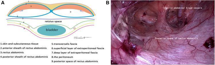
The visual is a two-part educational material focused on the surgical anatomy of the abdominal wall and preperitoneal spaces. Panel A is a cross-sectional anatomical diagram illustrating the layers of the lower abdominal wall below the arcuate line. It identifies several numbered structures: skin/subcutaneous tissue (1), anterior rectus sheath (2), rectus abdominis muscle (3), posterior rectus sheath (4), transversalis fascia (5), and extraperitoneal fascia layers (6-7) overlying the peritoneum (8) and bladder. It clearly delineates the Retzius space and the posterior space of the rectus abdominis (9). Panel B is a laparoscopic still-frame of the posterior space of the rectus abdominis during a surgical procedure (likely TEP inguinal hernia repair). The image shows the reddish-pink, striated fibers of the rectus abdominis muscle and the moist, glistening texture of the posterior sheath. Labels identify the 'posterior space of rectus abdominis' and 'inferior abdominal blood vessels,' demonstrating the vascular supply and the surgical plane necessary for safe dissection above the transversalis fascia.
Abdominal Wall Layers - Detailed Anatomy
Complete Transverse Section - All Layers
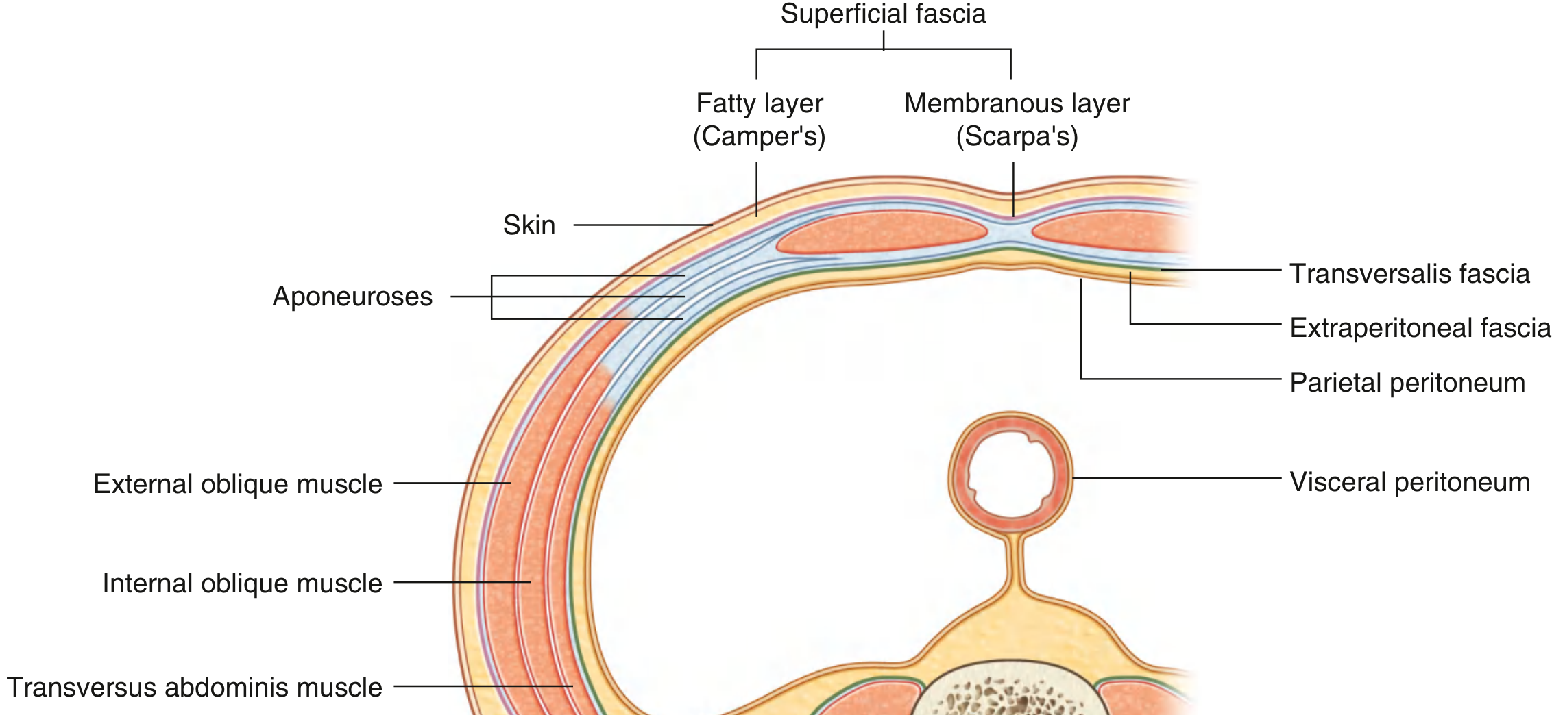
Layer-by-Layer Breakdown (Superficial to Deep)
1. Skin
2. Superficial Fascia (Two Sub-layers)
- Also called the panniculus adiposus
- Contains varying amounts of adipose tissue
- Houses the superficial epigastric vessels and superficial circumflex iliac vessels (tributaries of the femoral vessels)
- Lymphatics drain inferiorly to the inguinal lymph nodes
- Continuous inferiorly with the outer perineal fascia and contains dartos muscle fibers of the scrotum
- A compressed fibrous component of the superficial fascia
- Forms the fundiform ligament of the penis (suspensory ligament of the clitoris in females)
- Fuses inferiorly with the fascia lata of the thigh at the inguinal ligament, sealing the space beneath it
- Clinically relevant: urine can track under Scarpa's fascia but not into the thigh - Mulholland & Greenfield's Surgery, p. 3633
3. External Oblique Muscle (+ its Aponeurosis)
- Most superficial of the three flat lateral muscles
- Arises from the posterior aspects of the lower 8 ribs, interdigitating with serratus anterior and latissimus dorsi
- Fibers run inferomedially ("hands in pockets" direction)
- Aponeurosis forms:
- The anterior rectus sheath (throughout its entire length)
- The inguinal ligament (where inferolateral fibers fold under themselves)
- The superficial inguinal ring (split into medial and lateral crura)
- The lacunar ligament is a fan-shaped medial extension attaching to the pectin pubis - Mulholland & Greenfield's Surgery, p. 3634-3635
4. Internal Oblique Muscle (+ its Aponeurosis)
- Originates from the thoracolumbar fascia, iliac crest, and lateral inguinal ligament
- Fibers run superolaterally (perpendicular to external oblique)
- Its aponeurosis is bilaminar - this is key:
- Above the arcuate line: splits to contribute to BOTH anterior and posterior rectus sheaths
- Below the arcuate line: all fibers go to the anterior rectus sheath only
- Lower fibers arch over the spermatic cord to form the conjoint tendon (inguinal falx) with transversus abdominis
- Lower free arched edge, together with transversus, gives rise to the cremaster muscle
5. Transversus Abdominis Muscle (+ its Aponeurosis)
- Deepest of the three flat lateral muscles
- Originates from the costal margin, lumbar fascia, iliac crest, and lateral inguinal ligament
- Fibers run horizontally (transversely)
- Aponeurosis contribution:
- Above the arcuate line: contributes to the posterior rectus sheath
- Below the arcuate line: swings anterior to contribute to the anterior rectus sheath only
- The linea semilunaris marks its lateral border - Schwartz's Principles of Surgery, p. 960
Rectus Abdominis (Midline Muscle - Not a Lateral Layer)
- Paired longitudinal muscles running from the pubic symphysis/crest to the xiphoid process and 5th-7th costal cartilages
- Divided by the linea alba in the midline
- Has 3-4 tendinous intersections along its length
- Enclosed in the rectus sheath formed by the aponeuroses of the three flat muscles
- A small pyramidalis muscle lies anterior to the lower rectus, attaching to the linea alba
6. Transversalis Fascia
- A continuous fascial layer lining the entire inner surface of the transversus abdominis muscle
- Below the arcuate line it is the only posterior covering of the rectus abdominis (the posterior rectus sheath disappears here)
- Forms the deep inguinal ring (an opening for the spermatic cord)
- A relatively weak fibrous layer - this is why hernia mesh reinforcement targets this plane
- "The final common denominator in all groin hernias is failure of the transversalis fascia to retain the intra-abdominal contents" - Mulholland & Greenfield's Surgery, p. 3814
7. Extraperitoneal (Preperitoneal) Fat / Fascia
- Also called the extraperitoneal fascia
- A layer of connective tissue containing variable amounts of fat between the transversalis fascia and the peritoneum
- On the anterior side it is called preperitoneal fat/fascia; on the posterior side, retroperitoneal fat
- Contains the urachus and remnants of the umbilical arteries (forming peritoneal folds)
- Surgically important: this space is accessed in laparoscopic hernia repair (TEP) - Gray's Anatomy for Students, p. 345
8. Parietal Peritoneum
- The innermost layer - a thin serous membrane lining the abdominal cavity
- Forms the peritoneal folds on the internal surface of the anterior abdominal wall:
- Median umbilical fold - contains the obliterated urachus (midline, unpaired)
- Medial umbilical folds (paired) - contain obliterated umbilical arteries
- Lateral umbilical folds (paired) - contain the inferior epigastric vessels
- Between these folds are three fossae on each side: supravesical fossa, medial inguinal fossa (Hesselbach's triangle), and lateral inguinal fossa (site of the deep inguinal ring) - THIEME Atlas of General Anatomy, p. 233
The Arcuate Line - Key Structural Change
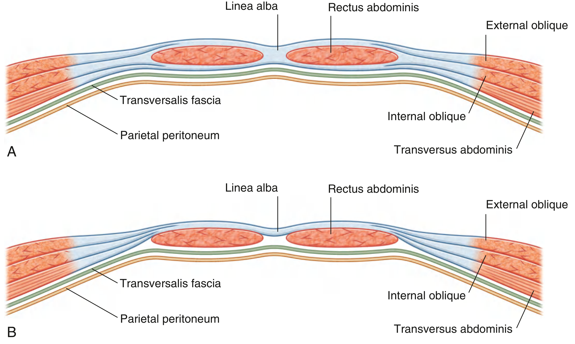
| Feature | Above Arcuate Line | Below Arcuate Line |
|---|---|---|
| Anterior rectus sheath | External oblique + anterior lamella of internal oblique | ALL three aponeuroses (ext. oblique + int. oblique + transversus) |
| Posterior rectus sheath | Posterior lamella of internal oblique + transversus abdominis | Absent |
| Posterior covering of rectus | Posterior rectus sheath | Only transversalis fascia |
Axial CT Correlation
Abdominal Wall Layers and Their Spermatic Cord Derivatives
| Abdominal Wall Layer | Spermatic Cord/Testis Covering |
|---|---|
| Skin + membranous superficial fascia | Scrotal skin + tunica dartos |
| External oblique fascia | External spermatic fascia |
| Internal oblique muscle + fascia | Cremaster muscle + cremasteric fascia |
| Transversalis fascia | Internal spermatic fascia |
| Peritoneum | Tunica vaginalis (parietal + visceral) |
Summary Table
| # | Layer | Key Contents / Notes |
|---|---|---|
| 1 | Skin | Dermatomes T7-T12, L1-L2 |
| 2a | Camper's fascia | Superficial fat, epigastric vessels |
| 2b | Scarpa's fascia | Membranous, fuses with fascia lata |
| 3 | External oblique | Aponeurosis → ant. sheath, inguinal lig., sup. inguinal ring |
| 4 | Internal oblique | Bilaminar aponeurosis; cremaster origin |
| 5 | Transversus abdominis | Horizontal fibers; deepest flat muscle |
| 6 | Transversalis fascia | Deep inguinal ring; weak "final barrier" |
| 7 | Extraperitoneal fat | Preperitoneal space; TEP hernia repair plane |
| 8 | Parietal peritoneum | Umbilical folds; innermost layer |