
Blood supply of long bone , anatomy short note
Reading File
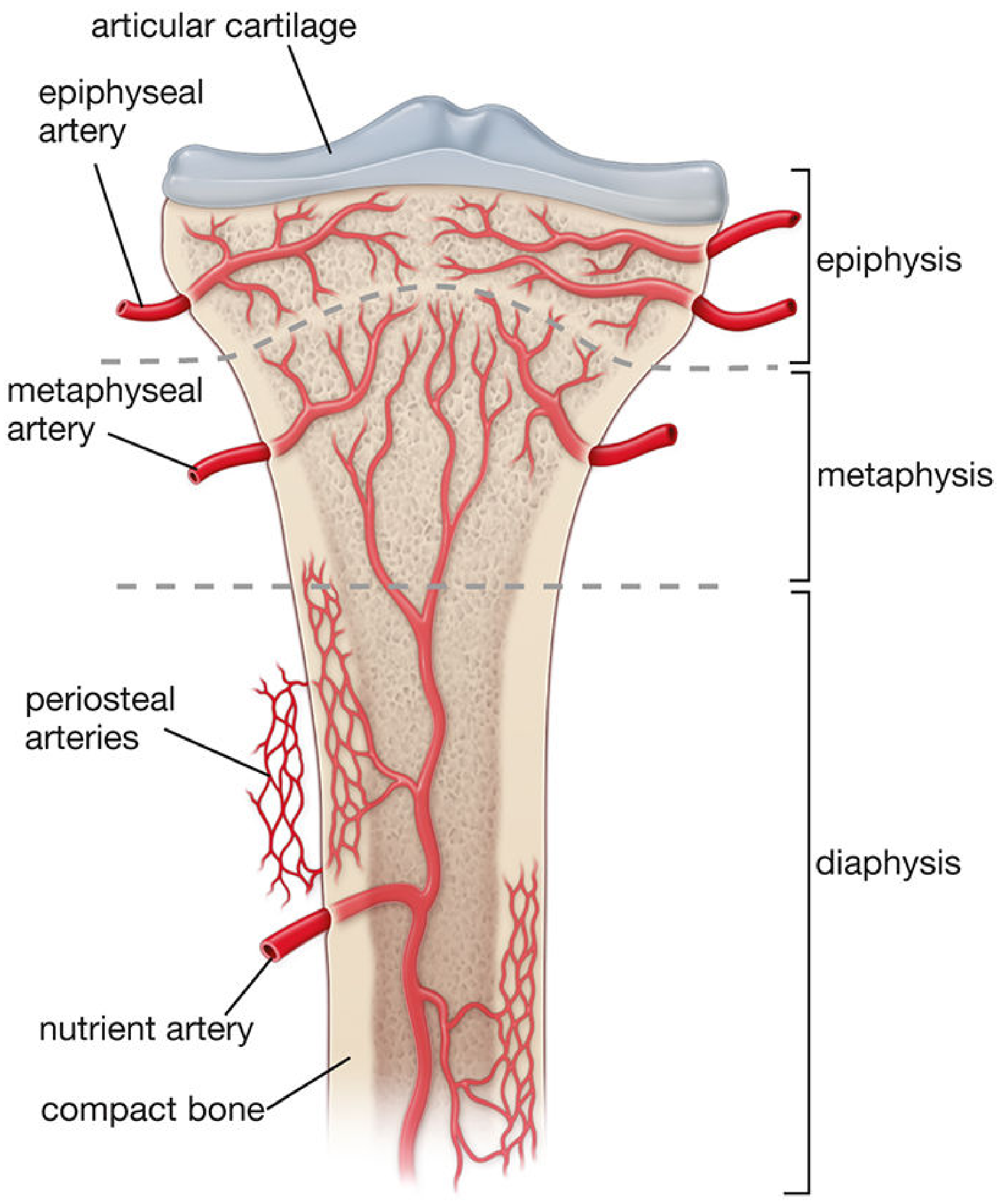
The blood supply of a long bone comes from three main sources: the nutrient artery (main diaphyseal supply), metaphyseal/epiphyseal vessels, and periosteal vessels. The nutrient artery enters through the nutrient foramen, divides into ascending and descending branches inside the medullary cavity, and supplies the inner two-thirds of the cortex. Periosteal vessels supply the outer one-third.
Searching sources...
Finding Sources
Finding Sources
Reading File
Reading File
Reading File
Finding Sources
Reading File
Reading File
Now let me fetch the key diagram from Miller's Review of Orthopaedics and the histology atlas:
Loading Image
Loading Image
Excellent - I have comprehensive, well-sourced content from multiple authoritative orthopaedic and histology textbooks. Here is the complete short note:
Blood Supply of Long Bone - Anatomy Short Note
Overview
Long bones receive approximately 5-10% of cardiac output. Three distinct arterial systems supply them, each responsible for a specific region.
Sources of Blood Supply
1. Nutrient Artery System (Diaphyseal/Medullary System)
- Pressure: High-pressure system
- Supplies: Inner two-thirds of the diaphyseal (cortical) compact bone
- Origin: Branch from a systemic/regional artery (e.g., the humeral nutrient artery arises from the brachial artery; the tibial nutrient artery from the posterior tibial artery)
- Entry: Enters via the nutrient foramen (an oblique canal in the diaphysis), directed away from the growing end in most bones
- Course: Enters the medullary cavity, divides into ascending and descending medullary arteries, which branch into arterioles supplying the endosteal cortex and inner 2/3 of compact bone via the Haversian system
- Significance: Accounts for ~60% of cortical bone vascularization
2. Metaphyseal-Epiphyseal System
- Origin: Arises from the periarticular vascular plexus (e.g., geniculate arteries around the knee; circumflex arteries at the shoulder/hip)
- Epiphyseal arteries enter through nutrient foramina at the epiphysis and supply the trabecular (spongy) bone of the epiphysis
- Metaphyseal arteries arise from periosteal vessels incorporated into the metaphysis during bone growth (widening). They supplement diaphyseal supply and are especially important in the growing bone
- In the growing skeleton, the physis (growth plate) acts as a barrier - epiphyseal and metaphyseal circulations are largely separate, making the metaphysis vulnerable to hematogenous infection (osteomyelitis)
3. Periosteal System
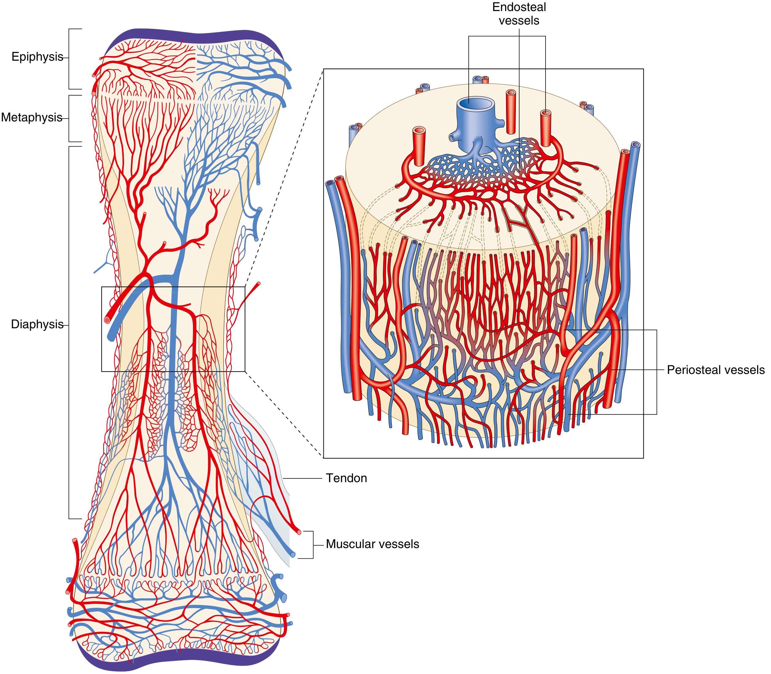
- Pressure: Low-pressure system
- Supplies: Outer one-third of the diaphyseal compact bone
- Consists mostly of capillaries that penetrate the outer cortex via Volkmann canals
- These vessels communicate with Haversian canals to form an anastomotic network
- Also drains lymphatic vessels (the periosteum is the only part of bone with lymphatics - bone tissue itself lacks lymphatics)
Direction of Blood Flow
| State | Arterial Direction | Venous Direction |
|---|---|---|
| Mature bone (normal) | Centrifugal (inside → outside) | Centripetal (outside → inside) |
| Immature bone / post-fracture | Centripetal (periosteal predominates) | - |
- In the mature bone, arterial flow is centrifugal - driven by the net effect of the high-pressure nutrient artery overcoming the low-pressure periosteal system
- When a fracture disrupts the nutrient artery or in immature/growing bone, the periosteal system pressure predominates and flow reverses to centripetal
Venous and Lymphatic Drainage
- Veins exit via the nutrient foramina and through bone tissue of the shaft, continuing out through the periosteum - venous flow is centripetal
- Lymphatic vessels are present only in the periosteum - the bone tissue itself has no lymphatics
Intracortical Vascular Architecture
- Within compact bone, blood moves through Haversian canals (run parallel to the long axis) - each contains a single arteriole + venule or capillary
- Volkmann canals run perpendicular to Haversian canals and provide the major entry route for vessels into compact bone from the periosteum
- These two canal systems form an interconnected network throughout the cortex
Regulation of Bone Blood Flow
- Influenced by metabolic, humoral, and autonomic inputs
- Hypoxia, hypercapnia, and sympathectomy increase blood flow
- Vessels have vasoactive receptors: β-adrenergic, muscarinic, thromboxane/prostaglandin
- Bone blood flow is the primary determinant of fracture healing:
- Initially decreases after fracture
- Increases within hours-to-days (regional acceleratory phenomenon)
- Peaks at ~2 weeks
- Returns to normal by 3-5 months
Bones with Tenuous Blood Supply (Clinically Important)
Certain bones have limited collateral supply and are prone to avascular necrosis (AVN) after fracture:
- Scaphoid (proximal pole supplied retrogradely)
- Talus (body supplied retrogradely)
- Femoral head (retinacular vessels; disrupted in intracapsular neck fractures)
- Odontoid process (C2 dens)
- Lunate (Kienböck disease)
Summary Table
| System | Region Supplied | Pressure | Key Vessels |
|---|---|---|---|
| Nutrient artery | Inner 2/3 of diaphysis | High | Medullary arteries via nutrient foramen |
| Metaphyseal-epiphyseal | Metaphysis + epiphysis | Moderate | Periarticular plexus branches |
| Periosteal | Outer 1/3 of diaphysis | Low | Periosteal capillaries via Volkmann canals |
Sources: Miller's Review of Orthopaedics 9th Ed., p. 25-26 | Histology: A Text and Atlas (Wheater), p. 589 | Imaging Anatomy: Bones, Joints, Vessels and Nerves, p. 172 | Rosen's Emergency Medicine, p. 2720
Give me exam style written answer for 6 marks
Blood Supply of Long Bone
Introduction
A long bone receives its blood supply from three arterial systems that work together to nourish the cortex, medullary cavity, epiphysis, and periosteum.
1. Nutrient Artery System
- The principal source of blood supply to the diaphysis
- Arises from a regional systemic artery and enters the bone obliquely through the nutrient foramen, directed away from the growing end
- Passes through the nutrient canal into the medullary cavity, where it divides into ascending and descending branches
- These branch into arterioles that supply the inner two-thirds of the cortex via Haversian canals
- Supplies ~60% of cortical bone
2. Metaphyseal-Epiphyseal System
- Arises from the periarticular vascular plexus surrounding each joint
- Epiphyseal arteries enter through small foramina at the bone ends and supply the epiphyseal trabecular bone
- Metaphyseal arteries supplement supply at the flared ends of the diaphysis
- In growing bone, the growth plate (physis) separates these two circulations - making the metaphysis vulnerable to hematogenous osteomyelitis due to sluggish sinusoidal blood flow
3. Periosteal System
- Low-pressure system supplying the outer one-third of the cortex
- Small periosteal capillaries enter compact bone via Volkmann canals (perpendicular) and communicate with Haversian canals (longitudinal)
Direction of Flow
- Normal mature bone: Arterial flow is centrifugal (medulla → cortex → periosteum), driven by the high-pressure nutrient artery overcoming the low-pressure periosteal system
- After fracture / immature bone: Nutrient artery is disrupted; periosteal system predominates → flow becomes centripetal
- Venous drainage is always centripetal (outside → inside), exiting via nutrient foramina
Clinical Significance
- Bones with retrograde or tenuous supply (femoral head, scaphoid, talus, odontoid) are prone to avascular necrosis (AVN) after fracture
- Unreamed intramedullary nails preserve endosteal blood supply better than reamed nails
- Bone blood flow peaks at ~2 weeks post-fracture and is the major determinant of fracture healing
This is a shared conversation. Sign in to Orris to start your own chat.