
Rectovaginal fistula
"rectovaginal fistula"[MeSH Terms] AND management
rectovaginal fistula anatomy diagram classification
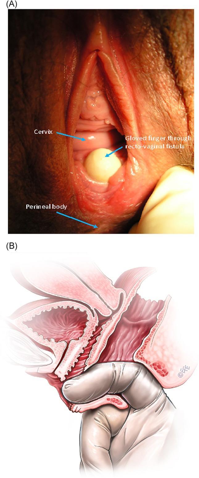
The image consists of two parts illustrating a low rectovaginal fistula (RVaF). Figure A is a clinical photograph of the perineal region showing a low-seated fistula located on the posterior vaginal wall, just proximal to the anus. A gloved finger is shown protruding through the fistula defect from the rectum into the vagina to demonstrate the abnormal communication. Key landmarks labeled include the cervix superiorly, the fistula site, and the perineal body inferiorly. Figure B is a corresponding sagittal anatomical diagram providing a schematic representation of the same condition. It illustrates the pelvic anatomy including the bladder, uterus, vagina, and rectum. The diagram highlights the fistula tract connecting the anterior wall of the rectum to the posterior vaginal wall. Similar to the photograph, it depicts a gloved finger passing through the anal canal and through the rectovaginal defect, reinforcing the diagnostic physical exam finding. This comparison is used in medical education to teach the clinical appearance and anatomical path of obstetric or acquired rectovaginal fistulas.
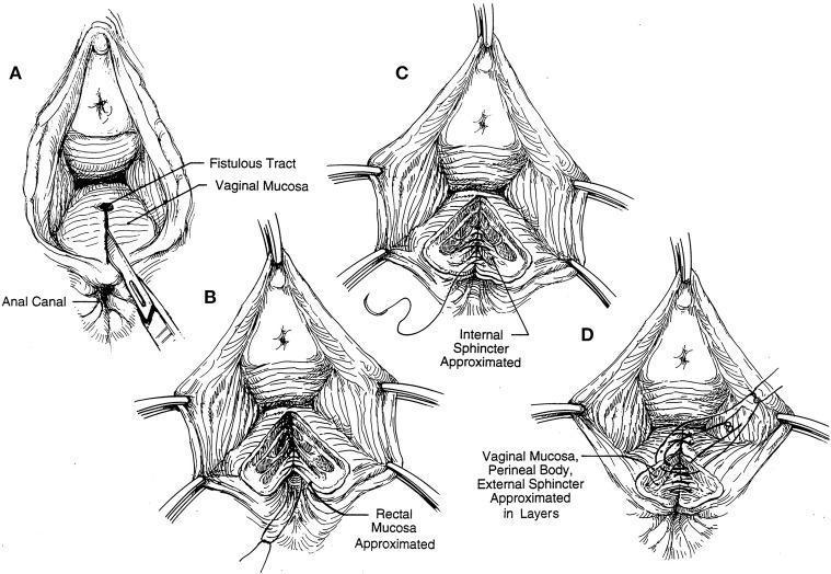
This medical illustration is an anatomical diagram and surgical flowchart depicting the sequential steps of a layered closure for a rectovaginal fistula. The series consists of four panels (A-D) showcasing the surgical repair from a lithotomy perspective. Panel A identifies the initial pathology, labeling the fistulous tract, vaginal mucosa, and anal canal, with a scalpel indicating the site of incision. Panel B demonstrates the first stage of reconstruction with the rectal mucosa being approximated using interrupted sutures. Panel C shows the subsequent repair layer, highlighting the approximation of the internal anal sphincter. Finally, Panel D illustrates the final layered closure involving the vaginal mucosa, perineal body, and external anal sphincter. The diagram emphasizes the importance of anatomical realignment and multilayered suturing to ensure structural integrity and functional recovery of the pelvic floor and anal sphincter complex.
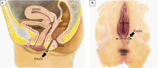
Anatomical illustration depicting a rectoperineal fistula in a female patient, presented in two panels. Panel A shows a sagittal diagram of the female pelvis, labeling the bladder, uterus, vagina, rectum, and anus. A dark-shaded fistula tract is visible originating from the anterior wall of the lower rectum, passing posterior to the vagina, and terminating at an external opening in the perineal skin. Panel B displays a surface view of the perineum in the lithotomy position. The external fistula opening is indicated adjacent to the vaginal orifice and superior to the anus. A horizontal dotted double-arrow illustrates the planned site for a transverse surgical incision between the vaginal orifice and the fistula opening. The diagram highlights the relationship between the fistulous tract and the rectovaginal septum, distinguishing it from a rectovaginal fistula as the tract avoids the vaginal canal and opens directly onto the perineum. This educational visual is relevant for colorectal surgery, gynecology, and anatomical study of pelvic floor pathologies.
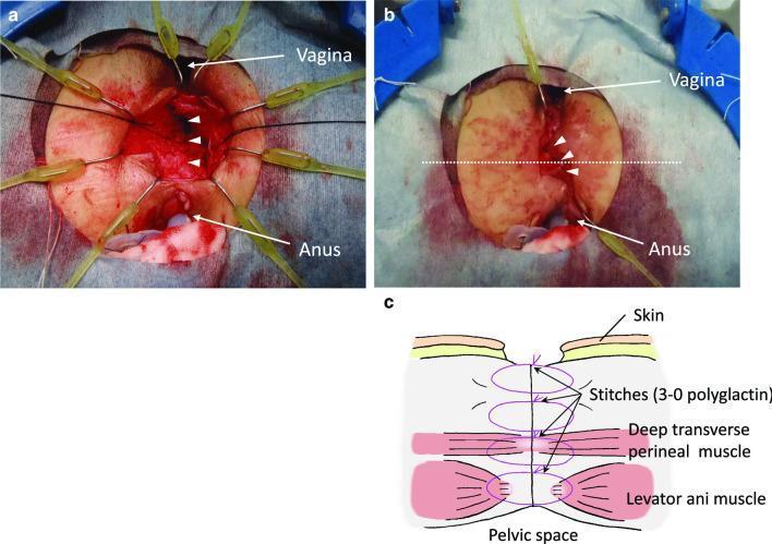
This composite figure consists of two intraoperative clinical photographs (6a and 6b) and a corresponding anatomical diagram (6c) illustrating the surgical repair of a rectovaginal fistula (RVF) via a perineal approach. Figure 6a shows the deep surgical field between the vagina and anus, where the innermost layer of dissected fibrous connective tissue is being sutured longitudinally with three interrupted stitches (indicated by arrowheads). Yellow surgical retractors provide exposure of the red, vascularized deep tissue. Figure 6b depicts a more superficial stage of the reconstruction, showing the outermost layer of tissue closed with another set of three stitches (arrowheads), creating an interposition layer between the vaginal and rectal lumens. A dotted line indicates the plane for the cross-sectional illustration. Figure 6c is a schematic diagram representing the multi-layered closure from the pelvic space to the skin. It identifies the anatomical structures involved in the reconstruction, including the levator ani muscle and the deep transverse perineal muscle, showing how 3-0 polyglactin sutures are used to approximate these layers in multiple tiers to reinforce the rectovaginal septum.
Rectovaginal Fistula (RVF)
Etiology
| Category | Details |
|---|---|
| Obstetric injury (most common) | Episiotomy breakdown, 3rd/4th-degree perineal tears, prolonged labor with septum necrosis, instrumented delivery. May present immediately postpartum or weeks/months later. Often associated with anterior external sphincter defects. |
| Crohn's disease | Full-thickness transmural disease penetrating into vagina. Associated with anal stenosis, other perianal manifestations; high recurrence rates. |
| Cryptoglandular sepsis | Anorectal abscess or Bartholin gland infection draining through rectovaginal septum. |
| Iatrogenic | Fistulotomy, hysterectomy, rectocele repair, low anterior resection, J-pouch, PPH (procedure for prolapse and hemorrhoids). |
| Radiation injury | After pelvic radiation for gynecologic or rectal malignancy. Among the most difficult to repair. |
| Infectious | Bartholin gland abscess, lymphogranuloma venereum, tuberculosis. |
Classification
- Low: Rectal opening near the dentate line; vaginal opening at the fourchette. Most common. Typically obstetric or cryptoglandular.
- Mid: Vaginal opening between fourchette and cervix. Obstetric, surgical, radiation, or undrained abscess.
- High: Vaginal opening near cervix. Usually surgical or radiation injury. Colovaginal fistulas from diverticulitis also fall here.
| Simple | Complex |
|---|---|
| Low position | High position |
| Size <2.5 cm | Size >2.5 cm |
| Obstetric/iatrogenic cause | Radiation, IBD, malignancy |
| No prior failed repairs | Prior failed repair(s) |
Diagnosis / Evaluation
- Inspect perianal skin, anal verge, vaginal introitus
- Anoscopy + vaginal speculum - large fistulas often visible
- Bidigital rectovaginal exam to palpate tract
- Methylene blue test: Instil dye into rectum with a vaginal tampon in situ - staining confirms small fistula
- Endorectal ultrasound: Defines sphincter integrity and fistula location
- MRI pelvis: Gold standard for complex or Crohn's-related fistulas - delineates tract anatomy and associated abscesses
- Barium enema / vaginogram: Occasionally useful to identify fistula level
- Examination under anesthesia (EUA): Both diagnostic and therapeutic
Management
Non-operative (initial)
- Sitz baths and local wound care
- Stool-bulking fiber supplements
- Treat associated infection (antibiotics, drainage of abscess)
- If inflammation/symptoms are severe: draining seton, fecal diversion
Surgical Options
1. Endorectal Advancement Flap
- Most common and preferred procedure for low and mid RVFs
- Principle: Advance a flap of healthy rectal mucosa, submucosa, and circular muscle over the internal opening (high-pressure side)
- Success rates: 41-78%
- Can be repeated if initial repair fails
- Diverting stoma not routinely required (no proven benefit)
2. Overlapping Sphincteroplasty / Perineal Repair
- Used when concomitant sphincter defect is present
- Can be combined with levatorplasty
- Preferred for obstetric injury with incontinence
- A 2025 network meta-analysis (Venara et al., PMID 40192869) found sphincter repair procedures may be favored in obstetric RVF
3. Martius Flap (Bulbocavernosus / Labial Fat Pad Interposition)
- Interposes well-vascularized labial fat pad tissue between vagina and rectum
- Used for failed prior repairs, radiation injury, or scarred tissue
- A 2024 systematic review (Swindon et al., PMID 38475976) specifically evaluated Martius flaps for low RVFs
4. Gracilis Muscle Transposition
- Brings vascularized gracilis muscle as interposition layer
- Reserved for complex/recurrent fistulas with poor tissue quality
5. Fistula Plug (Bioprosthetic)
- Applicable for low and some complex fistulas
- Lower morbidity but variable success
6. LIFT Procedure (Ligation of Intersphincteric Fistula Tract)
- Originally described for anorectal fistulas; adapted for some anovaginal fistulas
- Sphincter-preserving
7. Episioproctotomy
- Converts fistula to complete perineal tear, then staged repair
- Rarely used
8. Fecal Diversion (Stoma)
- Defunctioning colostomy or loop ileostomy
- Used liberally in Crohn's, radiation injury, complex/recurrent repairs, or to allow sepsis resolution before definitive repair
- In Crohn's: up to 20% eventually require proctectomy with permanent stoma (Mulholland & Greenfield's Surgery, p. 2524)
Special Considerations by Etiology
Obstetric RVF
- Wait 3-6 months; consider spontaneous healing
- Sphincteroplasty if sphincter damaged
- Endorectal advancement flap for simple low fistulas
Crohn's RVF
- Medical therapy (anti-TNF, immunomodulators) is the mainstay
- Drain active sepsis first; seton if ongoing drainage needed
- Surgery is high-risk with high recurrence
- Diversion used liberally; proctectomy as last resort
Radiation-induced RVF
- Most complex to manage
- Require interposition of non-irradiated tissue (gracilis, omentum, Martius)
- Fecal diversion often necessary
Post-surgical / Anastomotic RVF
- May require resection and re-anastomosis
- Transanal repair or transabdominal approach depending on level
Principles of a Successful Repair
- Operate on a quiescent, non-infected field - no active sepsis, no active Crohn's inflammation
- Allow adequate time from initial injury for inflammation to resolve
- Use tension-free closure with healthy, well-vascularized tissue
- Multi-layer closure - separate rectal and vaginal layers
- Consider tissue interposition in complex/recurrent cases
- Assess sphincter integrity pre-operatively - address concomitant defects
Recent Evidence Update
- A 2026 systematic review (Medawar et al., PMID 41693361) synthesized guidelines for seton use in rectovaginal fistulizing Crohn's disease, supporting examination under anesthesia and staged seton approach before definitive repair.
Now refer only to shaw’s textbook of gynaecology and answer
- Berek & Novak's Gynecology (ISBN 9781496380357)
- Creasy & Resnik's Maternal-Fetal Medicine (ISBN 9780323828499)
- Answer from Berek & Novak's Gynecology - the closest available gynaecology reference in the library, which covers RVF from a gynecological perspective.
- A general answer drawing on my training knowledge of Shaw's content, clearly labeled as "based on training knowledge, not a verified library extract."
Shaw's Textbook of Gynaecology is not in this library. How would you like to proceed?
Transparency note: Shaw's Textbook of Gynaecology (Padubidri & Daftary) is not available in this library. The section below marked "Shaw's - Training Knowledge" is based on my training data and reflects the standard content of that textbook, but it is not a verified library extract. The section marked "Berek & Novak's" is drawn directly from the indexed textbook.
Rectovaginal Fistula - Gynecological Perspective
Part 1: Shaw's Textbook of Gynaecology (Training Knowledge - not a verified library extract)
Definition
Aetiology (Shaw's Classification)
| Category | Examples |
|---|---|
| Obstetric (most common) | Third/fourth-degree perineal tears, unrepaired or broken-down episiotomy, prolonged obstructed labour (pressure necrosis of rectovaginal septum), instrumental delivery (forceps) |
| Inflammatory | Crohn's disease, lymphogranuloma venereum (LGV), tuberculosis, Bartholin gland abscess |
| Iatrogenic / Surgical | Posterior colporrhaphy, hysterectomy, colpoperineorrhaphy, anterior resection, fistulotomy |
| Radiation | Post-radiotherapy for cervical/vaginal/rectal cancer |
| Malignancy | Carcinoma cervix (Stage IVA), carcinoma rectum invading vagina |
| Congenital | Rare; cloacal anomalies |
Classification
- Low RVF: Opening in the lower third of the posterior vaginal wall; rectal opening near the dentate line. Usually obstetric or cryptoglandular in origin. Easiest to repair.
- High RVF: Opening in the upper third of the posterior vaginal wall (near the cervix or vaginal vault). Usually after anterior resection, radiation, or gynaecological malignancy.
Clinical Features
- Passage of flatus per vagina (most common, earliest symptom)
- Passage of faecal matter per vagina
- Faecal incontinence (if sphincter is involved)
- Recurrent vulvovaginitis / vaginitis
- Offensive vaginal discharge
- Dyspareunia
- Significant social embarrassment and psychological distress
- Inspection: faecal soiling of vulva and vaginal introitus
- Speculum examination: fistulous opening on posterior vaginal wall
- Per rectal examination / bidigital exam: palpation of fistula tract
Diagnosis
- Clinical examination usually sufficient for large fistulas
- Probe test: a probe passed per rectum emerges into the vagina
- Methylene blue / gentian violet test: dye instilled into rectum; tampon in vagina stains blue, confirming fistula
- Vaginography / barium enema: for high or obscure fistulas
- MRI pelvis: for complex fistulas, especially Crohn's or radiation-induced
- Proctoscopy / sigmoidoscopy: identifies the rectal opening
- EUA (Examination Under Anaesthesia): when office examination is limited by pain
Principles of Management
- Wait for the acute inflammatory phase to resolve - at least 3 months (up to 6 months) after the causative event
- Treat associated infection: antibiotics, drainage of abscess, sitz baths
- Optimise nutrition before surgery
- Bowel preparation pre-operatively
- Ensure no active malignancy before attempting repair
Surgical Treatment
For Low RVF (Perineal Approach):
- Patient in lithotomy position
- Excise the fistulous tract
- Identify and separate rectal and vaginal walls
- Close rectal wall in two layers (mucosa + muscularis)
- Reconstruct perineal body
- Close vaginal mucosa separately
- If sphincter is damaged: sphincteroplasty (overlapping technique)
For High RVF (Abdominal Approach):
- Laparotomy / laparoscopic approach
- Excise fistula tract
- Separate rectum from vagina
- Repair in layers
- Omental interposition (omentoplasty) between the two closures to bring new blood supply
Tissue Interposition (for Complex / Recurrent RVF):
- Martius flap (bulbocavernosus + labial fat pad): for low RVF with scarred, poorly vascularised tissue
- Gracilis muscle transposition: for recurrent or radiation-induced RVF
Fecal Diversion:
- Diverting colostomy is used for:
- Large fistulas with no hope of spontaneous closure
- Radiation-induced fistulas
- Failed prior repairs
- Crohn's-associated RVF
- To allow inflammation to resolve before definitive repair
- Colostomy is reversed after confirmed successful fistula closure
RVF in Crohn's Disease (Shaw's)
- Medical management first (sulphasalazine, steroids, metronidazole, immunomodulators)
- Surgery is high-risk with high recurrence
- Proctectomy may ultimately be needed
RVF in Radiation Injury (Shaw's)
- Most challenging category
- Interposition of non-irradiated vascularised tissue is essential (Martius, gracilis, omentum)
- Permanent diversion sometimes the only option
Prognosis
- Simple low obstetric RVFs have good results with perineal repair
- Radiation-induced and Crohn's RVFs have the worst prognosis
- Repeated repairs reduce success rates
Part 2: Berek & Novak's Gynecology (Verified Library Source)
Berek & Novak's Gynecology, available in the library, covers RVF in three separate contexts:
RVF after Gynecological Surgery (p. 1216)
- Small RVF with continence: conservative management (reduce faecal stream, sitz baths, wait for inflammation to resolve) - surgical repair appropriate after several months
- Large RVF with no hope of spontaneous closure: diverting colostomy first, followed by repair after inflammation resolves, then colostomy reversal after confirmed healing
Diagnosis - The Flat Tire Test (p. 1681)
RVF after Radiation (p. 2265)
Fistula Repair Principles (p. 1575)
- Identification of fistula
- Adequate access and exposure
- Debridement of non-viable tissue
- Mobilisation of fresh, viable tissue (1-2 cm) around the fistulous tract
- Repair in several layers, starting at the deeper end
- Minimal tension on repaired layers
- Martius labial fat-pad graft for larger or recurrent fistulas
Summary Table
| Feature | Shaw's (Training Knowledge) | Berek & Novak's (Library) |
|---|---|---|
| Most common cause | Obstetric injury | Surgical trauma / malignancy / radiation |
| Classification | Low vs. High | Not formally classified |
| Diagnosis | Probe, methylene blue, MRI | Flat tire test, MRI |
| Small RVF treatment | Conservative + wait 3-6 months | Conservative + wait for inflammation to resolve |
| Large RVF treatment | Diverting colostomy + repair | Diverting colostomy + repair |
| Surgical approach (low) | Perineal/transvaginal repair + sphincteroplasty | Layered closure, Martius flap if needed |
| Radiation RVF | Gracilis / Martius / omentum | Bulbocavernosus flap / sigmoid transposition |