
Superolateral surface of brain
Reading File
The superolateral surface (also called the convex surface) of the brain is the largest surface of the cerebral hemisphere, facing outward and upward. It contains the major gyri and sulci and is organized into frontal, parietal, temporal, and occipital lobes.
Searching sources...
Finding Sources
Finding Sources
Finding Sources
Finding Sources
Finding Sources
Finding Sources
Finding Sources
Finding Sources
Finding Sources
Finding Sources
I found a key section. Let me read it and also fetch the relevant images.
Reading File
Reading File
Excellent! Let me now read more content and fetch the key images.
Reading File
Loading Image
Loading Image
I now have all the material needed for a comprehensive answer. Let me compile it.
Superolateral Surface of the Brain
The superolateral (convex) surface is the largest surface of each cerebral hemisphere. It faces upward, outward, and forward, and is the surface most visible when the brain is viewed from the side. It is covered by the calvaria of the skull and is related to the meninges and the venous sinuses above.
Overview of Lobes
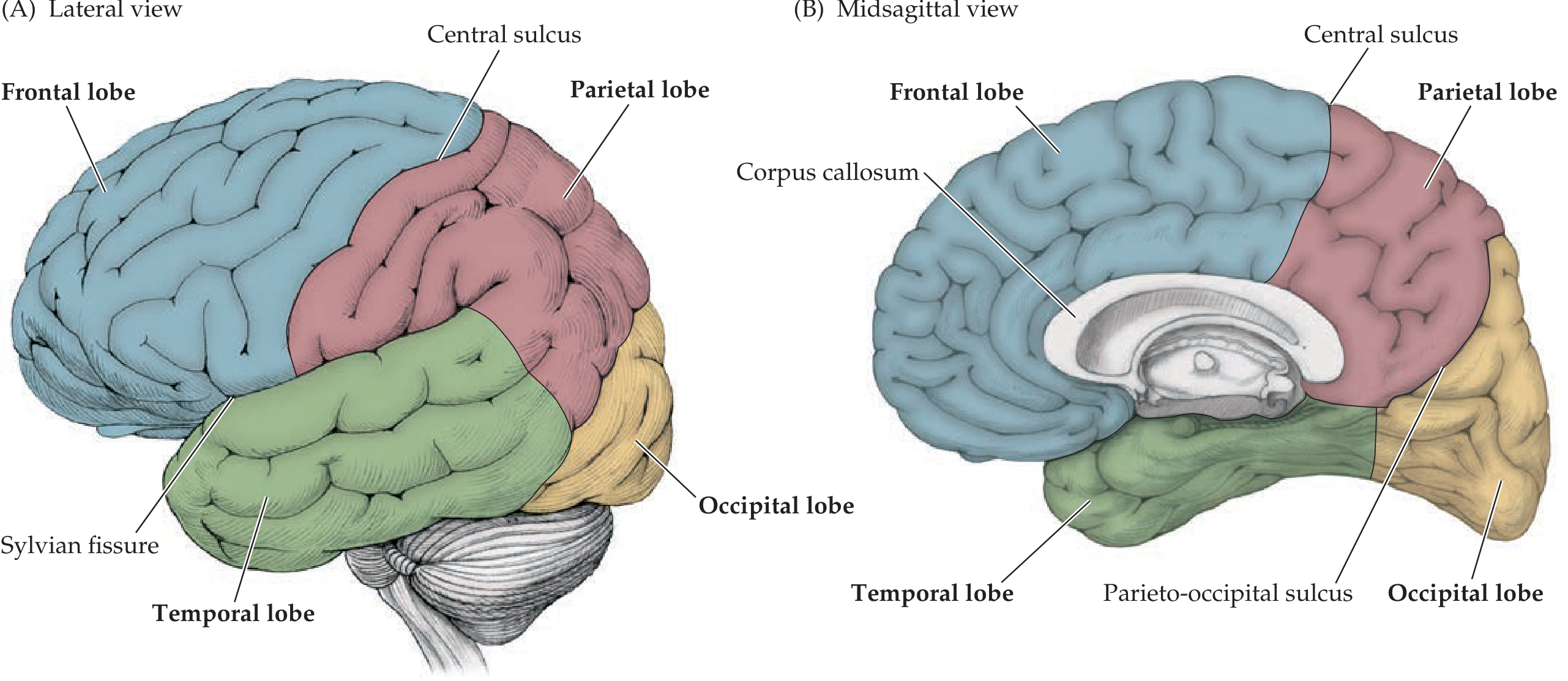
The superolateral surface is divided into four major lobes. Two key sulci define their boundaries:
| Sulcus / Fissure | Boundary it forms |
|---|---|
| Central sulcus (of Rolando) | Frontal lobe (anterior) from Parietal lobe (posterior) |
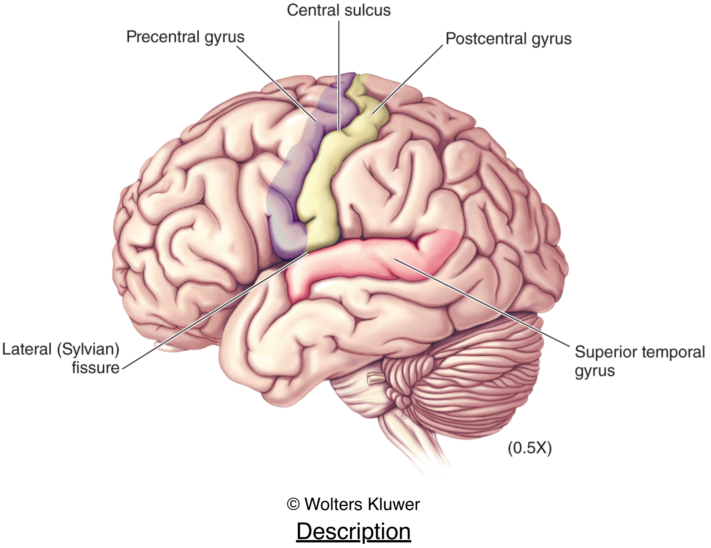
| Lateral (Sylvian) fissure | Frontal/Parietal lobes above from Temporal lobe below |
| Parieto-occipital sulcus | Parietal from Occipital lobe (visible best on medial surface) |
1. Frontal Lobe (Lateral Surface)
Bounded posteriorly by the central sulcus and inferolaterally by the Sylvian fissure.
Gyri (anterior to posterior):
- Precentral gyrus - lies immediately anterior to the central sulcus; contains the primary motor cortex (area 4). Controls voluntary movement of contralateral body. The motor homunculus is here.
- Superior frontal gyrus - above the superior frontal sulcus
- Middle frontal gyrus - between superior and inferior frontal sulci
- Inferior frontal gyrus - below the inferior frontal sulcus; divided into three parts:
- Pars orbitalis (most anterior)
- Pars triangularis (between ascending rami of Sylvian fissure)
- Pars opercularis (posterior part, adjacent to precentral gyrus)
- In the dominant (usually left) hemisphere, pars triangularis + pars opercularis = Broca's area (area 44 and 45) - expressive speech
Key sulci: Superior frontal sulcus, inferior frontal sulcus, precentral sulcus
2. Parietal Lobe (Lateral Surface)
Bounded anteriorly by the central sulcus and antero-inferiorly by the Sylvian fissure. Has no sharp boundary with temporal or occipital lobes laterally.
Gyri:
- Postcentral gyrus - immediately posterior to the central sulcus; contains the primary somatosensory cortex (areas 3, 1, 2). Receives contralateral tactile, proprioceptive, and pain/temperature sensations.
- Superior parietal lobule - above the intraparietal sulcus; involved in visuospatial processing
- Inferior parietal lobule - below the intraparietal sulcus; contains:
- Supramarginal gyrus (area 40) - surrounds the upturned end of the Sylvian fissure
- Angular gyrus (area 39) - surrounds the upturned end of the superior temporal sulcus
- In the dominant hemisphere, these form Wernicke's area (along with the posterior superior temporal gyrus) - receptive/comprehension speech
Key sulci: Intraparietal sulcus (horizontal), postcentral sulcus
3. Temporal Lobe (Lateral Surface)
Lies below the Sylvian fissure on the lateral surface.
Gyri:
- Superior temporal gyrus - its upper surface (planum temporale) hidden in the Sylvian fissure contains Heschl's transverse gyri = primary auditory cortex (areas 41, 42). The posterior part of the superior temporal gyrus is part of Wernicke's area.
- Middle temporal gyrus - between superior and inferior temporal sulci; involved in semantic memory and language
- Inferior temporal gyrus - the most inferior gyrus visible on the lateral surface; involved in visual object recognition (face recognition)
Key sulci: Superior temporal sulcus, inferior temporal sulcus
4. Occipital Lobe (Lateral Surface)
The smallest lobe on the lateral surface; has no sharp boundary with parietal or temporal lobes laterally. The primary visual cortex (area 17, striate cortex) is mostly on the medial surface in the calcarine sulcus, but the lateral occipital gyri (areas 18, 19) for visual association are visible on the superolateral surface.
Important Landmarks - Summary Diagram
The Insula (Hidden from View)
A fifth area of cortex, the insular cortex, lies buried in the depths of the Sylvian fissure and is not visible on the superolateral surface without retracting the opercula. It is covered by:
- Frontal operculum (anteriorly)
- Parietal operculum (posteriorly)
Key Functional Areas on Superolateral Surface
| Area | Location | Brodmann Area | Function |
|---|---|---|---|
| Primary Motor Cortex | Precentral gyrus | Area 4 | Voluntary movement (contralateral) |
| Premotor Cortex | Anterior to precentral | Area 6 | Motor planning |
| Prefrontal Cortex | Anterior frontal lobe | Areas 9-12, 46, 47 | Executive functions, personality |
| Broca's Area (dominant) | Pars triangularis + opercularis | Areas 44, 45 | Expressive speech |
| Primary Somatosensory | Postcentral gyrus | Areas 3, 1, 2 | Tactile and proprioceptive sensation |
| Primary Auditory | Superior temporal (Heschl's) | Areas 41, 42 | Hearing |
| Wernicke's Area (dominant) | Posterior STG + SMG + AG | Areas 39, 40, 22 | Language comprehension |
| Visual Association | Lateral occipital | Areas 18, 19 | Higher visual processing |
Arterial Supply
- Frontal and parietal lobes (superolateral surface): branches of the middle cerebral artery (MCA) - the largest branch of the internal carotid artery, running in the Sylvian fissure
- Superior strip of frontal and parietal lobes near the midline: anterior cerebral artery (ACA)
- Occipital lobe and posterior temporal: posterior cerebral artery (PCA)
Sources: Neuroanatomy through Clinical Cases, 3rd Edition, pp. 50-51, 53 | Neuroscience: Exploring the Brain, 5th Edition, pp. 659-661
This is a shared conversation. Sign in to Orris to start your own chat.