marjolin ulcer

Reading File
Finding Sources
Finding Sources
Searching Images

Marjolin ulcer squamous cell carcinoma arising in chronic burn scar
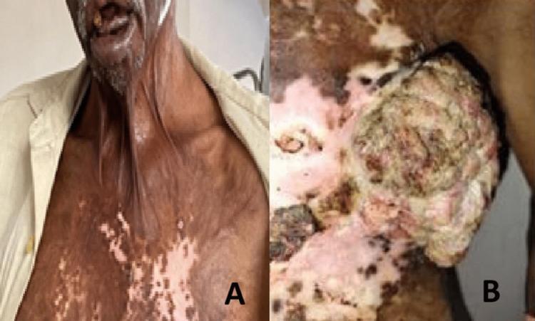
This clinical photograph consists of two panels (A and B) demonstrating long-term sequelae of severe burn injuries. Panel A is an anterior-view clinical photograph of a patient showing a significant post-burn neck contracture, characterized by vertical bands of tightened, shortened skin extending from the chest to the chin, causing eversion of the lower lip (ectropion). The chest skin displays extensive mottled dyspigmentation with areas of hyperpigmentation and hypopigmentation, consistent with an unstable chronic scar. Panel B is a close-up of the left chest wall and axillary region, revealing a large (approximately 15 cm x 10 cm), exophytic, fungating mass arising from the scarred tissue. The lesion presents a cauliflower-like or cobblestone texture with variegated colors indicating areas of necrosis, purulent discharge, and potential hemorrhage. This presentation is highly clinically significant for the development of a Marjolin ulcer—a squamous cell carcinoma arising within a site of chronic inflammation or previous thermal injury. The image serves as an educational tool for identifying late burn complications and malignant transformation in chronic scars.
This clinical photograph consists of two panels (A and B) demonstrating long-term sequelae of severe burn injuries. Panel A is an anterior-view clinical photograph of a patient showing a significant post-burn neck contracture, characterized by vertical bands of tightened, shortened skin extending from the chest to the chin, causing eversion of the lower lip (ectropion). The chest skin displays extensive mottled dyspigmentation with areas of hyperpigmentation and hypopigmentation, consistent with an unstable chronic scar. Panel B is a close-up of the left chest wall and axillary region, revealing a large (approximately 15 cm x 10 cm), exophytic, fungating mass arising from the scarred tissue. The lesion presents a cauliflower-like or cobblestone texture with variegated colors indicating areas of necrosis, purulent discharge, and potential hemorrhage. This presentation is highly clinically significant for the development of a Marjolin ulcer—a squamous cell carcinoma arising within a site of chronic inflammation or previous thermal injury. The image serves as an educational tool for identifying late burn complications and malignant transformation in chronic scars.

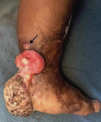
Clinical photograph of the medial and posterior aspects of a human foot showing a large, exophytic, fungating mass and extensive scarring. The mass is localized to the posterior heel and Achilles tendon region, measuring approximately 6x10x12cm. It exhibits a heterogeneous appearance with a reddish, fleshy, and friable proximal border, while the more distal portion on the plantar heel shows a rough, cauliflower-like texture with areas of yellow-brown discoloration and suspected necrosis. An arrow points to a smaller, elevated satellite lesion superior to the main mass. The surrounding integument of the foot and distal leg shows significant chronic scarring, dyspigmentation, and texture changes consistent with prior burn injuries. This clinical presentation is highly suggestive of a Marjolin ulcer, a squamous cell carcinoma arising from a chronic burn scar. The educational focus is on identifying malignant transformation in chronic wounds and understanding the visual morphology of aggressive skin malignancies.
Clinical photograph of the medial and posterior aspects of a human foot showing a large, exophytic, fungating mass and extensive scarring. The mass is localized to the posterior heel and Achilles tendon region, measuring approximately 6x10x12cm. It exhibits a heterogeneous appearance with a reddish, fleshy, and friable proximal border, while the more distal portion on the plantar heel shows a rough, cauliflower-like texture with areas of yellow-brown discoloration and suspected necrosis. An arrow points to a smaller, elevated satellite lesion superior to the main mass. The surrounding integument of the foot and distal leg shows significant chronic scarring, dyspigmentation, and texture changes consistent with prior burn injuries. This clinical presentation is highly suggestive of a Marjolin ulcer, a squamous cell carcinoma arising from a chronic burn scar. The educational focus is on identifying malignant transformation in chronic wounds and understanding the visual morphology of aggressive skin malignancies.

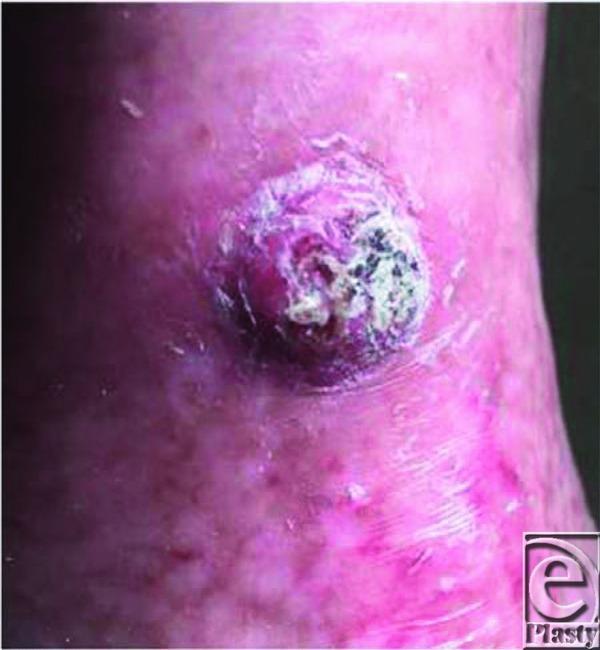
This clinical photograph displays a close-up view of a prominent exophytic lesion on the left tibial region. The lesion is approximately circular but possesses irregular, raised borders and a friable, non-uniform surface characterized by granulation tissue, focal ulceration, and crusting. The color is variegated, showing a mixture of erythematous (red/pink), dusky purple, and dark grey/black tones. The surrounding skin exhibits clear signs of chronic tissue change consistent with a previous burn injury and subsequent autografting, including altered pigmentation, shiny texture, and scarring. A localized zone of erythema and induration is visible immediately adjacent to the base of the mass. Clinically, this presentation is highly suggestive of a Marjolin's ulcer—a squamous cell carcinoma (SCC) arising within a chronic wound or scar. This image illustrates key morphological red flags for malignancy in chronic wounds, such as rapid growth, exophytic architecture, and heterogeneous coloration in an area of prior thermal trauma.
This clinical photograph displays a close-up view of a prominent exophytic lesion on the left tibial region. The lesion is approximately circular but possesses irregular, raised borders and a friable, non-uniform surface characterized by granulation tissue, focal ulceration, and crusting. The color is variegated, showing a mixture of erythematous (red/pink), dusky purple, and dark grey/black tones. The surrounding skin exhibits clear signs of chronic tissue change consistent with a previous burn injury and subsequent autografting, including altered pigmentation, shiny texture, and scarring. A localized zone of erythema and induration is visible immediately adjacent to the base of the mass. Clinically, this presentation is highly suggestive of a Marjolin's ulcer—a squamous cell carcinoma (SCC) arising within a chronic wound or scar. This image illustrates key morphological red flags for malignancy in chronic wounds, such as rapid growth, exophytic architecture, and heterogeneous coloration in an area of prior thermal trauma.

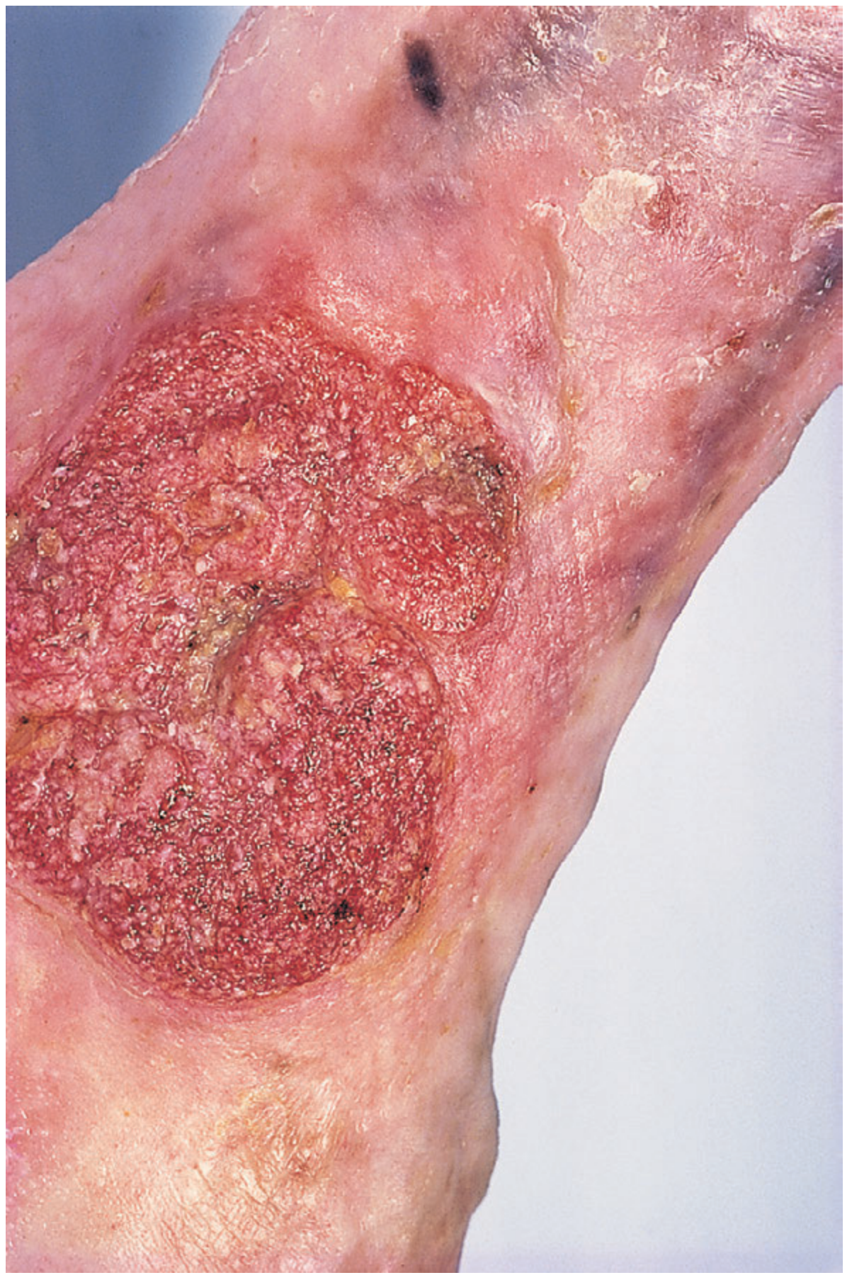
This clinical gross photography depicts a distal thigh- to above-knee amputation stump with a chronically scarred, ulcerated soft tissue lesion consistent with Marjolin's ulcer arising in a prior traumatic scar. The overlying skin shows hyperpigmented, leathery areas with focal induration and necrotic regions at the margin of the scar, transitioning to healthier surrounding tissue. The lesion is exophytic and crateriform, with irregular, raised borders and crusting, extending into the scarred subcutaneous tissue. In many Marjolin's ulcers, the histology reveals squamous cell carcinoma, typically well differentiated, characterized by nests of malignant keratinocytes with possible keratin pearl formation and invasion into deeper dermis and subcutaneous planes; clinically, this entity bears a latent interval of years to decades after initial injury. This image emphasizes the aggressive behavior and potential for local invasion and regional spread despite a relatively well-differentiated phenotype. Clinical relevance includes recognition in chronic wounds or burn scars, necessity of complete excision with wide margins, and evaluation for nodal metastasis. In resource-limited settings, delayed presentation is common; in developed regions, modern wound care reduces incidence but vigilance remains essential for long-standing scars. This gross finding informs surgical planning, prognosis assessment, and multidisciplinary oncologic management including possible radiotherapy or extended resection.

This clinical gross photography depicts a distal thigh- to above-knee amputation stump with a chronically scarred, ulcerated soft tissue lesion consistent with Marjolin's ulcer arising in a prior traumatic scar. The overlying skin shows hyperpigmented, leathery areas with focal induration and necrotic regions at the margin of the scar, transitioning to healthier surrounding tissue. The lesion is exophytic and crateriform, with irregular, raised borders and crusting, extending into the scarred subcutaneous tissue. In many Marjolin's ulcers, the histology reveals squamous cell carcinoma, typically well differentiated, characterized by nests of malignant keratinocytes with possible keratin pearl formation and invasion into deeper dermis and subcutaneous planes; clinically, this entity bears a latent interval of years to decades after initial injury. This image emphasizes the aggressive behavior and potential for local invasion and regional spread despite a relatively well-differentiated phenotype. Clinical relevance includes recognition in chronic wounds or burn scars, necessity of complete excision with wide margins, and evaluation for nodal metastasis. In resource-limited settings, delayed presentation is common; in developed regions, modern wound care reduces incidence but vigilance remains essential for long-standing scars. This gross finding informs surgical planning, prognosis assessment, and multidisciplinary oncologic management including possible radiotherapy or extended resection.

Searching PubMed

"Marjolin ulcer"

Reading File
Reading File
Reading File

Marjolin Ulcer

Definition: Marjolin ulcer is a malignancy (usually squamous cell carcinoma) that arises within a site of chronic scarring, most classically an old burn scar, but also chronic venous ulcers, osteomyelitis sinus tracts, pressure sores, and other longstanding wounds. It is essentially an umbrella term for malignant transformation occurring in a chronic scar - Fitzpatrick's Dermatology, p. 4831.

Key features

  • Histology: The vast majority are well-differentiated squamous cell carcinomas, though basal cell carcinoma, melanoma, sarcoma, and malignant fibrous histiocytoma have also been reported arising in scars - Fitzpatrick's Dermatology, p. 4835.
  • Behavior: Despite often being well-differentiated, these tumors show an aggressive growth pattern with a high propensity to metastasize and spread rapidly - K J Lee's Essential Otolaryngology, p. 1761; Fitzpatrick's Dermatology, p. 4837.
  • Timing:
    • Acute Marjolin ulcer - develops within 1 year of the original injury.
    • Chronic Marjolin ulcer - develops more than 1 year after the injury (classically a latency of years to decades is described).
  • Sites of predilection: extremities and areas overlying joints - Fitzpatrick's Dermatology, p. 4839.
  • Causes/settings: burn scars (about 2% of burn scars undergo malignant change), chronic venous/venous stasis ulcers, chronic sinuses/osteomyelitis tracts, pressure ulcers, and other non-healing wounds - Fitzpatrick's Dermatology, p. 4829; Bailey & Love's Short Practice of Surgery, p. 9928; Rockwood and Green's Fractures in Adults, p. 161-163.

Pathogenesis (proposed, not fully settled)

Chronic inflammation and repeated cycles of tissue destruction/repair in a scar are thought to drive malignant transformation, possibly through sustained inflammatory cytokine effects, impaired local immune surveillance (scar tissue is relatively avascular and lymphatic-poor, which may also explain why metastasis can appear late despite aggressive local behavior), and accumulated DNA damage in repeatedly regenerating epithelium - Rockwood and Green's Fractures in Adults, p. 161.

Clinical clues / when to suspect it

Any of the following in a chronic scar or non-healing ulcer should prompt biopsy:
  • New ulceration, erosion, or persistent non-healing area within an old scar
  • Exophytic, fungating, cauliflower-like mass with a friable, heterogeneous surface (necrosis, hemorrhage, purulent discharge)
  • Rolled or everted, irregular margins
  • Rapid growth or change in a previously stable scar
  • Foul odor or increasing pain/bleeding
Bailey & Love's specifically notes that any chronic longstanding venous ulcer that fails to respond to treatment or looks atypical should raise suspicion for Marjolin-type malignant change (SCC or BCC) - p. 9928.
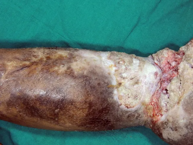
Here is a case illustrating a Marjolin ulcer arising from a chronic venous ulcer:
Marjolin's ulcer - a squamous cell cancer arising in a chronic venous ulcer
And an example arising in a chronic post-burn scar, showing the typical exophytic, fungating morphology:
Exophytic fungating mass arising from a chronic burn scar, consistent with Marjolin ulcer

Diagnosis

Biopsy is mandatory for any persistent, changing, or non-healing lesion in a scar - do not assume a new ulcer in an old scar is "just" recurrent breakdown. Once malignancy is confirmed, staging (imaging, regional lymph node assessment) is needed given the metastatic potential.

Management

  • Wide local excision with clear margins is the mainstay; amputation may be required for extensive extremity involvement.
  • Regional lymph node evaluation/dissection if nodal spread is suspected or confirmed.
  • Radiotherapy may be used adjuvantly in select cases.
  • Multidisciplinary oncologic management (plastic surgery, surgical oncology, radiation oncology) is recommended given aggressive behavior.

Prevention

Fitzpatrick's Dermatology emphasizes: good wound care, early skin grafting of burns, avoidance of chronic contractures, and early excision of any tissue showing degenerative/keratotic change in a scar - p. 4843.

Recent evidence

A 2023 meta-analysis and systematic review (PMID: 36729819, Plastic and Reconstructive Surgery) used machine learning techniques to identify prognostic factors for Marjolin ulcers, focusing on predictors of lymphatic metastasis and outcomes - this is a good source if you want more granular data on prognosis and risk stratification beyond what's in the standard textbooks.
If you want, I can go deeper into any specific angle - histopathology, differential diagnosis versus other chronic ulcer malignancies, or the burn-scar-specific literature.
This is a shared conversation. Sign in to Orris to start your own chat.