
a 45 year old women undergoes total thyroidectomy for multinodular goiter. on the first postoperative day she complains of hoarseness of voice in the hdu she denies breathing difficulty otherwise she is clinically stable
recurrent laryngeal nerve thyroid surgery anatomy
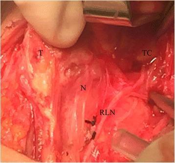
An intraoperative clinical photograph documenting an anatomical variation of the recurrent laryngeal nerve (RLN) during a thyroid surgery. The image shows the surgical field with the following landmarks labeled: the thyroid gland (T) medially retracted, the thyroid cartilage (TC) in the superior-lateral aspect, and the point of typical nerve entry (N). The recurrent laryngeal nerve (RLN) is visualized following an atypical trajectory, ascending along the medial margin of the superior thyroid pole and terminating beneath the superior cornu of the thyroid cartilage. This clinical case illustrates an anatomical variant that can lead to surgical complications if not properly identified. The surgical site displays typical tissue characteristics including erythematous vascularized structures and surgical retraction tools. This image serves as an educational resource for endocrine surgeons and medical students to understand neurovascular anatomy and the importance of intraoperative nerve monitoring in preventing iatrogenic vocal cord paralysis.
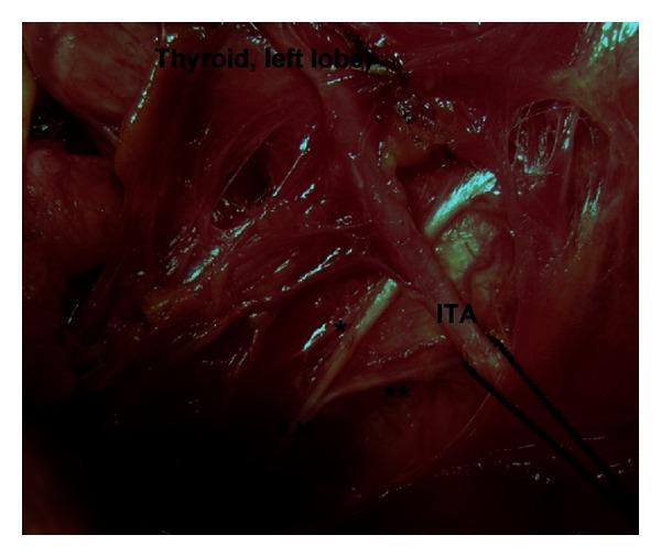
An intraoperative clinical photograph depicting the surgical anatomy of the left central neck compartment during thyroid surgery. The image highlights the anatomical relationship between the left inferior laryngeal nerve (ILN), also known as the recurrent laryngeal nerve, and the inferior thyroid artery (ITA). The photograph demonstrates a Type 4 variation, where the terminal division of the ILN into anterior (*) and posterior (**) branches occurs at the pre-arterial segment, proximal to its crossing with the ITA. The left lobe of the thyroid gland is visible in the upper portion of the surgical field. This visual is significant for endocrine surgery, illustrating extralaryngeal terminal bifurcation of the ILN, which is a critical landmark to identify to prevent iatrogenic nerve injury during thyroidectomy or parathyroidectomy. The image provides educational value regarding neurovascular variations and surgical risk regions in the cervical course of the laryngeal nerves.
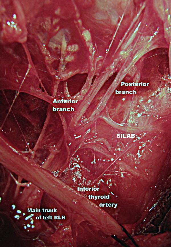
This magnified clinical operative photograph demonstrates the surgical anatomy of the left central neck compartment during thyroid surgery. The image highlights the variations in the course of the left recurrent laryngeal nerve (RLN) and its neurovascular relationships. The main trunk of the left RLN is visible in the lower quadrant, ascending and bifurcating into distinct anterior and posterior terminal branches. Medial to these branches, the inferior thyroid artery is identified, crossing the nerve path. A significant anatomical variation shown is the SILAB (Supra-Arterial Inferior Laryngeal Anastomotic Branch), a horizontal nerve anastomosis connecting the posterior branch of the RLN to adjacent neural structures near the laryngeal entry point. This photograph serves as an educational resource for identifying vulnerable neural structures during thyroidectomy and parathyroidectomy, emphasizing the clinical importance of meticulous dissection at the neurovascular crossing point to avoid iatrogenic nerve injury.
Post-Thyroidectomy Hoarseness: Day 1 Clinical Analysis
The Most Likely Diagnosis
Anatomy of the RLN and Why It Gets Injured
- The right RLN loops around the right subclavian artery and ascends obliquely in the tracheoesophageal groove
- The left RLN loops around the aortic arch (longer course), ascends more vertically in the tracheoesophageal groove
- The nerve crosses the inferior thyroid artery - a landmark used during surgery - and enters the larynx at the cricothyroid joint
Types of RLN Injury
| Type | Mechanism | Outcome |
|---|---|---|
| Neuropraxia (stretching/devascularization) | Traction, thermal spread, clips, edema | Temporary - resolves in weeks to 6 months |
| Axonotmesis | Crush or clamp injury, nerve intact | Partial/slow recovery |
| Neurotmesis (transection) | Divided nerve | Usually permanent |
Incidence
- Temporary RLN palsy: 4-10% of thyroidectomies
- Permanent RLN palsy: 0.5-2% of thyroidectomies
- Risk is up to 30% in reoperative thyroid surgery
- Transient hoarseness from any laryngeal cause: up to 80% of thyroidectomy patients at some point
Differential Diagnosis for Post-Thyroidectomy Hoarseness
| Cause | Features | How to Distinguish |
|---|---|---|
| Unilateral RLN palsy (most likely) | Hoarse, breathy voice; no dyspnea | Flexible laryngoscopy - unilateral cord immobility |
| EBSLN injury | Decreased pitch range, vocal fatigue | Laryngoscopy often normal; EMG needed |
| Intubation/ETT trauma | Laryngeal edema, granuloma | Symmetric findings on scopy |
| Hematoma compressing larynx | Neck swelling, possible dyspnea | Clinical exam, USS/CT; urgent if airway threatened |
| Bilateral RLN palsy | Stridor, dyspnea, respiratory distress | NOT this patient - she has no breathing difficulty |
| Arytenoid dislocation | Rare intubation complication | Laryngoscopy |
Immediate Management
- Rule out bilateral RLN injury (stridor, respiratory distress, SaO2 falling) - this is the emergency. This patient currently has NO respiratory distress, which is reassuring.
- Rule out neck hematoma - examine the wound; if expanding hematoma, open the wound at bedside immediately and take back to OR.
- Flexible nasolaryngoscopy should be performed urgently (within 24-48 hours) to:
- Directly visualize vocal cord movement
- Confirm unilateral vs bilateral palsy
- Document baseline (medicolegal importance)
- Guide prognosis and management
- Serum calcium and PTH (rule out concurrent hypoparathyroidism - the most common complication of thyroidectomy)
- Wound inspection for hematoma
Prognosis and Further Management
- If temporary (neuropraxia): Most transient RLN palsies resolve within 6 months. In some cases, recovery can take up to 2 years.
- If no recovery by 6-12 months: Likely permanent; consider voice therapy or surgical options (medialization laryngoplasty, vocal cord injection augmentation)
"Recurrent laryngeal nerve dysfunction can occur with an intact nerve that has been stretched or partially devascularized and may lead to postoperative hoarseness for up to six months. When laryngeal dysfunction persists longer than six months it is typically permanent; however, in some cases it may take up to 2 years for a recurrent laryngeal nerve palsy to resolve." - Fischer's Mastery of Surgery, 8th ed.
- Surgeon (follow-up laryngoscopy, counseling)
- ENT/Laryngologist
- Speech-language pathologist (voice therapy, swallowing assessment)
Other Complications to Monitor in This Patient
- Hypocalcemia / hypoparathyroidism - most common complication; check calcium and PTH. Rate of temporary hypocalcemia: 5-15%. Give calcium/Vitamin D supplements if PTH <10 pg/mL or calcium <8 mg/dL.
- Neck hematoma (~1% incidence) - if develops, open wound at bedside immediately to prevent airway compromise
- Thyroid hormone supplementation - all total thyroidectomy patients need levothyroxine replacement
Summary Answer for this Clinical Vignette
| Feature | Interpretation |
|---|---|
| Hoarseness day 1 | Unilateral RLN injury (neuropraxia most likely) |
| No breathing difficulty | Unilateral (not bilateral) palsy |
| Clinically stable | No hematoma, no airway compromise |
| Next step | Flexible nasolaryngoscopy + serum calcium/PTH |
| Prognosis | Most resolve within 6 months; counsel patient accordingly |
- Sabiston Textbook of Surgery, 21st ed. - Complications of Thyroidectomy (RLN injury rates, risk factors)
- Fischer's Mastery of Surgery, 8th ed. - Postoperative Hoarseness (mechanism, prognosis, MDT approach)
- Morgan & Mikhail's Clinical Anesthesiology, 7th ed. - Thyroidectomy complications overview
intrinsic muscle affect
intrinsic muscles of larynx diagram abduction adduction vocal cord
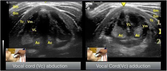
This diagnostic ultrasound image presents two side-by-side transverse views of the larynx at the level of the thyroid cartilage, demonstrating the functional anatomy of the vocal cords. The left frame illustrates vocal cord abduction, while the right frame shows vocal cord adduction. Key anatomical structures are labeled: the superficially located sternocleidomastoid muscles (Sm), the arch of the thyroid cartilage (Tc), the hypoechoic vocalis muscle (Vm), the hyperechoic vocalis ligament (VL), and the posteriorly situated arytenoid cartilages (Ac). In the abduction view, the arytenoid cartilages and vocal folds are positioned apart, creating an open glottic space. In the adduction view, the arytenoid cartilages move medially, bringing the hyperechoic vocal ligaments together at the midline. Small clinical inset photographs show the transverse transducer placement on the anterior neck over the thyroid cartilage. This comparison is used in clinical education to teach airway ultrasonography, specifically for assessing vocal cord mobility and verifying Laryngeal Mask Airway (LMA) or endotracheal tube positioning.
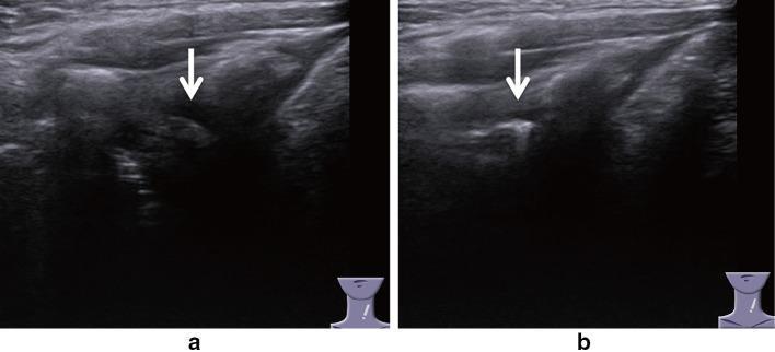
Side-by-side diagnostic ultrasound images demonstrating the laryngeal anatomy using a lateral vertical procedure. The images specifically focus on the visualization of the muscular process (MP) of the arytenoid cartilage to assess vocal cord dynamics. Panel (a) shows the larynx during abduction; at the white arrow, the muscular process is not visible as it has rotated out of the imaging plane. Panel (b) shows the larynx during adduction; the white arrow points to a distinct, hyperechoic structure representing the visible muscular process of the arytenoid cartilage. Surrounding soft tissue structures include the thyroarytenoid and lateral cricoarytenoid muscles. This imaging technique is utilized in otolaryngology to evaluate vocal cord movement and diagnose conditions such as vocal cord paresis or paralysis by observing the presence or absence of rhythmic arytenoid cartilage displacement. The lateral approach bypasses laryngeal air interference and thyroid cartilage calcification common in traditional transverse views.
Intrinsic Muscles of the Larynx Affected by RLN Injury
Complete List of Muscles Affected (RLN-innervated)
| Muscle | Action | Net Effect on Vocal Cord |
|---|---|---|
| Posterior cricoarytenoid (PCA) | Abducts the vocal cord - rotates arytenoid to open glottis | The ONLY abductor; loss causes cord to fall medially |
| Lateral cricoarytenoid (LCA) | Adducts and lowers the vocal cord | Adduction lost |
| Transverse arytenoid (unpaired) | Adducts vocal folds; closes posterior glottis | Note: receives bilateral RLN innervation - partially preserved even in unilateral palsy |
| Oblique arytenoid | Adducts, closes laryngeal inlet | Lost ipsilaterally |
| Thyroarytenoid (TA) / Vocalis | Shortens and tenses the vocal cord body; controls pitch | Cord becomes lax and flaccid |
The One Muscle SPARED
| Muscle | Nerve Supply | Action | Why Important |
|---|---|---|---|
| Cricothyroid | External branch of SLN (EBSLN) | Elongates and tenses the vocal cord for high-pitch sounds | Intact in pure RLN injury - can tense the paralyzed cord somewhat, explaining why voice is hoarse but not completely aphonic |
What Happens to the Vocal Cord After Unilateral RLN Injury?
- The PCA (abductor) is paralyzed - the cord cannot open
- The LCA and arytenoids (adductors) are also paralyzed - the cord cannot actively close
- Net position: the cord falls to the paramedian (cadaveric) position - neither fully open nor fully closed
- The cricothyroid (EBSLN) continues to pull the cord anteriorly, keeping it taut in the paramedian position
- Result: hoarse, breathy voice because the opposite healthy cord cannot fully meet the paralyzed cord at the midline
Bilateral RLN Injury - The Emergency
- All abductors (PCA bilaterally) are lost
- The cricothyroids (EBSLN intact) pull both cords to the paramedian position
- Glottis is nearly closed at rest - stridor and life-threatening airway obstruction
- Requires emergency intubation or tracheostomy
Key Summary
RLN injury → loses ALL intrinsic muscles EXCEPT cricothyroid
           → PCA (abductor) paralysis is most dangerous
           → Unilateral: cord paramedian → hoarseness
           → Bilateral: both cords paramedian → airway emergency
- K.J. Lee's Essential Otolaryngology, Nerve Supply section
- Scott-Brown's Otorhinolaryngology, Table 58.2 - Intrinsic Muscles
- Fischer's Mastery of Surgery, 8th ed.