
speaking valve tolrating
speaking valve tracheostomy tolerance assessment criteria
"speaking valve" tracheostomy tolerance
Passy-Muir speaking valve tracheostomy
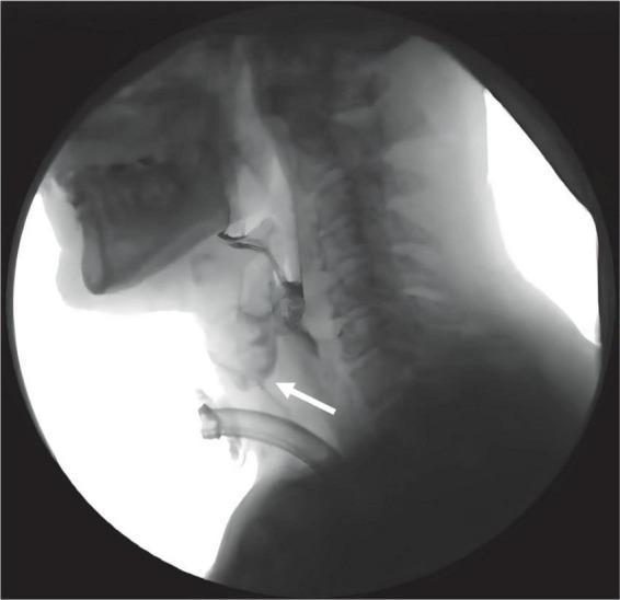
This diagnostic image is a lateral view from a Videofluoroscopic Swallowing Study (VFSS) captured using X-ray radiography. The anatomical focus includes the oral cavity, pharynx, larynx, and cervical spine. A radiopaque tracheostomy tube is visible in the lower neck region. A radiopaque contrast bolus (iohexol) is present in the oral cavity and pharyngeal space. A white arrow highlights a clear aspiration event, showing the contrast bolus deviating from the normal swallowing pathway and entering the laryngeal vestibule and upper airway above the level of the tracheostomy tube. This demonstrates a failure of the normal airway protection mechanisms during deglutition, resulting in aspiration. This clinical image is typical for assessing dysphagia and swallowing biomechanics in patients following acquired brain injury (ABI) to determine the Penetration-Aspiration Scale (PAS) score and the efficacy of interventions like the Passy Muir Valve (PMV).
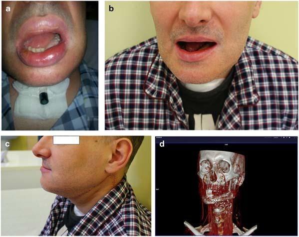
A multi-panel medical image series documenting mandibular reconstruction for advanced oral squamous cell carcinoma. Panel (a) shows a clinical photograph one week post-surgery, featuring significant perioral edema and a tracheostomy tube with a speaking valve secured by a neck dressing. Panels (b) and (c) provide frontal and lateral views of the patient two months post-operatively, demonstrating favorable aesthetic outcomes, restoration of facial symmetry, and healed surgical scars following partial mandibular resection and fibular free flap reconstruction. Panel (d) displays a 3D CT reconstruction of the head and neck, highlighting the surgical hardware including a contoured titanium reconstruction plate and screws stabilizing the osseous fibular graft to the remaining mandible. The reconstruction integrates 3D model printing technology to restore the mandibular contour and floor of the mouth. The series illustrates the clinical evolution from the immediate post-operative phase to functional recovery in complex maxillofacial reconstructive surgery.
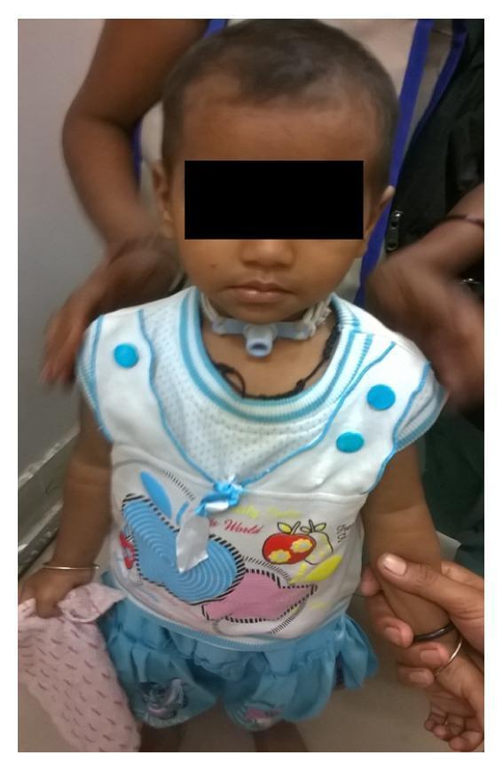
Clinical photograph of a 10-month-old female patient showing a long-term tracheostomy in situ. The infant is depicted in a front-facing view, with a standard pediatric tracheostomy tube secured around the neck using a soft white tie or strap. The tube's flange is visible against the skin, and a blue three-way valve or heat and moisture exchanger (HME) is attached to the tracheal opening. The image illustrates post-acute management of airway obstruction, following complications from lithium battery ingestion and subsequent bilateral vocal cord palsy. The anatomical focus is the anterior neck region where the surgical airway is maintained. The photograph serves to demonstrate clinical follow-up and the use of medical devices for chronic airway management in pediatric patients with vocal cord dysfunction.
Speaking Valve - Tolerating (Clinical Guide)
What Is a Speaking Valve?
- Phonation (speech)
- Improved swallowing by restoring subglottic pressure
- Reduced aspiration risk
- Better secretion management
- Olfaction (sense of smell) - often lost with a trach
Prerequisites Before Trialing a Speaking Valve
| Prerequisite | Detail |
|---|---|
| Cuff deflation | The trach cuff MUST be fully deflated - air cannot escape upward if the cuff is inflated. A speaking valve should NOT be used with an inflated cuff. |
| Patent upper airway | Sufficient airflow must be able to pass through the glottis and out the nose/mouth on exhalation |
| Adequate alertness | Patient must be awake, able to follow commands, and cooperative |
| Secretion management | Secretions must not be so excessive that the valve cannot be tolerated |
| SLP involvement | Early involvement of a Speech-Language Pathologist (SLP) to assess candidacy |
Signs of Tolerating the Speaking Valve
- Maintains stable SpO2 (no significant desaturation)
- Stable respiratory rate and heart rate - no distress
- Can phonate - produces voice on exhalation
- No signs of increased work of breathing (use of accessory muscles, tracheal tug)
- Comfortable - no agitation, panic, or requests to remove
- Manages secretions adequately
- Maintains tolerance for an increasing duration (minutes, then hours)
Signs of Not Tolerating (Contraindications to Continued Use)
- SpO2 drops / hypoxia
- Respiratory distress, tachypnea, increased accessory muscle use
- Significant anxiety or distress
- Audible upper airway obstruction (suggesting stenosis or poor upper airway patency)
- Patient unable to exhale through upper airway (e.g., severe laryngeal/subglottic stenosis)
- Excessive secretions blocking airflow
- Poor or absent voice despite valve in place
Tolerance Progression Protocol
- First trial - supervised by SLP and/or respiratory therapist (RT)
- Short duration (5-15 minutes) initially
- Gradual increase as tolerated (30 min, then hours)
- Never worn during sleep
- Never used with the cuff inflated
- If on a ventilator - ventilator settings may need adjustment (the valve changes exhalation mechanics)
Special Considerations
- Small infants may not tolerate the valve and will often blow it off by coughing
- Many children learn compensatory maneuvers (e.g., flexing the neck to occlude the tube) to aid phonation
- Must be used under supervision and never while sleeping
- Best guided by speech therapy input (Scott-Brown's Vol 2)
- In-line speaking valve placement is possible but requires careful coordination with RT
- Ventilator settings (especially PEEP and pressure support) may need modification
- If patient cannot tolerate cuff deflation, discuss with the medical team about tube changes (e.g., Portex Blue Line Ultra Suctionaid tube)
- Large suprastomal granulations can make speaking valve use impossible - must be addressed first
- Patients with laryngeal stenosis may not have adequate upper airway for valve use; ability to tolerate a speaking valve is a key assessment point in the workup (Scott-Brown's Vol 2 - History section)
Valve Types
| Valve | Mechanism |
|---|---|
| Passy-Muir Valve (PMV) | Closed-position default - opens only on inspiration; encourages use of normal airway even in quiet breathing |
| Rusch speaking valve | Open at rest - closes only during significant expiration |
Role of MDT
- SLP - assesses voice, swallowing, secretion management, and communication
- Respiratory Therapist (RT) - manages ventilator interface and adjustments
- Nursing - daily trial support, education of family
- Physician - orders trials, addresses underlying barriers to tolerance