
Rickettsial infection diagnosis and treatment
"Rickettsia" AND treatment
Rickettsial infection diagnosis treatment doxycycline guidelines
Rocky Mountain spotted fever petechial rash
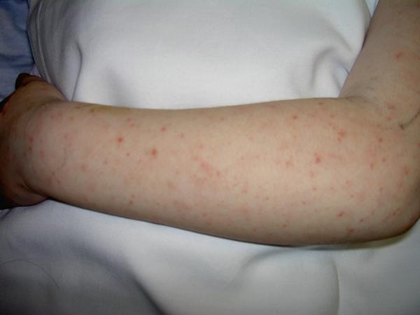
This clinical photograph displays the upper extremity of a patient, focusing on the ventral surface of the forearm and the dorsal aspect of the hand. The skin demonstrates an extensive, widespread eruption consisting of small, discrete, erythematous lesions. These lesions are primarily characterized as blanching macules and non-blanching petechiae, approximately 1-3 mm in diameter. The distribution is relatively uniform across the limb, with some areas showing higher density or mild clustering. This type of petechial and macular rash is a significant clinical finding in various systemic conditions, including infectious vasculitides such as Rocky Mountain Spotted Fever (RMSF), meningococcemia, or other febrile illnesses associated with thrombocytopenia. The image serves as a teaching tool for recognizing non-confluent exanthems and differentiating between inflammatory macular rashes and hemorrhagic petechial lesions in an acute clinical setting.
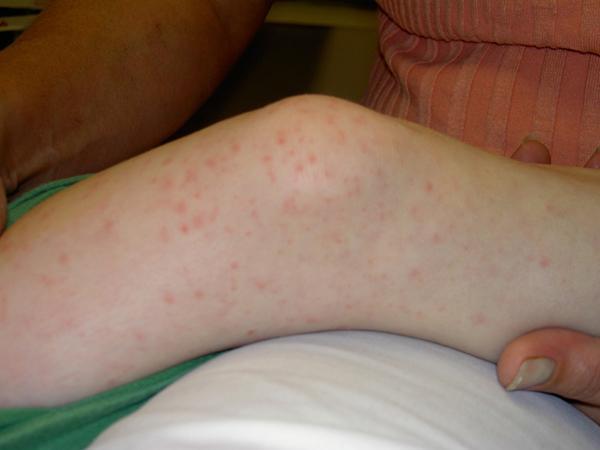
This clinical photograph displays the lower extremity of a pediatric patient, specifically focusing on the knee and proximal lower leg. The skin exhibits a generalized, non-confluent exanthem. The rash is composed of multiple small, discrete, erythematous pinkish-red macules and occasional fine papules. The distribution is widespread and random across the visible cutaneous surface, with no distinct linear or dermatomal clustering. The individual lesions are small, round, and flat, consistent with a macular morphology. The surrounding skin appears pale and non-icteric. This visual finding is characteristic of various systemic infectious or inflammatory conditions, such as viral exanthems, early Kawasaki disease, or rickettsial infections like Rocky Mountain Spotted Fever, where blanching macular and petechial rashes often involve the extremities.
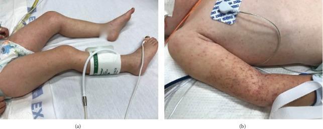
Two clinical photographs demonstrate a diffuse, nonblanching rash in a pediatric patient. Figure (a) shows the lower extremities with scattered, reddish-purple maculopapular lesions and petechiae extending to the feet. A blood pressure cuff is visible on the right lower leg. Figure (b) provides a detailed view of the upper extremity and trunk, highlighting a high density of petechiae and purpura, particularly concentrated in the area previously compressed by a blood pressure cuff (positive Rumpel-Leede phenomenon). The lesions are small, erythematous to violaceous, and distributed across the trunk and arm. The clinical presentation is highly suggestive of systemic vasculitis or severe infection, such as Rocky Mountain Spotted Fever or meningococcemia, characterized by microvascular injury and capillary fragility. The educational focus is on identifying petechial morphology and the provocation of lesions by localized pressure in critically ill patients.
Rickettsial Infection: Diagnosis and Treatment
Main clinical entities
| Group | Organism | Vector | Disease |
|---|---|---|---|
| Spotted fever group | R. rickettsii | Dermacentor/Rhipicephalus ticks | Rocky Mountain spotted fever (RMSF) |
| Spotted fever group | R. conorii | Tick | Mediterranean/boutonneuse fever |
| Typhus group | R. prowazekii | Body louse | Epidemic typhus |
| Typhus group | R. typhi | Flea | Murine (endemic) typhus |
| Scrub typhus | Orientia tsutsugamushi | Trombiculid mite (chigger) | Scrub typhus |
| Ehrlichiosis/Anaplasmosis | Ehrlichia/Anaplasma spp. | Tick | Human monocytotropic ehrlichiosis / granulocytotropic anaplasmosis |
| Q fever | Coxiella burnetii | Aerosol from livestock (not typically arthropod) | Acute/chronic Q fever |
Diagnosis
- History/exam: tick or mite exposure, travel/geography, fever + headache + myalgia, followed by a rash (maculopapular, petechial, or an eschar at the bite site in scrub typhus/spotted fevers). RMSF classically starts on wrists/ankles and spreads centrally; a third of patients have no rash early on.
- Serology (indirect immunofluorescence assay, IFA) is the mainstay for confirmation, but antibodies typically aren't detectable until 7-10 days into illness - a 4-fold rise in acute-to-convalescent titers is diagnostic, not useful acutely (Jawetz, Melnick & Adelberg's Medical Microbiology, Ch. 24).
- Weil-Felix test (Proteus cross-agglutination) is historical and no longer recommended - insensitive and nonspecific (Medical Microbiology 9e).
- PCR of blood, eschar swabs, or skin biopsy allows detection before a serologic response develops and has largely supplanted older methods; real-time/multiplex PCR can distinguish species (Fitzpatrick's Dermatology; Jawetz).
- Skin biopsy with immunohistochemistry on a petechial lesion can demonstrate rickettsiae directly and remains useful, especially early in RMSF; antirickettsial treatment for <24 hours doesn't significantly reduce biopsy sensitivity (Rosen's Emergency Medicine).
- Q fever is distinct: chronic infection (e.g., endocarditis) is diagnosed by a high phase I antibody titer (IFA ≥1:800) rather than acute serology (Henry's Clinical Diagnosis).
- Supportive labs: thrombocytopenia, hyponatremia, mildly elevated transaminases, and normal-to-low WBC are common but nonspecific clues in RMSF and typhus.
Treatment
- Adults: doxycycline 100 mg orally or IV twice daily.
- Children (any age, including <8 yr): doxycycline 2.2 mg/kg (up to 100 mg) orally or IV twice daily.
- Duration: at least 5-7 days, continued until 72 hours after fever resolves and clinical improvement is evident. Boutonneuse fever/African tick typhus may need only 3 days and is often self-limited.
- Scrub typhus alternative regimen (per DHR-ICMR guidelines): azithromycin 500 mg once daily for 5 days (10 mg/kg in children) is an accepted alternative, particularly in areas with possible doxycycline resistance.
- Severe/complicated disease (ARDS, renal failure, meningoencephalitis, multiorgan dysfunction): IV doxycycline 100 mg twice daily, or IV azithromycin, with supportive ICU-level care; total duration extended to 7-15 days.
- Chloramphenicol is a second-line alternative, reserved for situations where tetracyclines are contraindicated (e.g., true doxycycline hypersensitivity), because of the risk of fatal bone marrow toxicity (Goodman & Gilman's Pharmacology, Ch. 56).
- Q fever: acute disease responds to doxycycline; chronic Q fever/endocarditis requires prolonged combination therapy, classically doxycycline plus hydroxychloroquine for 18 months or longer.
- Response is typically rapid - defervescence within 24-48 hours of starting doxycycline in RMSF and boutonneuse fever. Failure to improve after appropriate doxycycline therapy should prompt reconsideration of the diagnosis.