Degenoniru injury

"Degenoniru injury" isn't a term I recognize - it looks like it may be a typo or autocorrect mangling of another term. Could you clarify which of these you mean, or type it again?
Asking for Preferences

Which injury/condition did you mean by 'Degenoniru injury'?

"Degenoniru injury" isn't a term I recognize - it looks like it may be a typo or autocorrect mangling of another term. Could you clarify which of these you mean, or type it again?

Degeoning injury

Reading File
Finding Sources
Finding Sources
Searching the Web

degloving injury management classification

Searching PubMed

"degloving injury"[Title] AND management

Searching Images

degloving injury extremity clinical photo

Reading File
Reading File
Degloving injury (I've read this as "degloving" - let me know if you meant something else).

What it is

Degloving is the traumatic avulsion/separation of skin and subcutaneous fat from the underlying deep fascia, muscle, or bone - the tissue is sheared off much like pulling off a glove. It disrupts the perforating vessels and lymphatics that supply the skin, which is why the overlying skin often becomes non-viable even when it looks intact at first glance - Bailey and Love's Short Practice of Surgery, p. 54; Rockwood and Green's Fractures in Adults, p. 638.

Open vs closed

  • Open degloving: skin is torn away and underlying structures are visible (e.g. a finger avulsion injury with skin loss).
  • Closed degloving: skin remains intact over the top but is sheared from the fascia beneath, usually from a shearing/torsion force (classic mechanism: motor vehicle run-over or wheel entrapment). This is easy to underestimate because the skin surface can look deceptively normal.
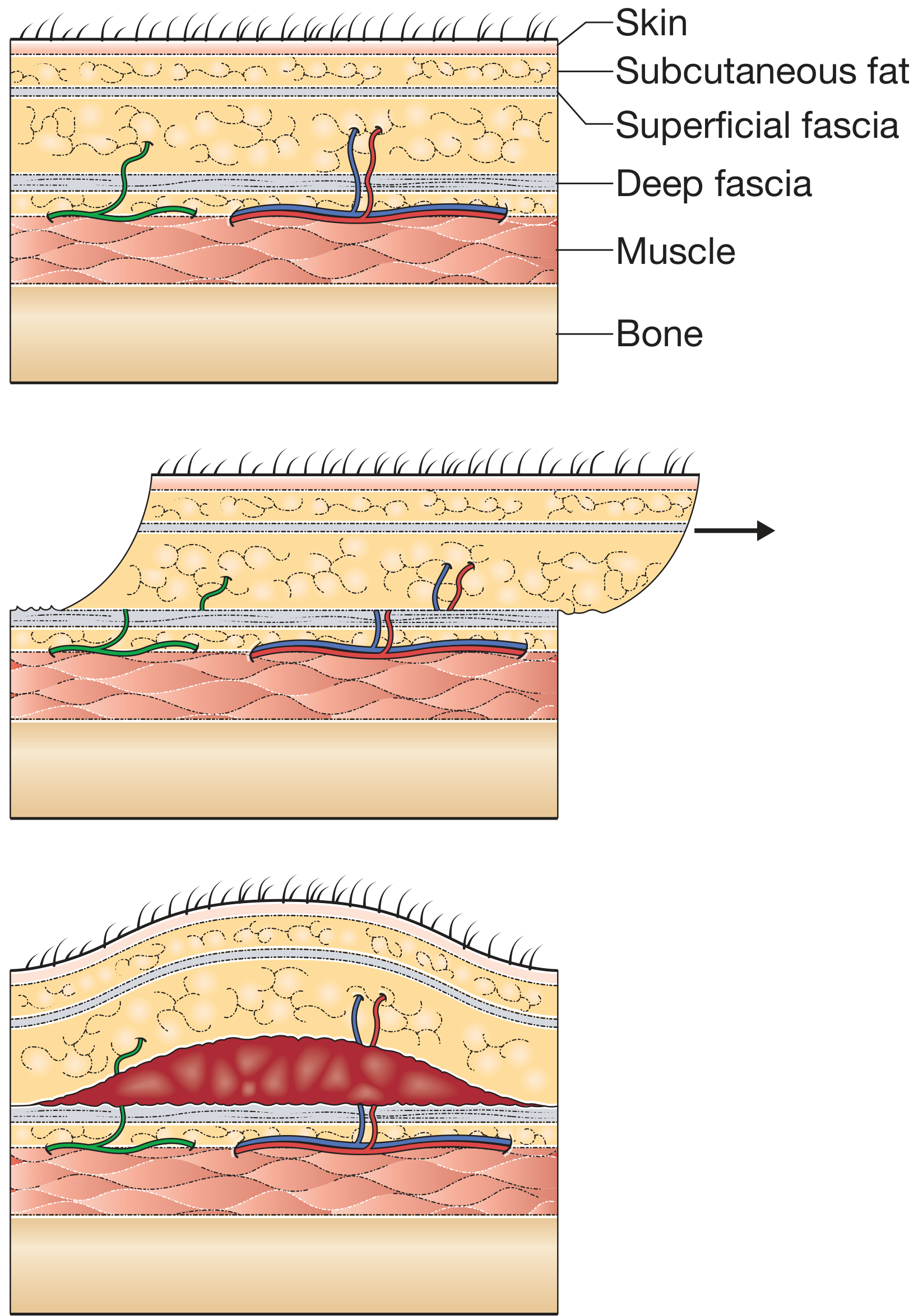
  • When a closed shearing injury creates a fluid-filled potential space (blood + lymph) between fascia and subcutaneous fat, it's called a Morel-Lavallée lesion - classically over the greater trochanter/hip and thigh, but now described at other sites too.

Classification (limb trauma)

  1. Limited degloving with abrasion or avulsion
  2. Non-circumferential degloving
  3. Circumferential, single-plane degloving
  4. Circumferential, multiplanar degloving
Single-plane degloving occurs between subcutaneous fat and deep fascia; multiplanar injury extends within muscle groups and between bone and muscle - the more extensive and multiplanar the injury, the higher the likelihood the skin is non-viable - Rockwood and Green's Fractures in Adults, p. 638.

Assessing tissue viability

Signs of non-viable skin:
  • Fixed staining (discoloration that does not blanch with digital pressure)
  • Thrombosis of subcutaneous veins
  • Failure of punctate dermal bleeding on incision
Because viability is often hard to judge on initial inspection, some centers use a "second look" at 24-48 hours, though most trauma literature now favors early definitive management (ideally within 72 hours) over serial staged debridement when the extent of injury is clear. Intravenous fluorescein or indocyanine green fluorescence angiography can help delineate dead tissue but require specialist equipment.

Management principles

  • Resuscitate and treat as a high-energy polytrauma patient until proven otherwise (associated fractures, vascular injury are common).
  • Serially excise clearly non-viable skin until punctate dermal bleeding confirms viable tissue at the margins.
  • Circumferentially degloved skin is almost always non-viable and typically requires excision.
  • Reconstructive options once debrided: meshed split-thickness skin graft (if bone/fracture not exposed) or flap coverage (local/free myocutaneous flap) if bone or fracture site is exposed. Negative-pressure wound therapy (NPWT) is often used short-term to help graft take, especially over irregular wound beds.
  • Special/high-risk sites: degloving over bony prominences (e.g. malleoli) or the heel/calcaneus is particularly problematic because there's little tissue reserve and specialized skin (heel pad) cannot be well replaced.
  • In upper-limb degloving specifically, options include salvage of the degloved segment via microsurgical revascularization (direct arterial anastomosis or AV shunting) versus reconstruction of the unsalvageable segment.
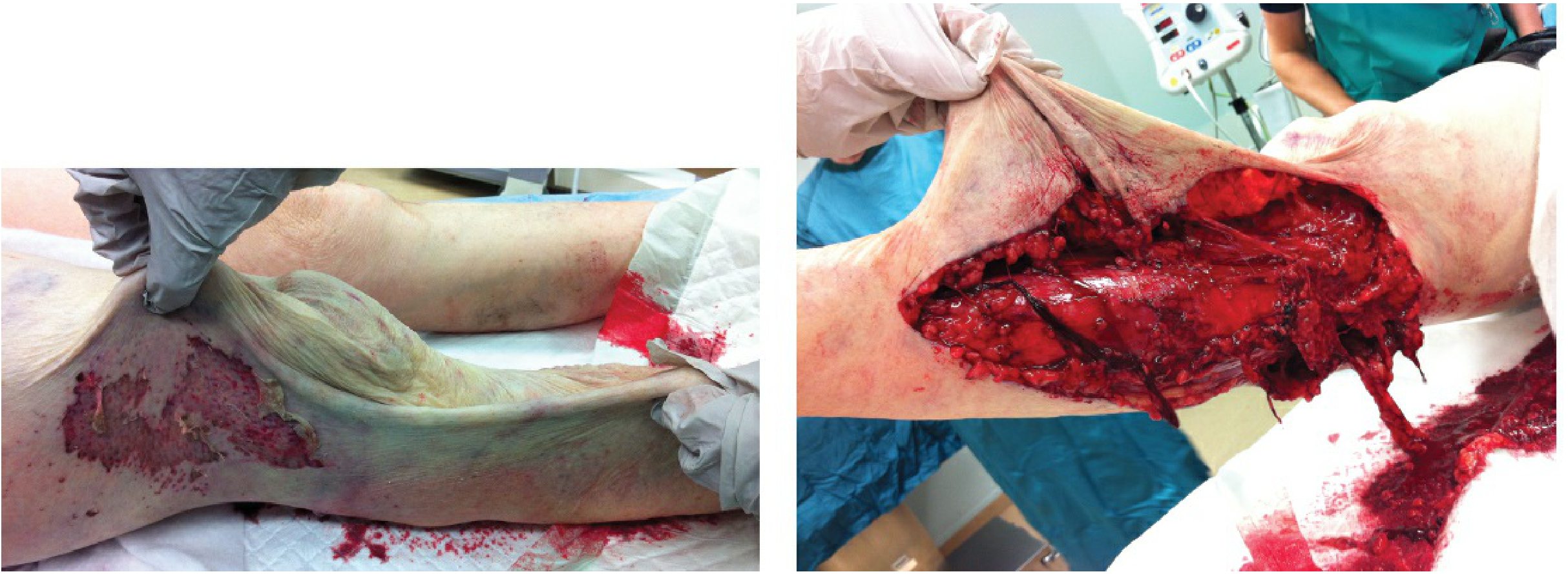
Below is an image showing how closed degloving allows the soft tissue to be lifted away from the underlying fascia:
Closed degloving injury of the leg showing soft tissue lifted from fascia
And the mechanism of a Morel-Lavallée lesion (shearing of perforating vessels creating a hemolymphatic collection):
Diagram of Morel-Lavallée lesion mechanism showing shearing of perforating vessels between fascia and subcutaneous tissue

Evidence check

A targeted PubMed search for recent systematic reviews/meta-analyses on degloving injury management did not return directly relevant results (returned unrelated wound-care topics), so there's no recent high-level evidence I found that contradicts the textbook approach above. Broader literature (e.g. Latifi et al., J Emerg Trauma Shock 2014; Yan et al., J Trauma Acute Care Surg 2013) supports the same classification and staged-debridement/reconstruction approach summarized here.
If you actually meant a different term (De Quervain's tenosynovitis, a knee/"genou" injury, or something else), let me know and I'll pull the specific details.

Definition

Degloving is the traumatic avulsion (separation) of skin and subcutaneous fat from the underlying deep fascia, muscle, or bone. The shearing force strips the skin envelope away from the deeper tissue layer much like pulling a glove off a hand - hence the name.
Key points:
  • It disrupts the perforating blood vessels and lymphatics that run between fascia and skin, so the skin often becomes non-viable even if it looks intact on the surface.
  • It can be open (skin torn away, underlying structures visible - e.g. a finger avulsion with skin loss) or closed (skin surface stays intact but is sheared off the fascia beneath, typically from a shearing/torsion mechanism such as a motor vehicle run-over or wheel entrapment).
  • When a closed shear injury creates a blood/lymph-filled space between the fascia and subcutaneous fat, that specific entity is called a Morel-Lavallée lesion.
Source: Bailey and Love's Short Practice of Surgery, p. 54; Rockwood and Green's Fractures in Adults, p. 638.

What are the main classification types of degloving injuries?

There are two classification systems commonly used for degloving injuries, based on plane and extent of tissue separation:

1. Limb trauma degloving classification (4 grades)

  1. Limited degloving with abrasion or avulsion
  2. Non-circumferential degloving
  3. Circumferential, single-plane degloving
  4. Circumferential, multiplanar degloving
  • Single-plane degloving occurs between the subcutaneous fat and the deep fascia.
  • Multiplanar degloving extends further, occurring within muscle groups and between bone and muscle.
  • Skin non-viability risk increases progressively from grade 1 to grade 4 - circumferential and multiplanar injuries carry the highest risk of non-viable skin because they disrupt more of the perforating vascular supply.
(Source: Bailey and Love's Short Practice of Surgery, p. 54; Rockwood and Green's Fractures in Adults, p. 638 - based on the Arnez classification of soft-tissue degloving in limb trauma.)

2. Open vs. closed (overarching category)

  • Open degloving: skin is torn away and the underlying structures are visible (e.g. a finger avulsion injury with skin loss).
  • Closed degloving: the skin surface remains intact, but is sheared away from the underlying fascia - typically from torsion, avulsion, or crush forces (classic mechanism: motor vehicle run-over, wheel entrapment). This type is more dangerous because the extent of non-viable tissue is easy to underestimate on visual inspection.
A special closed variant is the Morel-Lavallée lesion: a shearing injury that creates a hemolymphatic (blood + lymph) collection in the potential space between fascia and subcutaneous fat, classically over the greater trochanter/thigh but now recognized at other sites too.

Related site-specific classification (fracture context)

In closed fracture skin assessment (used alongside fracture grading, e.g. in Campbell's Operative Orthopaedics), degloving severity is graded from no lesion up through circumferential and extensive closed degloving, alongside contusion and necrosis - reflecting that degloving severity is also documented as part of overall soft-tissue injury grading in orthopedic trauma.

Abhijit Kamble 33 urss

That doesn't seem related to degloving injuries or anything else we've discussed - it looks like it might be a name, age, and an abbreviation ("urss"?), but I'm not sure what you're asking.
Could you clarify what you'd like to know? For example, is this a patient case you want help with, or did this get sent by mistake?

Abhijit Kamble 33 yrs male resident of beed had RTA on 18/7/26 in Pune due to collision of truck got injury in right leg so went to susun hospital was admitted there for 8 to 10 days and was treated and operated there reports not available but patient did not have have improvement and was having pain soo patient decided to goo to kem soo visited kem emergency there he was investigated and suggested to get admit and then suggested for amputation for which he was operated on 31/7/26 and was reported on 7/8/26 give present medical history

Present Medical History
Patient: Abhijit Kamble Age/Sex: 33 years / Male Residence: Beed
History of Present Illness:
The patient sustained an injury to the right lower limb (right leg) on 18/07/2026 at Pune, following a road traffic accident (RTA) involving collision with a truck.
Following the accident, the patient was taken to Sassoon Hospital, Pune, where he was admitted for approximately 8-10 days. During this admission he underwent surgical treatment/operative management of the right leg injury; however, the treatment records and operative reports from this admission are not available.
Despite the treatment received, the patient did not show clinical improvement and continued to experience persistent pain in the right leg. In view of the lack of improvement, the patient subsequently presented to the Emergency Department, KEM Hospital for further evaluation and management.
At KEM Hospital, the patient was investigated and, based on clinical and investigative findings, was advised hospital admission. Following assessment, the treating team advised amputation of the affected right lower limb, indicating that the limb was assessed as non-salvageable at that stage.
The patient underwent amputation surgery on 31/07/2026 at KEM Hospital.
The case was subsequently reported/reviewed on 07/08/2026.
Summary timeline:
DateEvent
18/07/2026RTA in Pune - collision with truck; right leg injury sustained
18/07/2026 (approx.)Admitted at Sassoon Hospital, Pune; treated/operated (records unavailable); stayed 8-10 days
Post-dischargeNo improvement; persistent right leg pain
~19-30/07/2026Presented to KEM Hospital Emergency; investigated; advised admission
31/07/2026Underwent amputation surgery (right lower limb) at KEM Hospital
07/08/2026Case reported
Note on gaps in the record: The specific diagnosis from Sassoon Hospital (e.g. type of fracture/soft-tissue injury, whether a degloving or vascular injury was present), the exact surgical procedure performed there, the level of amputation at KEM, and the indication documented for amputation (e.g. non-viable/gangrenous limb, uncontrolled infection, failed vascular repair) are not specified in the information provided. If this history is being prepared for a medico-legal certificate or insurance/disability report, these details should be obtained from the KEM Hospital case papers and the Sassoon Hospital discharge summary to make the record complete and defensible.
This is a shared conversation. Sign in to Orris to start your own chat.