
Hydrocele
hydrocele scrotal swelling diagram anatomy
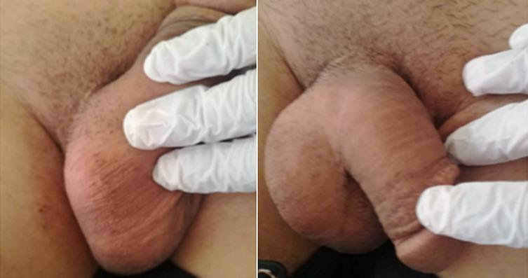
Clinical photograph showing a side-by-side comparison of the male external genitalia during a physical examination. The images depict significant scrotal enlargement and distension, characteristic of a bilateral hydrocele or varicocele. The scrotal skin appears erythematous and stretched, with visible wrinkling and rugosity. Gloved hands are shown palpating the structures to assess for fluid collection, masses, or spermatic cord abnormalities. This visual serves as a clinical example of scrotal swelling in the context of multi-system disease, such as Langerhans Cell Histiocytosis (LCH), where inguinal and genital involvement may occur. The educational focus is on the visual inspection and manual examination of the scrotum to distinguish between normal anatomy and pathological swelling or fluid accumulation.
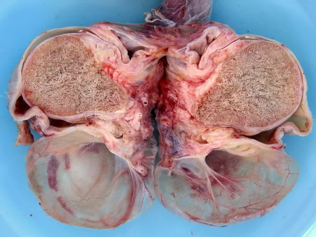
Macroscopic photograph of a scrotal hydrocele specimen measuring 5.0 cm in greatest dimension. The image shows an excised or opened scrotal sac consisting of two large, thin-walled hydrocele envelopes encasing the testicular tissue, with clear straw-colored serous fluid visible in the lumen. The walls appear translucent and delicate, lined by a single layer of mesothelial cells (cuboidal to flattened). The hydrocele forms when fluid accumulates between the visceral and parietal layers of the tunica vaginalis, often secondary to inflammation, trauma, hernia, or neoplastic processes, and congenital hydrocele results from persistent communication with the peritoneal cavity via a patent processus vaginalis. The surrounding tunica albuginea and testicular parenchyma appear normal, though edema and mild vascular congestion may be noted due to surgical manipulation. This specimen demonstrates the classic pathophysiology: serous fluid accumulation with a simple mesothelial lining and no solid tumor or pus, distinguishing it from inflammatory or infectious orchitis or malignant testicular lesions. Clinically, hydroceles may present as progressive scrotal swelling; etiologies include irritation of the mesothelial lining by hernia, epididymo-orchitis, trauma, or paratesticular tumors. Management ranges from watchful waiting in small, asymptomatic cysts to hydrocelectomy or aspiration in larger or symptomatic cases. This educational image aids recognition of hydrocele morphology and supports differential diagnosis in scrotal swellings.
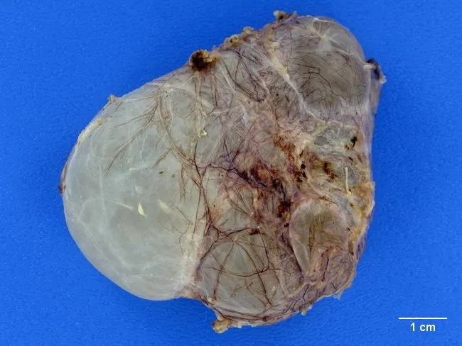
This gross pathology photograph depicts a scrotal adnexal cystic structure consistent with a spermatocele arising from dilatation of efferent ductules near the rete testis or the head of the epididymis. The specimen shows a thin-walled, translucent (glass-like) cyst attached to posterior testicular/epididymal tissue, with variable lobulation indicating possible multilocular configuration. A single or multiple cysts may be present, filled with clear, straw-colored fluid. The fluid composition may include motile or non-motile spermatozoa, a hallmark distinguishing spermatocele from hydrocele, which contains serous fluid without sperm. The surrounding testicular parenchyma appears unremarkable; vascular and stromal tissues may show marginal adhesions. The scale bar indicates 1 cm for size estimation; overall dimensions correspond to a small to moderate cystic lesion, typically a few millimeters to a few centimeters in diameter. Pathophysiology involves dilation of efferent ductules or proximal epididymal ducts with preserved testicular architecture, absence of malignant features, and minimal inflammatory infiltrate. Clinically, spermatocele presents as a painless scrotal swelling, often anterior or superior to the testis, distinct from hydrocele by fluid content. This image supports education in gross anatomy of the male reproductive tract, differential diagnosis of scrotal cystic lesions, and correlates with radiologic and clinical findings used to guide conservative versus surgical management, including epididymal-sparing excision when indicated.
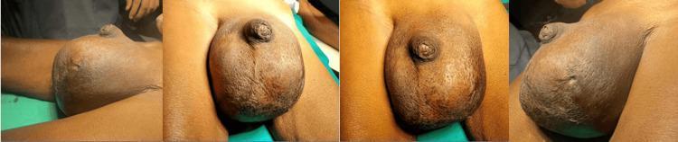
A series of four clinical photographs showing preoperative views of a massive, unilateral swelling of the left scrotum in an adult male. The swelling is approximately 25 x 15 cm, causing significant distortion and displacement of the penile shaft. The skin over the affected area is tense and shiny with a notable loss of normal scrotal rugosities (wrinkles), indicating severe distension. A visible cutaneous scar is present on the inferior aspect of the left hemiscrotum, consistent with a history of previous aspiration. The swelling is well-circumscribed, and the overlying skin appears hyperpigmented compared to the surrounding inguinal and thigh regions. This image illustrates the clinical presentation of chronic massive scrotal swelling, requiring differential diagnosis between giant hydrocele, pyocele, or a testicular neoplasm. The educational focus is on the physical examination findings of scrotal pathology, including changes in skin texture and topographical anatomy.
Hydrocele
Definition
Embryology & Pathophysiology
Classification
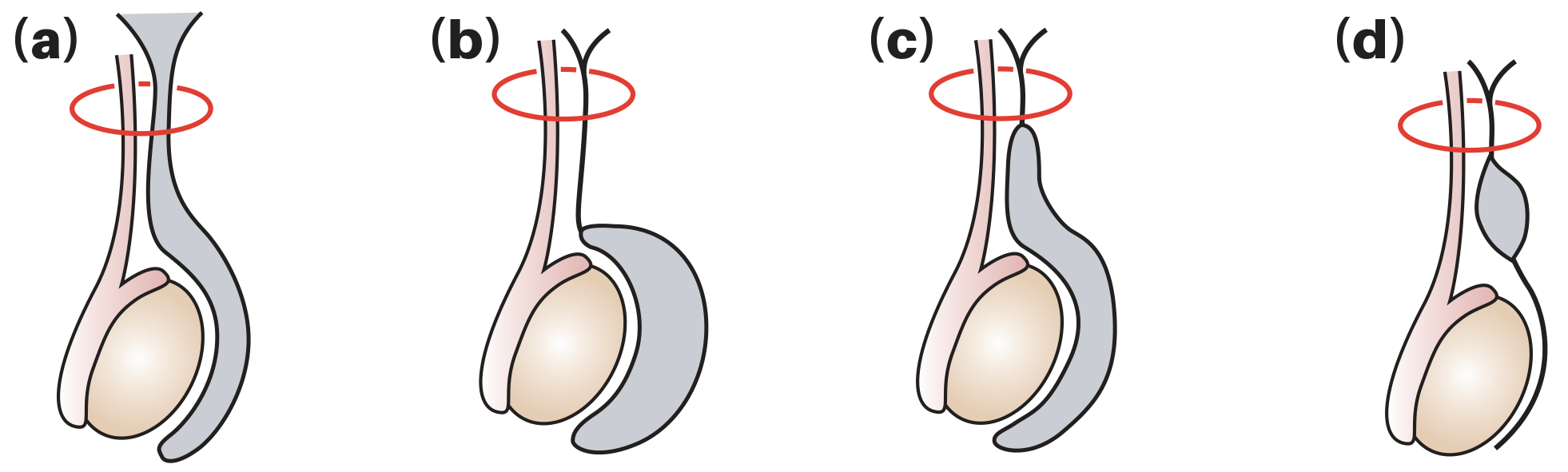
| Type | Mechanism |
|---|---|
| 1. Communicating (congenital) | Patent processus vaginalis allows free flow of peritoneal fluid into the tunica vaginalis; associated with indirect inguinal hernias |
| 2. Non-communicating (vaginal / primary) | Processus closed; fluid accumulates due to excessive production or defective lymphatic absorption by the tunica vaginalis; most common type globally |
| 3. Infantile | Distal processus closes correctly, mid-portion remains patent with open proximal end communicating with the tunica |
| 4. Hydrocele of the cord | Mid-portion of processus remains patent but both ends are closed, forming an isolated cystic swelling along the spermatic cord |
- Local injury (including testicular torsion)
- Infection (epididymo-orchitis)
- Neoplasm (testicular tumour)
- Radiotherapy
Important: If a tumour is suspected, the hydrocele must NOT be punctured due to risk of malignant needle-track implantation.
Clinical Features
- Can you get above the swelling? - If not, consider an inguinoscrotal hernia. A hydrocele allows palpation of a normal spermatic cord above it.
- Are the testis and epididymis palpable? - A hydrocele encloses both structures, making them impalpable.
- Does the swelling transilluminate? - Hydroceles are typically translucent and transilluminate brightly.
Clinical photograph and ultrasound

Investigations
- Ultrasound scrotum - First-line in all cases; excludes underlying testicular pathology
- Urine NAAT or culture - If infection is suspected
- Tumour markers (AFP, β-hCG, LDH) - If malignancy is suspected
Treatment
Conservative
Surgical (main treatment options)
| Operation | Indication / Notes |
|---|---|
| Herniotomy (ligation of patent processus vaginalis) | Congenital hydroceles that do not resolve spontaneously |
| Lord's operation (plication) | Suitable when the sac is thin-walled; minimal dissection, reduced risk of haematoma; a series of interrupted absorbable sutures plicates the redundant tunica |
| Jaboulay's procedure (eversion) | The hydrocele sac is everted around the testis and anchored with sutures; risk of scrotal haematoma from bleeding at the cut edge |
| Excision of the sac | For thick-walled, acquired hydroceles (common in secondary hydroceles) |
Aspiration (less preferred)
- Fluid invariably reaccumulates within a week or so
- Reserved for men unfit for surgery
- Risk: bleeding into the sac causing haematocele
- Sclerosant injection (e.g., tetracycline, polidocanol) can be effective but is painful
Do NOT aspirate if testicular malignancy is suspected.
Filarial Hydrocele and Chylocele
Differential Diagnosis of Scrotal Swelling
| Feature | Hydrocele | Inguinal Hernia | Epididymal Cyst | Varicocele |
|---|---|---|---|---|
| Get above swelling | Yes | No | Yes | Yes |
| Transillumination | Bright | No | Yes (clear fluid) | No |
| Testis palpable separately | No | Yes | Yes | Yes |
| Reducible | No (communicating may partially empty) | Yes | No | On lying down |
| Ultrasound | Anechoic fluid around testis | Bowel/omentum in sac | Separate from testis | Dilated veins >3mm |
Summary (Key Points)
- A hydrocele is a collection of fluid within the tunica vaginalis
- Hydroceles surround the testis and transilluminate brightly
- Ultrasound is essential, especially when the testis is impalpable
- Conservative management is appropriate for small, asymptomatic cases
- Surgery (Lord's or Jaboulay's) is the mainstay for large, symptomatic hydroceles
- Testicular malignancy must be excluded by ultrasound before any intervention
Testicular torsion
testicular torsion bell clapper deformity anatomy diagram
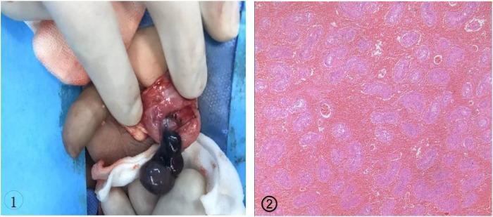
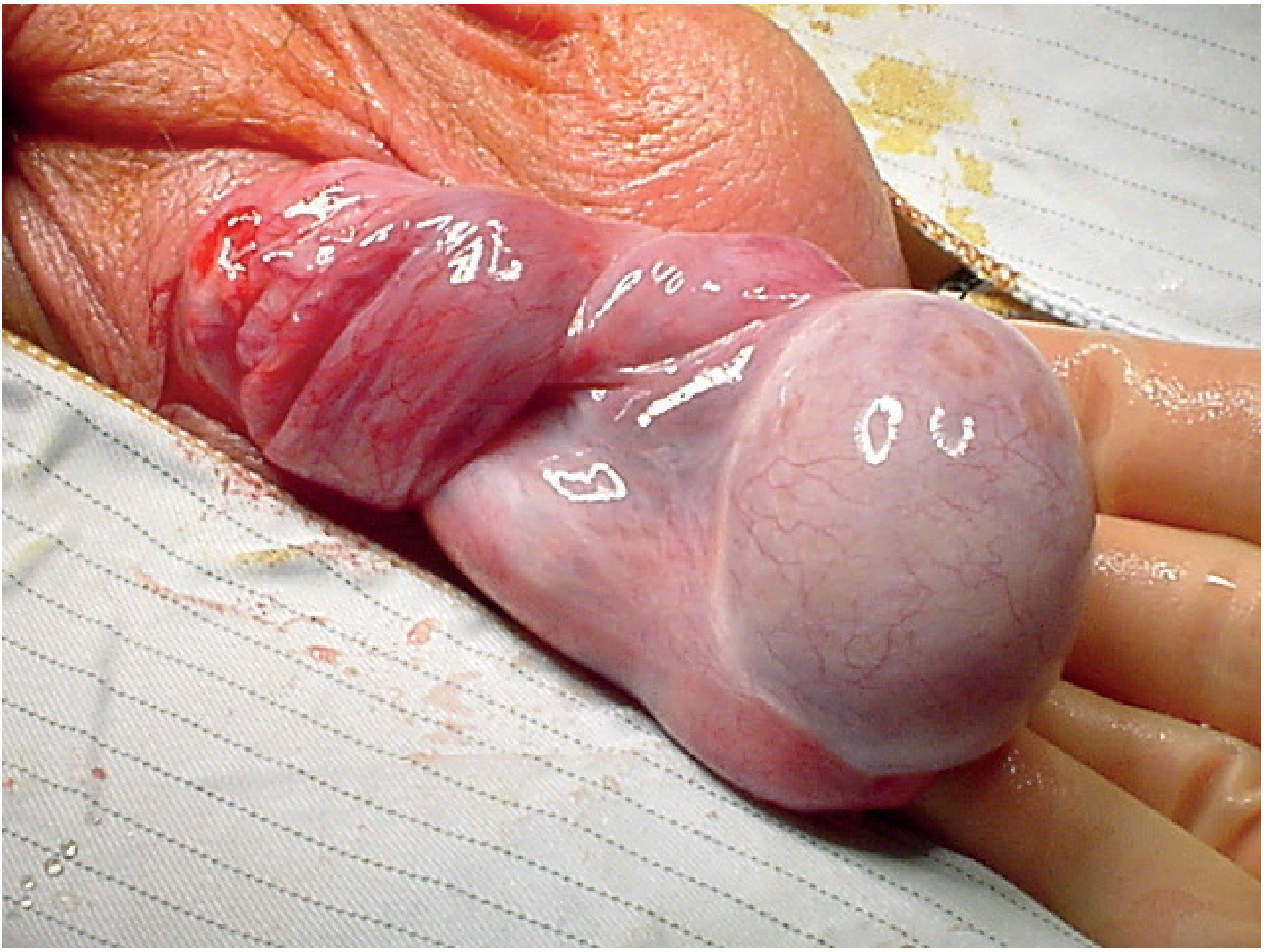
This dual-panel image demonstrates the surgical and pathological findings of testicular torsion associated with a bell clapper deformity. Panel 1 is an intraoperative clinical photograph of an inguinal/scrotal exploration. It shows a dark, cyanotic, and necrotic-appearing testis and epididymis held by gloved fingers. The tunica vaginalis is seen completely encircling the testis and distal spermatic cord, which characterizes the bell clapper deformity and allows the testis to rotate freely within the sac, leading to intravaginal torsion. Panel 2 is a histopathological micrograph (H&E stain) of the testicular tissue. The image reveals extensive coagulative necrosis of the seminiferous tubules and significant interstitial hemorrhage, indicated by the diffuse pink staining and loss of cellular and nuclear detail. This content illustrates a critical urologic emergency and is intended for medical professionals and students to recognize the gross and microscopic manifestations of non-viable testicular tissue following severe torsion.
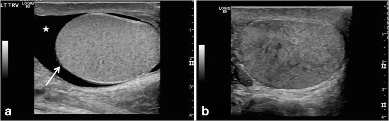
Two-panel gray-scale scrotal ultrasound images demonstrating sonographic features of testicular torsion in adolescent patients. Image (a) is a transverse view of the left scrotum showing a globular, enlarged testis with an abnormal horizontal lie (indicated by a white arrow). A surrounding anechoic area represents a reactive hydrocele (marked with a star). This orientation is characteristic of the 'bell clapper' deformity predisposing to torsion. Image (b) is a longitudinal view of a different patient's right testis showing a markedly heterogeneous parenchymal echotexture with mixed hyperechoic and hypoechoic areas. Such significant heterogeneity is a critical diagnostic sign of late-stage torsion, suggesting tissue congestion and potential nonviability. These diagnostic images illustrate key morphological and parenchymal changes used in pediatric radiology to differentiate testicular torsion from other causes of acute scrotum, such as epididymitis.
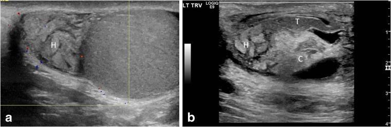
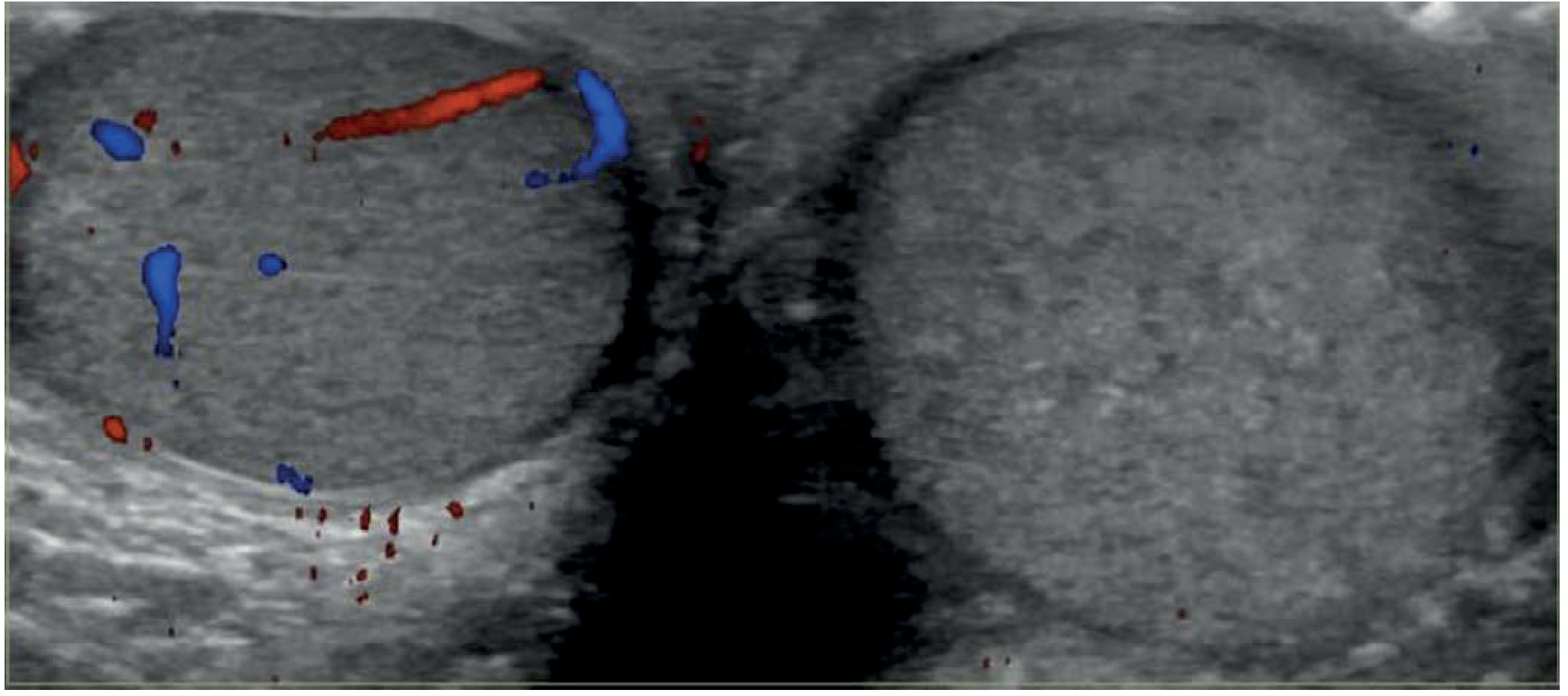
This dual-panel diagnostic image presents longitudinal ultrasound (US) scans of the scrotum, illustrating sonographic markers of testicular torsion. Figure A is a color Doppler US showing an enlarged epididymal head (labeled H) with a swollen, lobular appearance. Notably, there is a complete absence of intrinsic color flow within the epididymal head (avascularity) and decreased intratesticular flow, findings that help differentiate torsion from hyperemic epididymitis. Figure B is a gray-scale US image detailing the epididymal-cord complex. It demonstrates an edematous epididymal head (H) and a tangled, echogenic spermatic cord (C), characteristic of a 'torsion knot' or 'boggy pseudomass.' The epididymal tail (T) remains relatively uninvolved. These images highlight the importance of evaluating the extratesticular structures and the epididymal-cord complex in pediatric patients presenting with acute scrotal pain to diagnose complete or partial testicular torsion, often associated with the 'bell clapper' deformity.
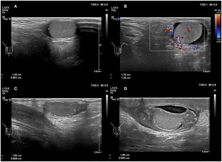
This diagnostic imaging panel consists of four scrotal ultrasound images (A-D) demonstrating comparison between normal and torsed pediatric testicles. Images A and C serve as controls, showing a normal testicle with homogeneous parenchymal echotexture and a vertical axial orientation. Images B and D display clinical findings of testicular torsion. Image B is a Color Doppler ultrasound from a 6-year-old boy showing a horizontal change in the testicular axial direction with preserved internal blood flow, suggesting incomplete or early torsion. Images C and D focus on an 8-year-old boy; image D illustrates advanced torsion with a distinct change in the axial orientation of the testicle and testicular mediastinum, accompanied by heterogeneous parenchymal echogenicity and an anechoic peritesticular fissure (reactive hydrocele). These visual markers—specifically horizontal lie (bell-clapper deformity) and altered echotexture—are critical diagnostic indicators for testicular torsion in pediatric patients, even when blood flow is intermittently detected.
Testicular Torsion
Definition
Types
1. Intravaginal Torsion (most common)
- Peak incidence: peripubertal boys (12-18 years), but can occur at any age
- The anatomical defect is bilateral - the contralateral testis is at equal risk
2. Extravaginal Torsion
- ~70% occur prenatally (in utero) - salvage is nearly nonexistent
- ~30% occur postnatally (within the first 30 days of life) - salvage rate up to 44%
- Postnatal torsion presents with new-onset scrotal erythema and swelling with a tender testis; prenatal torsion presents with a firm, non-tender testis fixed to the scrotal wall
Risk Factors
- Bell-clapper deformity (underpins most cases)
- Undescended testis
- Testicular tumour
- Cold weather (cremasteric muscle contraction)
- History of intermittent scrotal pain (previous episodes of partial torsion)
Clinical Features
| Finding | Significance |
|---|---|
| High-riding testis | Spermatic cord shortens as it twists; testis drawn upward |
| Absent cremasteric reflex | Normally brisk in children; loss is a strong indicator of torsion |
| Horizontal lie of testis | Due to bell-clapper deformity |
| Tender, swollen scrotum | Oedema and erythema appear later |
| Prehn's sign negative | Elevation of testis does not relieve pain (distinguishes from epididymo-orchitis, though not fully reliable) |
Intermittent torsion may produce a history of recurrent, self-resolving episodes of scrotal pain.
Painless torsion is a recognised (though uncommon) entity.
Investigations
Doppler Ultrasound
- First-line imaging when clinically available
- Shows absent or decreased intratesticular blood flow on the affected side vs. normal flow on the contralateral side
- Grey-scale features: enlarged, heterogeneous testis; reactive hydrocele; visible torted cord ("whirlpool sign")
- Whirlpool sign (twisting of spermatic cord on colour Doppler) has sensitivity ~92%, specificity ~99%
- A swollen, hypoechoic testis on US is usually not salvageable; a normal-appearing testis usually is
- Normal blood flow does NOT completely exclude torsion - clinical suspicion must drive the decision
Key principle: If ultrasound is not promptly available, clinical diagnosis is sufficient to proceed directly to surgical exploration. Do not delay surgery to obtain imaging.
Testicular Salvage Rates - Time is Testis
| Time from onset to surgery | Salvage rate |
|---|---|
| < 6 hours | ~80-90% |
| 6-12 hours | ~50% |
| 12-24 hours | ~20% |
| > 24 hours | Very low (<10%) |
Differential Diagnosis of Acute Scrotum
| Condition | Key distinguishing features |
|---|---|
| Testicular torsion | Sudden onset, vomiting, absent cremasteric reflex, high-riding testis, afebrile |
| Epididymo-orchitis | Gradual onset, fever, dysuria, positive cremasteric reflex, Prehn's sign positive |
| Torsion of appendix testis | Less severe pain, cremasteric reflex present, blue-dot sign, normal Doppler flow |
| Incarcerated hernia | Unable to get above swelling, bowel sounds in scrotum |
| Idiopathic scrotal oedema | Painless diffuse oedema, spreads to perineum, normal testis |
Torsion of the Appendix Testis (Appendage Torsion)
- Most common cause of acute scrotal pain in children aged 7-14 years
- Mild to severe pain, usually without vomiting
- Blue-dot sign - a dark infarcted appendage visible through the skin just above the upper pole of the testis
- Doppler US can help exclude testicular torsion
- Treatment: analgesia and rest in most cases; exploration reserved for severe or equivocal cases
Management
Emergency: Surgical Exploration
- Midline (median raphe) or bilateral transverse scrotal incisions
- Open the tunica vaginalis and identify the torted cord
- Detorse the testis - note the direction of torsion
- Assess viability - wrap in warm saline-soaked gauze for 5-10 minutes; observe for return of colour. Confirm with intraoperative Doppler or incise the tunica vaginalis to observe tissue bleeding
- If viable: three-point fixation to the dartos fascia with non-absorbable sutures (medial, lateral, and inferior/dependent pole)
- If non-viable (clearly gangrenous): orchidectomy to prevent later abscess formation. Only clearly necrotic testes should be excised - some severely compromised testes survive, and even those that atrophy are not harmful
- Contralateral orchidopexy ALWAYS: The bell-clapper deformity is bilateral. The contralateral testis must be fixed at the same operation to prevent asynchronous torsion
Manual Detorsion (temporary measure)
- Can be attempted while awaiting theatre
- Torsion is usually medial (the testis rotates inward), so manual detorsion is performed by rotating the testis outward (like opening a book)
- Successful detorsion gives partial pain relief; does NOT replace surgical fixation
Ultrasound Images
Neonatal Torsion - Special Considerations
| Feature | Prenatal torsion | Postnatal torsion |
|---|---|---|
| Timing | In utero | First 30 days of life |
| Presentation | Non-tender, firm testis fixed to scrotal wall; discoloured scrotum | New scrotal erythema/swelling; tender testis |
| US | Hyperechoic, avascular testis | Avascular ± heterogeneous |
| Salvage rate | Nearly zero | Up to 44% |
| Management | Contralateral orchidopexy (urgent but not emergency) | Immediate surgical exploration |
Summary (Key Points)
- Testicular torsion is a surgical emergency - time to theatre is the single most important factor
- The bell-clapper deformity is the underlying anatomical cause in adolescents and is bilateral
- Classic features: sudden-onset pain, vomiting, high-riding testis, absent cremasteric reflex
- If the diagnosis is clinically suspected, proceed to theatre - do not delay for imaging
- Doppler US is useful but normal flow does not exclude torsion
- At surgery: detorse, assess viability, fix both testes with non-absorbable sutures
- Only clearly gangrenous testes are excised; viable and borderline testes are preserved
- The contralateral testis must always be fixed at the same operation
Varicocele
varicocele pampiniform plexus dilated veins scrotum diagram
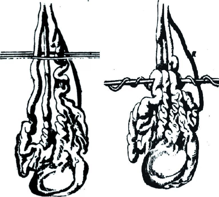
This historical medical illustration depicts 'Vidal's technique' (Vidal de Cassis), an early surgical procedure for the treatment of varicocele. The diagram shows the male reproductive anatomy, specifically the pampiniform plexus and scrotal sac. The visual is divided into two parts illustrating the progression of the technique. Elongated, tortuous varicose veins are shown suspended within the scrotum. A horizontal iron pin is passed behind the venous cluster, while silver wires are passed in front of them. The second part of the diagram demonstrates the mechanical objective of the procedure: the wire is progressively twisted around the pin to compress the dilated veins. This historical approach sought to cause the ulceration and subsequent obliteration of the varicoceles through pressure-induced necrosis and inflammatory adhesion. The illustration serves as a comparative reference for the evolution of urological surgery, highlighting the shift from traumatic mechanical compression to modern microsurgical varicocelectomy and ligation techniques.
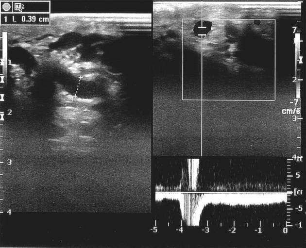
This diagnostic ultrasound image demonstrates a Color Doppler Sonography (CDS) study of the scrotum in a patient with clinical varicocele. The left panel shows a B-mode grayscale image of the pampiniform plexus, revealing multiple dilated, anechoic tubular structures consistent with enlarged testicular veins. Calipers measure a vein diameter of 0.39 cm (3.9 mm), which exceeds the typical upper threshold for normal testicular vein size. The right panels display Duplex mode imaging combining real-time B-mode with spectral Doppler analysis. The spectral waveform at the bottom illustrates the hemodynamic response to a Valsalva maneuver. A significant negative deflection below the baseline is visible, representing retrograde flow (venous reflux) with a duration exceeding 2 seconds. The velocity scale ranges from approximately -7 to 7 cm/s. This combination of venous dilation and Valsalva-induced reflux flow is characteristic of grade III varicocele and is used preoperatively to assess venous insufficiency and the severity of the condition within the field of urology and radiology.
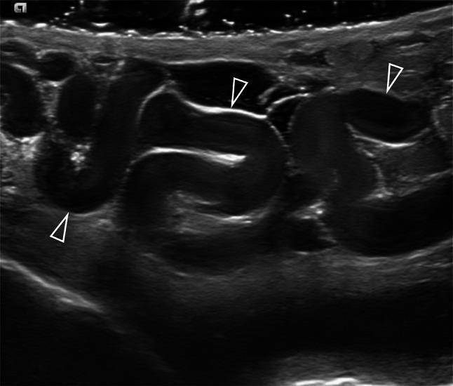
This diagnostic grayscale ultrasound image displays the classic sonographic findings of a varicocele within the pampiniform plexus of the scrotum. The image reveals multiple, markedly dilated and serpiginous (tortuous) venous structures, indicated by white arrowheads. These vessels are hypoechoic compared to the surrounding soft tissue and measure larger than the typical 3 mm diagnostic threshold. Within the lumen of the dilated veins, low-level internal echoes are visible, which may represent slow-moving blood flow (spontaneous contrast) or altered blood dynamics characteristic of venous insufficiency and reflux. The image demonstrates the structural abnormalities of the spermatic cord veins, providing a key educational example of urological diagnostic imaging for male infertility and scrotal pathology. This modality is essential for differentiating clinical and subclinical varicoceles by assessing morphology, diameter, and internal echogenicity before proceeding to Doppler analysis.
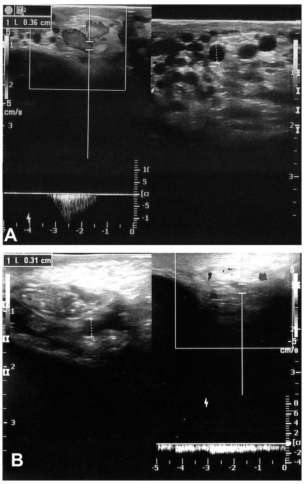
Diagnostic Color Doppler Sonography (CDS) of the scrotum in two patients (A and B) evaluated 6 months post-surgery for varicocele. Image A shows a grayscale ultrasound of the pampiniform plexus with multiple dilated, anechoic, round-to-ovoid venous structures. A caliper measurement indicates a vein diameter of 0.36 cm. The corresponding color Doppler and spectral waveform demonstrate significant retrograde flow (reflux) induced by the Valsalva maneuver, characteristic of recurrent varicocele. Image B displays a similar examination in a different patient, showing a measured venous diameter of 0.31 cm. Compared to A, the color Doppler signal in B is more sparse and intermittent, though still indicating persistent reflux. These images illustrate the use of ultrasound to identify clinical recurrence of varicoceles by measuring venous diameter and documenting the duration and velocity of Valsalva-induced reflux in the spermatic veins.
Varicocele
Definition
Epidemiology
| Population | Prevalence |
|---|---|
| General adult male population | 10-20% |
| Adolescent boys (<10 years) | Rare |
| Men with abnormal semen analysis | ~25-40% |
| Infertile men (bilateral) | Up to 20% |
| Healthy men (bilateral) | <10% |
- ~90% are left-sided (most consistent finding)
- If a left varicocele is identified, there is a 30-40% probability it is bilateral
- An isolated right-sided varicocele is extremely rare and warrants imaging to exclude a retroperitoneal mass or renal tumour
- Varicoceles do not spontaneously regress
Anatomy & Pathophysiology
Three Theories for Why Varicoceles Form
- Valvular incompetence - Absence or failure of the antireflux valve at the junction of the testicular vein with the left renal vein (or IVC on the right), causing retrograde blood flow
- Nutcracker effect - The left testicular vein is compressed between the superior mesenteric artery and the aorta, causing obstruction and venous congestion
- Angulation - Abnormal angle of the left testicular vein at its junction with the left renal vein increases resistance
How Varicocele Damages the Testis
- Reflux of renal or adrenal metabolites (cortisol, catecholamines) into the spermatic vein
- Increased oxidative stress and sperm DNA fragmentation
- Testicular hypoperfusion and hypoxia from venous stasis
- Pituitary-gonadal hormonal dysfunction
- Progressive testicular atrophy - in longstanding cases, the affected testis becomes smaller and softer
Grading (Clinical Classification)
| Grade | Finding |
|---|---|
| Subclinical | Not palpable or visible; detected only by Doppler US or thermography |
| Grade 1 | Palpable only during Valsalva manoeuvre; not visible |
| Grade 2 | Palpable at rest; not visible |
| Grade 3 | Visible and palpable at rest - classic "bag of worms" |
Clinical Features
- Dragging scrotal discomfort - typically on the left, worse on prolonged standing or at the end of the day, relieved by lying down
- The scrotum on the affected side hangs lower than normal
- "Bag of worms" - the classic description on palpation in the erect position; the plexus decompresses on lying supine
- There may be a cough impulse
- Longstanding cases: ipsilateral testicular atrophy (smaller, softer testis)
A varicocele that does not decompress in the supine position suggests obstruction of the testicular vein by a renal tumour or retroperitoneal pathology.
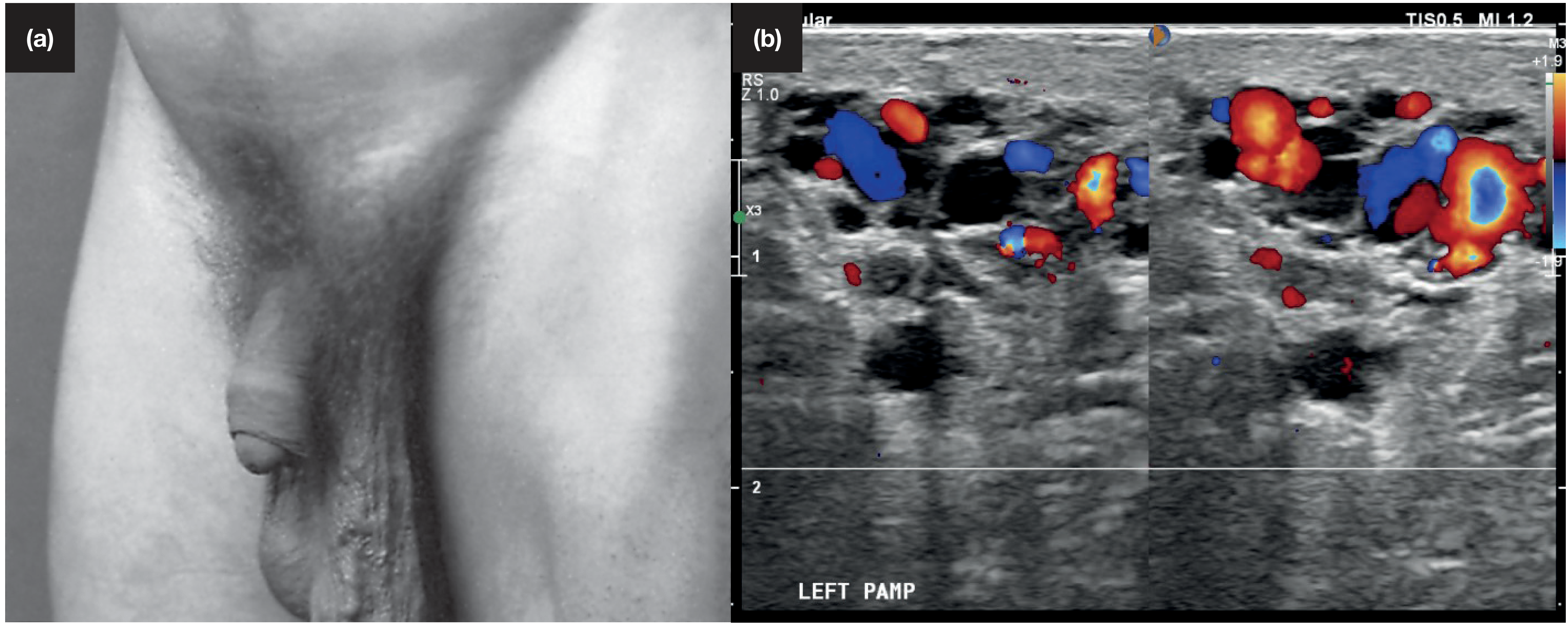
Clinical photograph and ultrasound
Investigations
Physical Examination (Gold Standard for Diagnosis)
- Examine standing; look and palpate
- Valsalva manoeuvre to reveal Grade 1
- Assess testicular size bilaterally (orchidometry)
Scrotal Doppler Ultrasound
- Indicated when clinical examination is equivocal, or for subclinical varicocele
- Diagnostic threshold: tortuous veins >3 mm in diameter
- Doppler confirms retrograde flow (reflux) on Valsalva
- Spectral Doppler: retrograde flow lasting >2 seconds on Valsalva is diagnostic
When to Image the Kidneys
- Isolated right-sided varicocele
- Older men with apparently recent onset
- Varicocele that does not decompress on lying down
- → Renal ultrasound / CT to exclude renal cell carcinoma or retroperitoneal lymphadenopathy
Semen Analysis
- Typically shows reduced concentration, motility, and morphology
- Motility deficits are often the most profound finding
- Baseline semen analysis is standard before any intervention for infertility
Varicocele and Infertility
- Found in ~40% of infertile men vs. 15% of healthy men
- Associated with progressive testicular damage from adolescence onward
- Effects on semen: reduced sperm count, motility, morphology, and increased DNA fragmentation
- A 2012 Cochrane review concluded that treatment in men with clinical varicocele and abnormal semen parameters in couples with otherwise unexplained subfertility may improve chances of spontaneous pregnancy
- Treatment of subclinical varicocele or when semen parameters are normal is considered ineffective for improving pregnancy rates
- Varicocelectomy may also improve outcomes following assisted reproduction (IVF/ICSI) in men with abnormal sperm parameters
NICE guideline note: Men should not be offered surgery for varicoceles as a form of fertility treatment purely to improve pregnancy rates. However, this is balanced against evidence supporting treatment when semen is abnormal.
Treatment
Indications for Intervention
- Significant scrotal discomfort
- Clinical varicocele + abnormal semen analysis + infertility (with normal female partner evaluation or in unexplained infertility)
- Adolescent with documented ipsilateral testicular atrophy (to prevent progressive damage)
- Bilateral clinical varicocele with impaired fertility
Options
| Method | Notes |
|---|---|
| Percutaneous embolisation | First-line treatment; day-case procedure via femoral or right jugular vein approach; selective catheterisation of testicular vein; venography confirms reflux; embolisation coils placed distally to proximally; ~20% recurrence rate |
| Microsurgical varicocelectomy (subinguinal) | Gold standard surgical technique; lowest recurrence and complication rates; use of operating microscope identifies and preserves the testicular artery and lymphatics; minimises risk of hydrocele formation and testicular artery injury |
| Open surgical ligation (inguinal/Palomo) | Older technique; ligates testicular veins at inguinal or retroperitoneal level; higher hydrocele formation rate (~7-33% without microscopy); recurrence possible |
| Laparoscopic varicocelectomy | Performed less often now due to refinements in subinguinal microsurgical technique; reasonable alternative in experienced hands |
Key Complications of Surgical Treatment
- Hydrocele formation (most common after non-microscopic varicocelectomy; 3-33%; caused by lymphatic obstruction) - largely eliminated by microsurgical lymphatic preservation
- Testicular artery injury - risk of testicular atrophy; minimised by microsurgery ± intraoperative Doppler
- Varicocele recurrence - 0.6-45% depending on technique; lowest with microsurgical approach
Summary (Key Points)
- Varicocele = dilated pampiniform plexus due to venous valvular incompetence; 90% left-sided
- Most common surgically correctable cause of male infertility
- Most are asymptomatic; symptomatic cases present with dragging discomfort and a "bag of worms"
- Grade 1-3 grading based on palpability/visibility; subclinical detected by Doppler only
- Doppler US: veins >3mm + Valsalva-induced reflux confirms diagnosis
- Isolated right-sided or non-decompressing varicocele → exclude renal tumour
- Treatment indicated for discomfort or infertility with clinical varicocele + abnormal semen
- Microsurgical subinguinal varicocelectomy is the most effective surgical technique
- Percutaneous embolisation is first-line minimally invasive treatment